Imaging - General Principles and Prework 1
1/58
There's no tags or description
Looks like no tags are added yet.
Name | Mastery | Learn | Test | Matching | Spaced | Call with Kai |
|---|
No analytics yet
Send a link to your students to track their progress
59 Terms
x ray beam source
patient
image receptor
What does production of a radiograph require?
Musculoskeletal imaging
the subspecialty of radiology concerned with the diagnostic evaluation of the musculoskeletal system
Radiograph
x-ray film containing an image of a part of a patient's body
X-ray
form of ionizing electromagnetic radiation, beam used to capture image, can not be seen
change in chemical properties of molecules in the tissues
How does tissue damage occur from radiation?
water molecule
What does radiation change to form a "free radical"?
DNA
What do free radicals have reactions with?
cell death
hereditary changes
cancer
What does damage to the DNA that cannot be readily repaired result in?
Attenuation
the degree to which the tissues absorbs or scatters the x-rays before they hit the recording medium (reductio in the number of x-ray photons in the x-ray beam as it passes through the body)
Fluoroscopy
dynamic/continuous real-time imaging
Computed radiography (CR)
Use of a phosphor screen (instead of film) to produce a digital image
Digital radiography
use an x-ray sensitive semiconductor material
Radiodensity
physical quality of tissue (or object) that determines how much an x-ray it absorbs
higher
If an object is thicker does it have a higher or lower radiodensity?
lower
If an object is thinner does it have a higher or lower radiodensity?
brighter
How do more radiodense objects appear in an image?
darker
How do less radiodense objects appear on an image?
Radiopaque
not easily penetrated by x-ray
Radiolucent
easily penetrated by x-ray
lighter
How do radiopaque objects appear on an image?
darker
How do radiolucent object sappear on an image?
black
What color is air on a radiograph?
gray-black
What color is fat on a radiograph?
gray
What color is water on a radiograph?
white
What color is bone on a radiograph?
trachea
lungs
stomach
digestive tract
Where is air normally seen on a radiograph?
around viscera and along muscle sheaths
Where is fat normally seen on a radiograph?
bright white outline
How does contrast media appear on a radiograph?
Arthrography
contrast enhanced image of a joint and soft tissue
Myelography
contrast enhanced image of spinal cord, nerve root and dura mater
projection/angle of the beam
What does form or shape of anatomic image depend on?
to have views in 3 dimensions
Why do you need more than one view of an image?
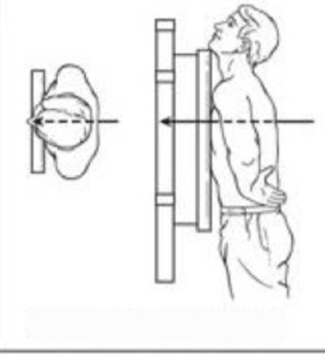
lateral
What projection path is this?
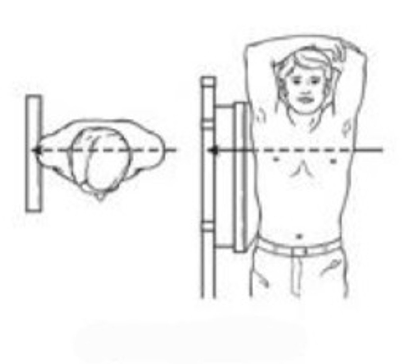
posteroanterior
What projection path is this?
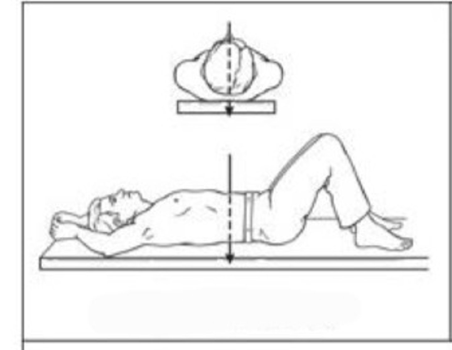
anteroposterior
What projection path is this?
Coronal/frontal plane
bisects the body from front to back dividing it into front and back sections
abduction and adduction
What movements occur in the frontal plane?
flexion and extension
What movements occur in the sagittal plane?
rotation
What movements occur in the transverse plane?
Sagittal plane
bisects the body from side to side, dividing it into left and right portions
Transverse/horizontal plane
divides the body horizontally into superior and inferior portions
Sagittal horizontal axis
Passes horizontally from posterior to anterior; formed by the intersection of the sagittal and transverse planes
Frontal horizontal axis
passes horizontally from left to right; formed by the intersection of the frontal and transverse planes
Vertical axis
passes vertically from inferior to superior (caudal to cranial); formed by the intersection of the sagittal and frontal planes
positive or negative for suspected clinical diagnosis
negative for suspected diagnosis, but raises index of suspicion for a different one
inconclusive
wrong
What are the potential results on a routine radiograph?
If you should take an image
What do the ottawa rules tell you?
What imaging is appropriate
What do the ACR appropriateness criteria tell you?
Upright position
seated or standing
Recumbent
lyding down in any position
Trendelenburg
Supine position with legs and feet elevated
Decubitis
both the body positioned on a horizontal surface and use of a horizontal x-ray beam
density
contrast
detail
distortion
What four factors indicate image quality on a radiograph?
air
What tissue is 1?

fat
What tissue is 2?

water
What tissue is 3?
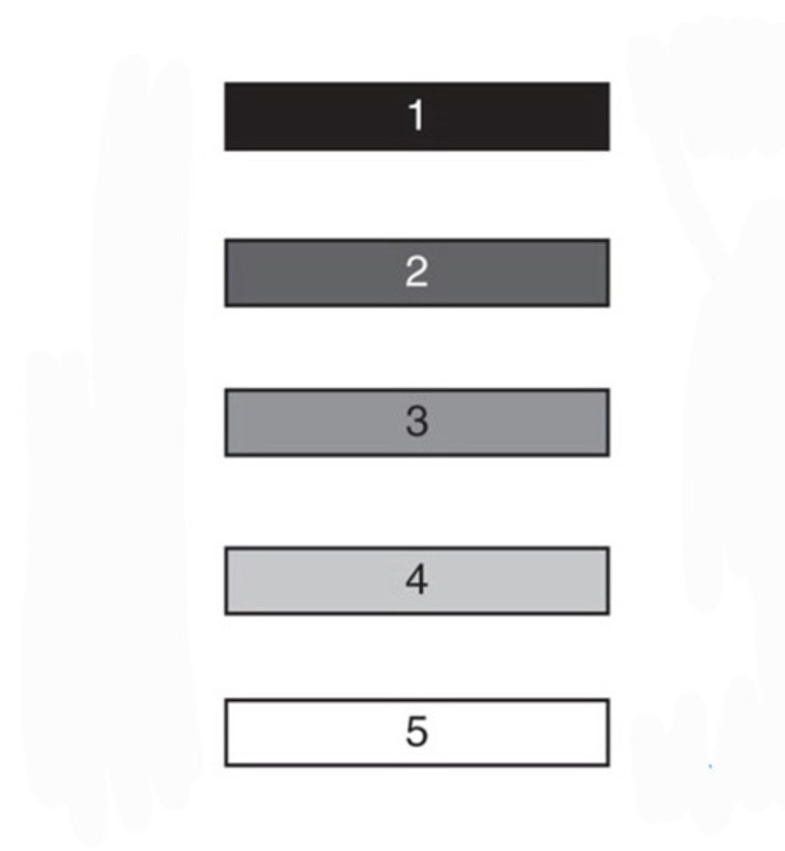
bone
What tissue is 4?

metal
What material is 5?

1
What number is the most radiopaque?

5
What number is the most radiolucent?
