Computed tomography CIT
1/29
There's no tags or description
Looks like no tags are added yet.
Name | Mastery | Learn | Test | Matching | Spaced | Call with Kai |
|---|
No analytics yet
Send a link to your students to track their progress
30 Terms
Tomography
Creating 2D images (slices) of a 3D object
CT scans
Measure the attenuation coefficients of X-rays as they pass through the body
Attenuation
X-rays are absorbed, scattered, or transmitted with lower energy as they pass through matter, resulting in a decrease in intensity
Types of attenuation
Compton scatter and photoelectric effect
3rd generation CT scanner
Wide fan beam with up to 1000 detector
Fast scanning - 0.5sec per slice
Back projection
process of mathematically mapping the attenuation pathway at every angle measured through a scan to locate where in a patient attenuation is occurring
If there is a dense object at a certain location in a patient then a projection at an angle in line with it will show us attenuation in this area because less photons will be arriving at the detector through this attenuation pathway
Slip ring technology
An electromechanical technology that allows transmission of power from a stationary to a rotating structure
Allow continuous gantry rotation
Helical scanning
Table moves continuously as the x ray tube rotates
Faster, smoother images, better 3D reconstructions
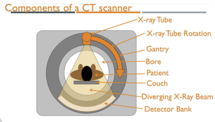
Components of a CT scanner
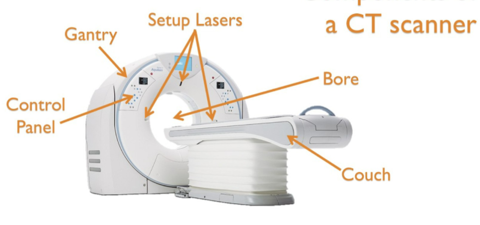
Components of a CT scanner
CT detectors
Measure X ray attenuation at multiple angles
Slice width is determined by size of detectors, a slice thickness of 3mm would need two 1.5mm detectors
Bowtie filters
Reduce radiation exposure by adjusting beam intensity for different body regions. Filters less in the centre and more around the edges
CT X ray tube
More powerful than conventional x-ray tubes
Operates at 80,100,120 and 140 kV
Needs replacing every 12 months
Exposure factors
kVp (Kilovolt peak): Controls x-ray penetration
mA (Milliamperes): Adjusts tube current based on body composition
Sequential (Axial) scanning (step and shoot)
X ray tube is off when the table moves
Moves in steps to capture each slice
Slower but precise
Advantages of helical scanning
Higher quality 3D imaging
Virtual procedures e.g bronchoscopy, colonography
Covers large areas quickly, reducing motion artefacts
Small pathologies unlikely to be missed
Better contrast enhancement
Reconstruct images at any angle
Pixels and voxels
CT images are 2D representations of 3D structures
Composed of pixels (2D) but represent voxels (3D, with depth)
Voxel depth corresponds to slice thickness
Voxels correspond to the location in computers, the brightness of each pixel correlates with the x-ray attenuation
Hounsfield units (HU)
Each voxel is assigned a value called HU or CT number
Water = 0 HU (calibration reference)
Tissues vary in HU based on density
Back projection
Raw CT data is reconstructed by summing back projected filtered attenuation profiles
Basis for image formation but needs filtering to remove blurring
Filtered back projection
Uses mathematical interpolation to reconstruct missing data
Smoothing is applied based on filtered rules
manipulation of data being reconstructed, which alters the value of a pixel and its neighbouring pixels relative to one another
FBP principles
if we want to see very sharp edges we would apply a sharpening filter, known as edge enhancement/high pass filter. This drives the value of each pixel away from the value of its neighbouring pixels.
Smooth filter, known as blurring or low pass filter does the opposite. Averaging pixels out with the value of its neighbouring pixels.
Iterative reconstruction
Repeats calculations multiple times to refine the image
Ensures image corresponds accurately to acquired projection data.
Advantage- reduces radiation dose up to 75%
Multiplanar reconstruction
Used to view the voxels in different angles
Axial , sagittal and oblique
Volume averaging is used to render the data
Signal to noise ratio
Signal= X-rays detected
Noise= variability in detector response, signal not supposed to be there gives a grainy appearance
High SNR= better image quality
Increasing tube current (mA) increases SNR
More tissue to go through= SNR is lower
Thicker slices= means higher SNR
High contrast resolution
Ability to distinguish tissues with small density differences
Major advantage of CT over x-ray
Improved by- Higher SNR, thicker slices (less noise, better contrast resolution)
IV contrast agents
Temporal resolution
Time needed to acquire data for a single image slice
Improved by faster gantry rotation time
Spatial resolution
Ability to distinguish fine details from the smallest distance. Two points the CT scanner can distinguish as individual objects.
Windowing in CT
CT images are displayed in grey scale (256 shades)
Window level (WL): Central HU value of displayed rannge
Window Width (WW): HU range displayed as greyscale
Examples of Windowing
Soft tissue (Abdomen) = 350/50 HU
Lung (Air density) = 1500/ -500 HU
Bone = 2000/250 HU
Multiplanar reconstruction (MPR)
CT images can be reconstructed in different planes-
Axial
Coronal
Sagittal