AUBF_Midterms_Lec: Microscopic examination
1/142
There's no tags or description
Looks like no tags are added yet.
Name | Mastery | Learn | Test | Matching | Spaced | Call with Kai |
|---|
No analytics yet
Send a link to your students to track their progress
143 Terms
Macroscopic screening: Color
Microscopic Correlations:
Microscopic Correlations: Blood
Macroscopic screening: Clarity
Microscopic Correlations:
Microscopic Correlations: Hematuria vs. hemoglobinuria/myoglobinuria
Macroscopic screening: Blood
Microscopic Correlations:
Microscopic Correlations: RBCs, and RBC Casts
Macroscopic screening: Protein
Microscopic Correlations:
Microscopic Correlations: Casts, Cells
Macroscopic screening: Nitrite
Microscopic Correlations:
Microscopic Correlations: Bacteria, WBCs
Macroscopic screening: Leukocyte esterase
Microscopic Correlations:
Microscopic Correlations: WBCs, WBS Cast, bacteria
Macroscopic screening: Glucose
Microscopic Correlations:
Microscopic Correlations: Yeast
Thomas Addis
he developed a quantitative method of examining urine sediment
Hemocytometer
•12-hr specimen
•RBCs, WBCs, casts and epithelial cells
Used to primarily monitor the course of diagnosed cases of renal disease
Preparation of the Urine Sediment, give criterias
Freshly voided urine
Midstream clean-catch specimen
10-15 mL (12 mL)
Centrifugation of urine
Centrifuge for 5mins @ 400 RCF
mL of urine sediment that was left after decantation
0.5-1.0mL
Explain examination of urine sediment
observe at 10 fields (both LPF and HPF)
Sternheimer-Malbin
Stain: WBC, Epithelial Cells, Casts
Sternheimer-Malbin Composition
Crystal Violet
Safranin O
Toluidine Blue
Stain:
enhances nuclear detail
differentiate WBC from RTE cells
2% Acetic Acid
Stain:
Lyses RBC and enhances nuclei of WBCs
Distinguish RBC from WBCs, yeast, oil droplets, and crystals
Lipid stains
Oil red O
Sudan III
Stain:
TAGs
Identify fat droplets and lipid-containing cells, and casts
Gram stain
Stain:
Diff. gram pos and gram neg bacteri
Identify bacterial casts
Hansel Stain
Stain:
uses Meth Blue and Eosin Y to stain eosinophilic granules
Acute Interstitial Nephritis (AIN)
This disease is determined thru the use of Hansel Stain
Prussian Blue stain
Stain:
identify yellow-brown granules of hemosiderin in cells and casts
Brightfield microscopy
common microsopic technique use droutine urinalssi
Phase-contrast microscopy
this microscopic technique is best used for low refractive indices
Hyaline cast
mixed cellular casts
mucus threads
Trichomonas vaginalis
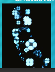
Polarized light microscopy
Cholesterol
microscopic technique to check form Maltese cross formation which indicates what metabolite
Type of microscopy used depends on (give 3)
Spx type
Refractive index of object
Imagery of unstained living cells
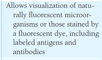
Fluorescence microsocpy
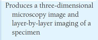
Interference-contrast
RBC Appearance in hypertonic urine
Crenated apperance
RBC Appearance in hypotonic urine
Ghost cells
Appearance of RBC with glomerular membrane damage
Dysmorphic appearance
Sources of identification error for RBCs
Yeast cells
Oil droplets
Air bubbles
RBCs
These sediments appear with conditions such as glomerulonephritis and strenuous exercise
Correlations with RBCs
Color
Reagents strip blood reaction
WBCs: in hypotonic urne
Glitter cells
WBC: Sources of identification error
Renal tubular epithelial cells
WBC: Urinalysis correlation
LE
Nitrite
Specific Gravity
pH
Squamous Epithelial cells
Largest cells that can be seen in microscopic exam
Squamous Epithelial cells
These are the cells used for point of reference whenever we do microscopic examination
increased no. of SEC inidcates what?
contam of urine specimen due to poor collection technique
RTE Cells
Leukocyte esterase and nitrite (pyelonephritis)
Color
Clarity
Protein Bilirubin (hepatitis)
Blood
Transitional cells: Urinalysis correlations
Clarity
blood (malignancy)
Transitional cells: Clinical significane
increased nos. indicate
UTI
Renal carcinoma
catheterization
RTE Cells: Appearance
Rectangular, columnar, round, oval, or cuboidal with an eccentric nucleus possibly bilirubin stained or hemosiderin laden
RTE Cells: Clinical significane
tubular necrosis
(to add more)
Oval Fat Bodies: Urinalysis correlations
Clarity
Blood
Protein
Free fat droplets/Fatty Casts
Oval Fat bodies: Clinical significance
Increased in glomerular damage by:
nephrotic syndrome,
tubular necrosis,
diabetes mellitus, long bone trauma
Bacteria: Appearance
Small spherical and rod-shaped structures
Bacteria: ID errors
Amorphous phosphates
Amorphous urates
How are Bacteria reported in urinalysis
R,F,Mod,Many (HPF)
note: Presence of WBCs may be required
Bacteria: Urinalysis correlation
pH
Nitrite
LE
WBCs
Yeast: Apperance
Small, oval, refractile structures with buds and/or mycelia
Yeast: ID Error
RBCs
Yeast: Correlation
Correlated with:
Glucose
LE
WBCs
Parasites (trichomonas)
Pear-shaped, motile, flagellated
Parasites: ID error
Errors:
WBCs
RTE cells
Parasites (Trichomonas): Correlation
Correlation
LE
WBCs
Enterobius vermicularis ova
This parasite is accidentally found due to fecal contamination
Spermatozoa: appearance
Tapered oval head with long, thin tail
not required
spermatozoa is (required/not required) to be reported based on lab protocols
Spermatozoa: correlation
Corrleation
Protein
Mucus threads
Single or clumped threads with a low refractive index
Mucus threads: ID Error
Error
Hyaline Casts
How can we differentiate RTE cells and WBC?
Based on the presence of protein in Chem exam
RTE Cells = (+) Proteins
WBC = (-) Proteins
Where are urine casts formed
Lumen of Distal Convoluted tubule and collecting duct
Casts are usually observe in what objective? Manner of reporting?
LPF detected around the cover slip edge
Tamm-Horsfall protein/Uromodulin
these are proteins that are responsible in forming casts
released by the cells in the Renal Tubules
•Substances that can become embedded in casts include (give 5)
Cells
Bacteria
Granules
Pigments
Crystals
Hyaline Cast
_:Appearance | Colorless homogenous matrix |
Hyaline Cast
_:ID Error | Mucus, fibers, hair, increased lighting |
Hyaline Cast
_: Correlation | • Protein -Blood (exercise) • Color (exercise) |
Hyaline Cast
_:Clinical significance | Glomerulonephritis Pyelonephritis Chronic renal disease Congestive heart failure (CHF) Stress and exercise |
RBC Cast
_:Appearance | Orange-red color, cast matrix containing RBCs |
RBC Cast
_:ID Error | RBC clumps |
RBC Cast
_: Correlation | A cast: • RBCs • Blood • Protein |
RBC Cast
_:Clinical significance | A cast: Glomerulonephritis Strenuous exercise |
WBC Cast
_:Appearance | Cast matrix containing WBCs |
WBC Cast
_:ID Error | A cast: WBC clumps |
WBC Cast
_: Correlation | A cast:
|
WBC Cast
_:Clinical significance | A cast:
|
Bacterial Casts
_:Appearance | A cast: Bacilli bound to protein matrix |
Bacterial Casts
_:ID Error | A cast: Granular casts |
Bacterial Casts
_: Correlation | A cast:
|
Bacterial Casts
_:Clinical significance | A cast:
|
Epithelial Cell Cast
_:Appearance | RTE cells attached to protein matrix |
Epithelial Cell Cast
_:ID Error | A cast: WBC cast |
Epithelial Cell Cast
_: Correlation | A cast: Protein RTE cells |
Epithelial Cell Cast
_:Clinical significance | A cast: Renal tubular damage |
Fatty Cast
_:Appearance | Fat droplets and oval fat bodies attached to protein matrix |
Fatty Cast
_:ID Error | A cast: Fecal debris |
Fatty Cast
_: Correlation | A cast:
|
Fatty Cast
_:Clinical significance | A cast:
|
Granular Cast
_:Appearance | Coarse and fine granules in a cast matrix |
Granular Cast
_:ID Error | A cast:
|
Granular Cast
_: Correlation | A cast:
|
Granular Cast
_:Clinical significance | A cast:
|
Waxy Cast
_:Appearance | Highly refractile cast with jagged ends and notches |
Waxy Cast
_:ID Error | A cast: Fibers and fecal material |
Waxy Cast
_: Correlation | A cast:
|
Waxy Cast
_:Clinical significance | A cast:
|