Physio. Ch.12 Muscles
1/58
There's no tags or description
Looks like no tags are added yet.
Name | Mastery | Learn | Test | Matching | Spaced | Call with Kai |
|---|
No analytics yet
Send a link to your students to track their progress
59 Terms
Intercalated discs
Junctions between cardiac muscle cells
Allows cardiac muscle cells to contract as one unit
Triad
Unit of a muscle
Made of T tubule + terminal cisternae
Transverse (T) tubule
Continuous with sarcolemma and penetrate into muscle fiber
Sarcolemma
Transparent tubular sheath that surrounds the fibers of skeletal muscles
Terminal cisternae
Enlargements of sarcoplasmic reticulum, store Ca
Neuromuscular Junction
Excitatory synapse between neuron and skeletal muscle
Comprised of
- Synaptic bulb of motor somatic neuron
- Synaptic cleft
- Motor end plate
NT used: ACh
Receptor used: Nicotinic
Motor Unit
A single motor neuron and all the muscle fibers it innervates
- Average 200 muscle fibers innervated by 1 neuron
Acts as a single functional unit
Types
- Small: few muscle fibers per motor neuron, used when fine muscle control needed
- Larger: high number of muscle fibers per motor neuron, used when large amounts of strength needed
Graded contractions
Varied contraction strength due to different numbers of motor units being stimulated
Sarcomere
Functional unit of muscle
Made of
- Thin myofilaments
- Thick myofilaments
- Titin filaments
- Cross bridges
- Z discs
- A band
- H band
Thin filament
Made of
- Actin: Building block
- Tropomyosin: Covers binding sites on actin
- Troponin complex: Moves Tropomyosin off of binding site
Thick filament
Has a bare sight in the middle of the filament
Made of
- Myosin: Building block
- Tail: Sight for myosin on myosin binding
- Head: Holds ATP and actin binding stie
- ATP site: Acts as binding sight for ATP
- Actin binding sight: Acts as binding sight for actin
Sliding Filament Model
Model of muscle contraction
Muscle contracts through filaments sliding past one another
- Thin filaments pulled towards middle of sarcomere, the M line
Powered by The Crossbridge Cycle
The Crossbridge Cycle
Cycle that powers sliding filament model
- Step 1: ATP binds to myosin causing myosin and actin unbind
- Step 2: Myosin turns ATP into ADP and phosphate, storing both
- Step 3: Myosin is in it’s high energy form and cocks back
- Step 4: Myosin and actin bind together
- Step 5: Phosphate is released from myosin which causes a power stroke
- Step 6: ADP is released
- Step 7: New ATP binds to myosin head
Excitation
Stimulation of a Muscle Fiber
Step 1: AP gets to motor neuron and causes the resale of ACh
Step 2: Nicotinic receptors receive ACh
Step 3: End plate potential generated, will always reach threshold
Excitation-Contraction Coupling
The sequence of events that links the action potential in a muscle cell to its contraction
T tubules and SR membranes are physically linked through DHP receptors and Ryanodine receptors
- Action potential in T tubules is able to affect Na channels in SR
DHP receptors
Receptors responsible for linking sarcolemma and t tubule
Located in T tubule
Connected to Ryanodine receptors
Ryanodine receptors
Receptors responsible for linking sarcolemma and t tubule
Calcium channel located in sarcolemma
Connected to DHP receptor
Calcium in muscle contraction
Bind to troponin complex
- Configuration changes, moving tropomyosin off of myosin binding sights
Ca must be actively transported back into sarcomere
Relaxation
Motor neuron stops stimulating muscle cell with action potential
Ca is actively transported back into the sarcolemma
Troponin and tropomyosin move to original positions, covering myosin binding sights
Muscle Twitch
Mechanical response of an individual muscle cell, motor unit, or whole muscle to a single action potential
Reproducible, all or nothing event
Varies from cell to cell
Phases
- Latent period
- Contraction phase
- Relaxation phase
Latent period
Phase of a muscle twitch
Delay between the muscle cell’s AP and the beginning of muscle contraction
Causes by events of excitation-contraction coupling needing to be done
- Calcium release & binding to troponin
Contraction phase
Phase of a muscle twitch
Time when muscle tension/force is increasing
Cross bridge cycle happens in this time
Relaxation phase
Phase of a muscle twitch
Time when muscle tension decreases back to zero
Ca2+ actively moved back into sarcolemma
Isotonic contraction
Muscle generates a constant tension just greater than any forces opposing it
Tonic” = tension
Subdivided into two forms
- Concentric
- Eccentric
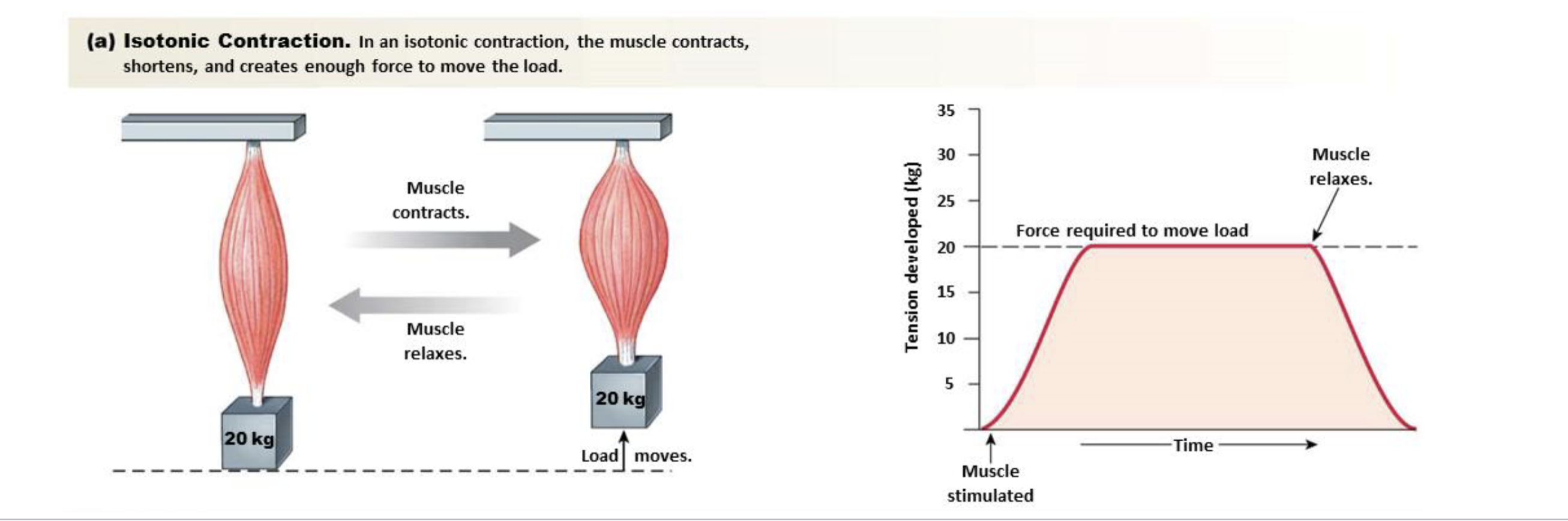
Concentric
Type of Isotonic contraction
Muscle length shortens
Eccentric
Type of Isotonic contraction
Muscle length increases
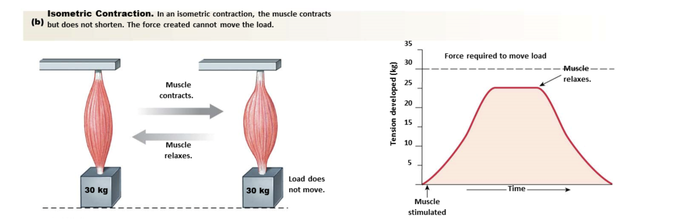
Isometric contraction
Muscle creates tension but maintains the same length
“Metric” = length
Load is slightly greater than force of muscle contraction
Force Generated by a Muscle
Determined in a muscle by the number of muscle fibers contracting
Determined in an individual fiber by number of myosin binding sites on actin that are exposed (active crossbridges)
- Frequency of stimulation determines number of active cross bridges
- Fiber diameter determines number of active cross bridges
- Changes in fiber length determines number of active cross bridges
Frequency of Stimulation
Rate of calcium release into cytosol exceeds rate of active transport back into SR
- More exposed myosin binding sites on actin allowing more crossbridges formation
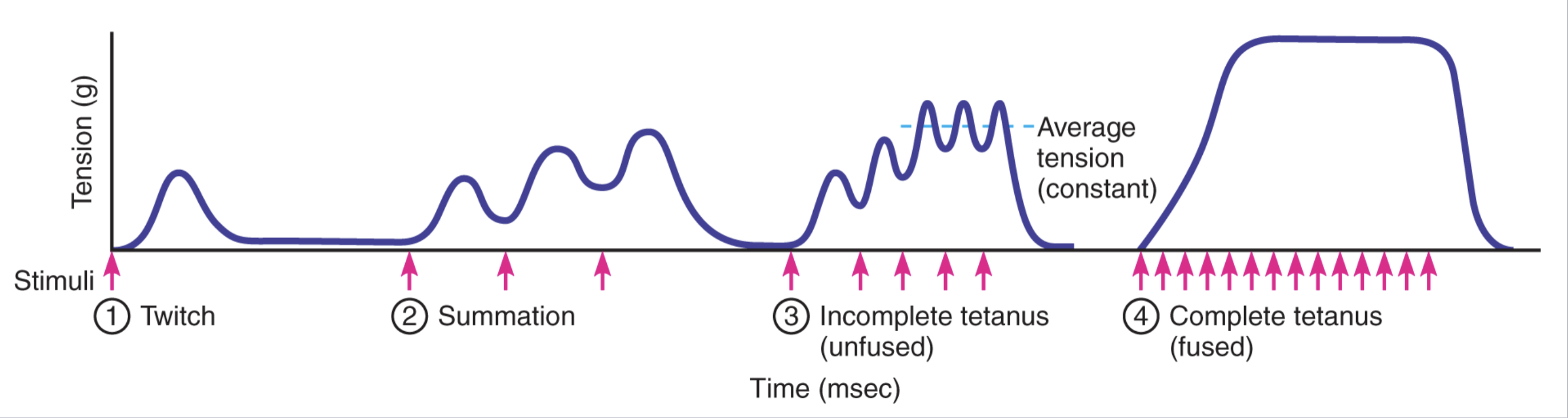
Summation
Twitches add together when a muscle is stimulated at a high frequency
Causes by multiple AP arrive before twitch complete
Tetanus
Peak summation, maximal sustained contraction
4x to 5x stronger than a twitch
Subdivided
- Incomplete Tetanus
- Complete Tetanus
Incomplete Tetanus
Brief periods of relaxation between twitches
- Peaks are when calcium levels saturate troponin
Complete Tetanus
A smooth, sustained contraction, no relaxation exist
- Enough calcium to continuously saturate troponin
Fiber Diameter
The diameter of a muscle fiber
Larger = more sarcomeres arranged parallel to one another
Creates a greater force
Changes in Fiber Length
Effects the force produced by one fiber
Optimal ranges between 100 and 120
- Muscles try to operate in this range for as long as possible
Regulation of Muscle Force
Amount of active motor units
- Primary way muscle force is regulated
Recruitment: act of increasing the number of active
motor units
Muscle cell energy generation
Constant ATP needed for muscle contraction
Sources of ATP
- Oxidative phosphorylation of ADP in mitochondria
- Phosphorylation of ADP by creatine phosphate
- Anaerobic glycolysis
Oxidative phosphorylation of ADP in mitochondria
Main source of ATP production
Fueled by glucose from muscle/liver or by fatty acids
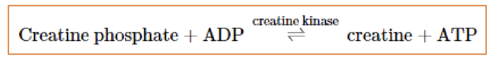
Phosphorylation of ADP by creatine phosphate
Secondary source of ATP production
Transfer of a high-energy phosphate to ADP
Active when muscle at rest
Can supply 40 ATP, 5x the normal 8 ATP stored in a muscle
Creatine produced by the liver and kidneys or obtained in the diet
Law of mass action
- Use of ATP drives the reaction to the right
- At rest, creatine phosphate is replenished
Anaerobic glycolysis
Secondary source of ATP production
Turns pyruvate to lactate
Light exercise energy production
Oxidative phosphorylation
Moderate & heavy exercise energy production
Cells first use up glycogen stores
Then use blood glucose (first ~30 minutes)
- As exercise intensity and duration increase, more GLUT-4 transporters are inserted into the sarcolemma
Then fatty acids
Anaerobic glycolysis is especially important in heavy exercise (pyruvate buildup is converted to lactic acid)
Classification of Skeletal Muscle Fibers
Two major categories
- Speed of contraction: time to reach peak tension
- Primary mode of ATP production
Speed of contraction
Classification of skeletal muscle fibers
Reffers to time to reach peak tension
Subdivided
- Slow switch: slow myosin
- Fast twitch: fast myosin
Primary mode of ATP production
Subdivision used to classify Skeletal Muscle Fibers
Subdivided
- Glycolytic
- Oxidative
- Myoglobin
Glycolytic
Fibers that primary energy source is this have high concentrations of glycolytic
enzymes (glycolysis), few mitochondria
Oxidative
Fibers that primary energy source are high in mitochondria concentration and capillaries
- Low low concentrations of glycolytic
- Contain myoglobin
Myoglobin
Oxygen binding protein
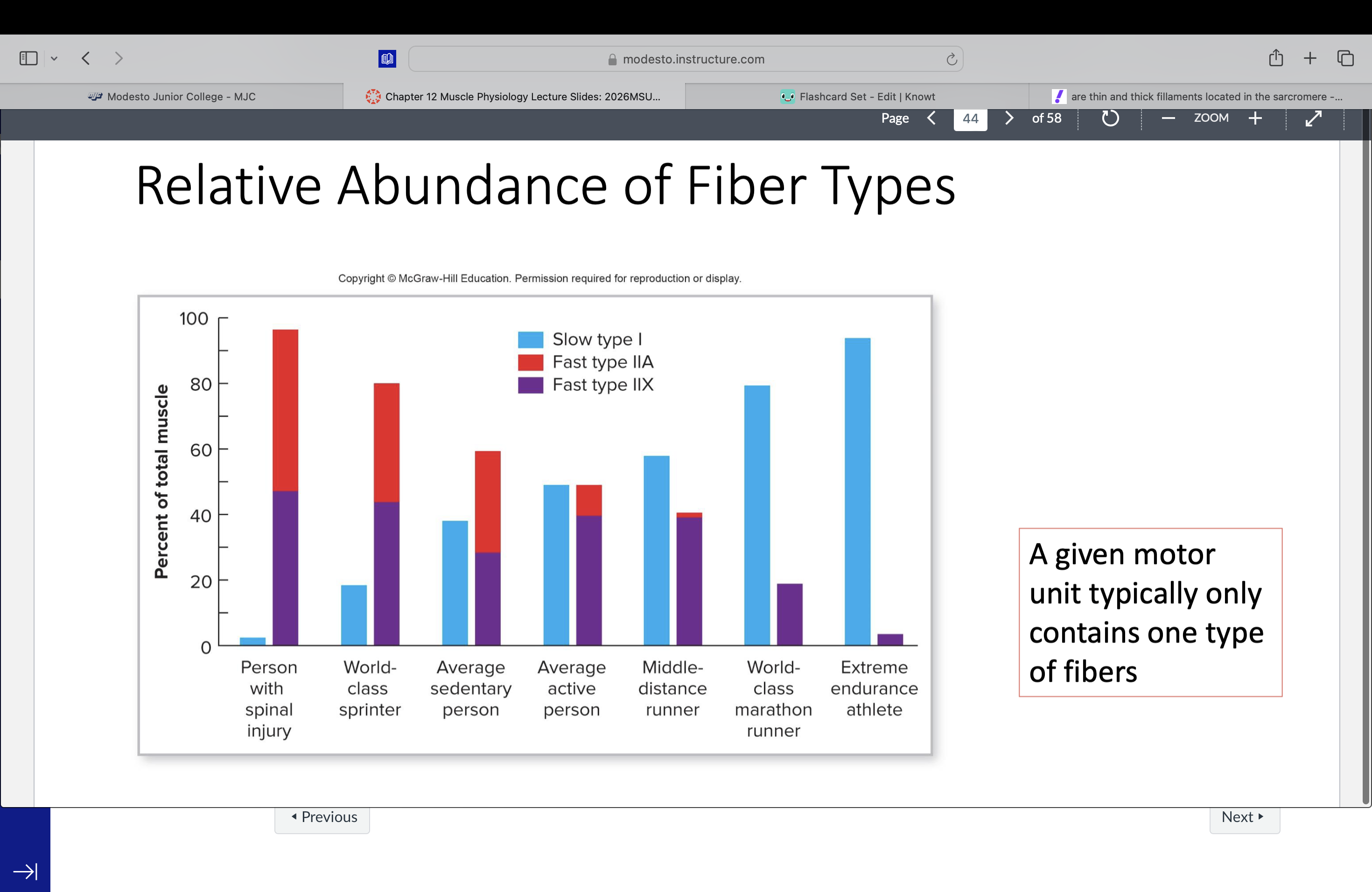
Major Types of Muscle Fibers
Slow oxidative
- slow myosin + produce most ATP by oxidative phosphorylation
- small diameter
Fast glycolytic
- fast myosin + produce most ATP by glycolysis
- large diameter
Fast oxidative (rare)
- ‘fast’ myosin + produce most ATP by oxidative phosphorylation
- Intermediate diameter
Muscle Fatigue
Decline in a muscle’s ability to generate force
Causes
- Depletion of stored glycogen
- Lactic acid accumulation (lowers pH → altered enzyme
activity)
- Interruption of blood flow due to strong contractions
- Accumulation of extracellular K+, reducing membrane
potential
- Neuromuscular fatigue: depletion of synaptic terminals of
ACh
- Fatigue of upper motor neurons in the CNS (Central fatigue)
Cardiac Muscle Cells
Seen in heart
Involuntary control
Striated like skeletal muscle
- Use sliding filament method like skeletal muscle
Connected by intercalated discs
Has gap junctions
- Allowed electrical synapses
Electrical synapses
Gap junctions allow AP to travel through entire cell network
Muscle fibers contract as one unit
Long AP durations stops summation when not wanted
Pacemaker controls how many AP are sent out
Smooth Muscle Cells
Involuntary control
Regulated by ANS
Short cells with one nucleus
Nonstriated, no sarcomere
- Contraction occurs along several axes
Electrical synapses present
Smooth Muscle Contractile Apparatus
Myosin filaments are stacked vertically and can form cross bridges with actin
Entire thick filament covered with myosin heads
Myofilament arrangement allows contraction even when greatly stretched
Excitation-Contraction Coupling Mechanism in Smooth Muscle
Step 1: Depolarization causing Voltage-gated Ca2+ channels in PM and SR open.
intracellular calcium level raise
Step 2: Calcium binds to calmodulin causing a conformation change
Step 3: Activates myosin light chain kinase (MLCK)
Step 4: MLCK phosphorylates myosin light
chains activate Myosin crossbridges and Crossbridge cycling starts
Step 5: Muscle contracts
Smooth Muscle Relaxation
Relaxation when Ca is pumped out by the ATP pump
Myosin phosphatase dephosphorylation myosin
Regulation of Smooth Muscle Contraction
Done by NE, ACh, paracrine or stretching of cell
Regulated by both branches of ANS
- Effects of either branch may be excitatory or inhibitory
Single-unit smooth muscle system
Multiple gap junctions that allows cells behave as a unit
Most smooth muscles
Multiple-unit smooth muscle system
Few or no gap junctions → cells require individual nerve innervation