Day 4: Visual Processing
 Call Kai
 Learn
Learn Practice Test
 Spaced Repetition
 Match
Match Flashcards
 Knowt Play
1/25

There's no tags or description
Looks like no tags are added yet.
Name | Mastery | Learn | Test | Matching | Spaced | Call with Kai |
|---|
No analytics yet
Send a link to your students to track their progress
26 Terms
Where are the photoreceptors located
what are photoreceptors and what do they do?
What is a process that they do?
in the retina
They are light-sensitive neurons that convert light energy into electrical energy in cells.
The conversion is called phototransduction.

Two types of photoreceptors in the retina
how many of each
What are they?
How do they respond to stimuli (what does the photoreceptors do next right after receiving stimuli)
Each retina contains ~6 million cones and 120 million rods.
Rods and cones are neurons, though they do not fire action potentials, but instead respond to stimuli with graded membrane potentials.
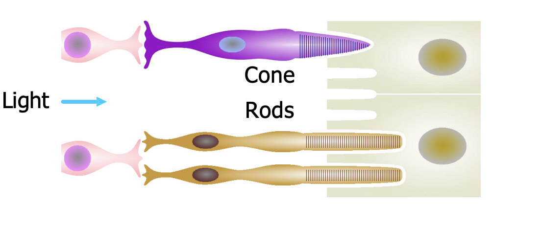
Basic structure of cones and rods
outer segment structure
In the outer segment, the membrane folds into disk-like layers which contain the visual pigments that respond to light.
In the inner segment are the nucleus and organelles for protein synthesis; and in a basal layer (base of the photoreceptor cell), a synapse that releases glutamate.
Both receptor types point toward the back of the eye.
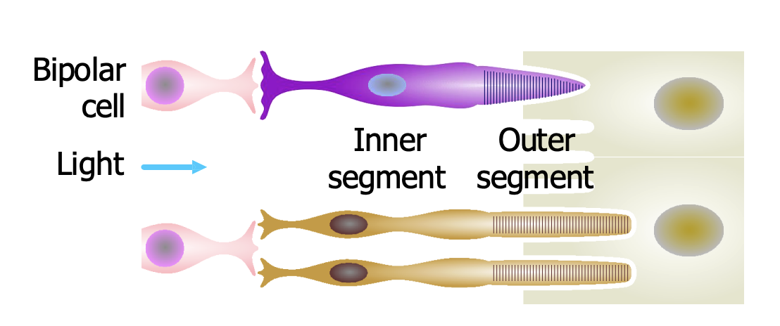
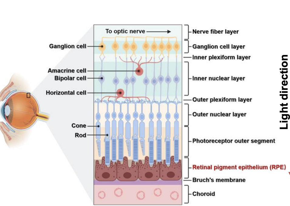
The retinal architecture of the inner layer of the eye
what do we have in the inner layer of the eye that prevents us from seeing a particular region in out vision
Explain why do we have this
What does it not have in this region
What are muller glial cells
What does it act like
Two functions it plays
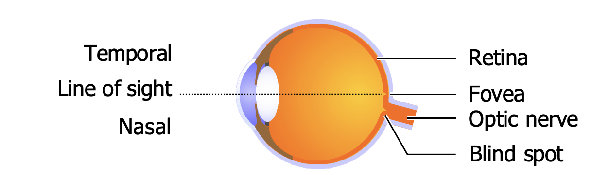
• Remember we do have a blind spot — a region where the optic nerve exits the eye and there are no photoreceptors.
• Müller glial cells play a crucial optical role. They span the entire retina and actually act like optical fibers, guiding light through the neural layers and reducing light scatter
Photoreceptors detect light using membrane-bound visual pigments
what happens when light hits the pigments
What happens to glutamate
When are photoreceptors more active
Number of type of pigments in rod vs cone
When light hits them, pigment molecules change shape, starting a chemical cascade that hyperpolarizes the cell, reducing its release of glutamate, i.e. photoreceptors are more active in darkness.
Each photoreceptor contains millions of molecules of its pigment, but each type of photoreceptor has just one type of pigment: rhodopsin in rods, 3 other pigments in 3 types of cone.
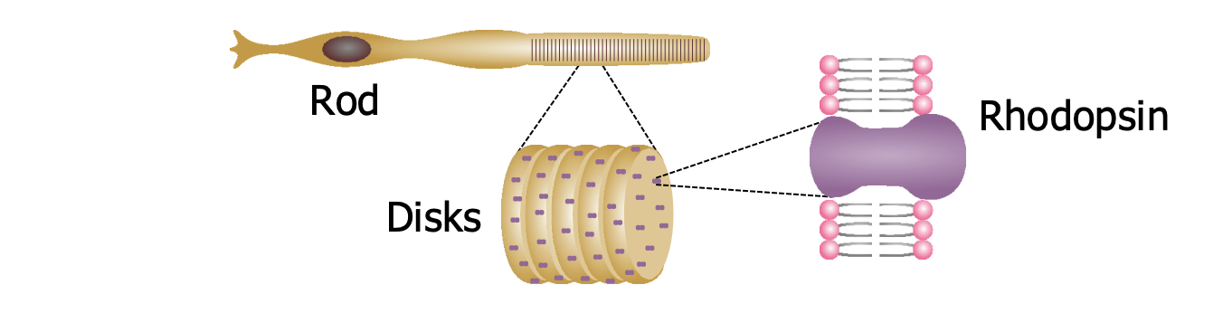
Type of pigment in rod
Rhodopsin
Phototransduction- Turning Light Into Signals
precursor for retinal
Why is it important for pigment
What is Nyctalopia
Vitamin A is the precursor for retinal.
Without it: rhodopsin cannot regenerate, rods remain inactive
This leads to nyctalopia (night blindness)- impaired vision in low-light conditions.
Dark vs light
when is cones used
when is rods used
Sensitivity level of both
How are each one deactivation in their opposite conditions
Cones are for bright light, rods for dim
Cones are less sensitive than rods; they are responsible for vision in bright light and for distinguishing colors, but they don’t operate in dim conditions.
Rods can detect single photons (more sensitive). But they operate only in low light: in daylight they are “bleached out” , i.e. their rhodopsin is broken down so they can’t sense light.
When the lights go dim the rods dark adapt, i.e. they rebuild their stores of rhodopsin over ~30 minutes.
How photoreceptors are distributed
where are most of them found (both cones and rods)
The general area - name it
The very dense spot in that area - name it
Blind spot - why does this exist
How com we dont notice it?
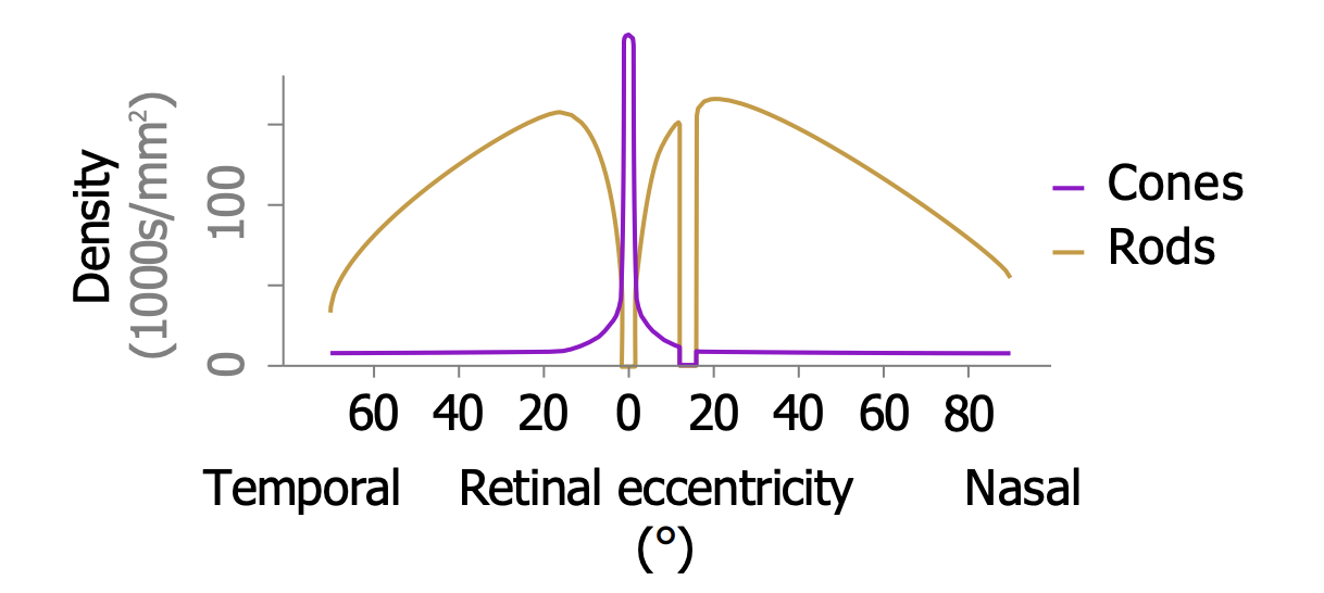
They are most densely packed in the macula, a central disk 5.5 mm across, and especially in its central pit called the fovea, 1.5 mm across (2° of visual angle — about as wide as 2 fingers viewed at arm’s length).
We use the fovea for detailed vision. 5° away from its center, acuity is quartered; at 20° it falls below the standard for legal blindness.
There are no receptors in the blind spot — the hole where axons carrying visual information exit the eyeball to form the optic nerve.
Normally you don’t notice your blind spots because objects in one eye’s blind spot aren’t in the other eye’s, and because the brain fills in the gaps.
Cones and rods are distributed differently
what does the fovea have more of
what receptors does the rest of the macula have more of?
The fovea contains almost exclusively cones. More-peripheral retina contains mainly rods.
That is one reason your peripheral retina is more sensitive to light than your fovea is; e.g. you can see a dim star better by looking slightly away from it.
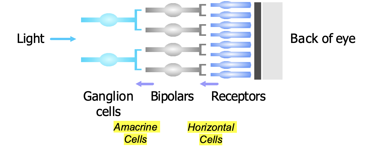
The 3 Layers of Neurons in the Retina
Receptors
Bipolar cells
Ganglion cells
Up to 45 photoreceptors may converge on a single bipolar cell. Bipolars in turn converge on ganglion cells, so in each eye the signal from 126 million receptors is condensed into 1 million ganglion cells.
Convergence is greatest in the peripheral retina and least in the fovea, where some receptors project 1:1 to bipolars.
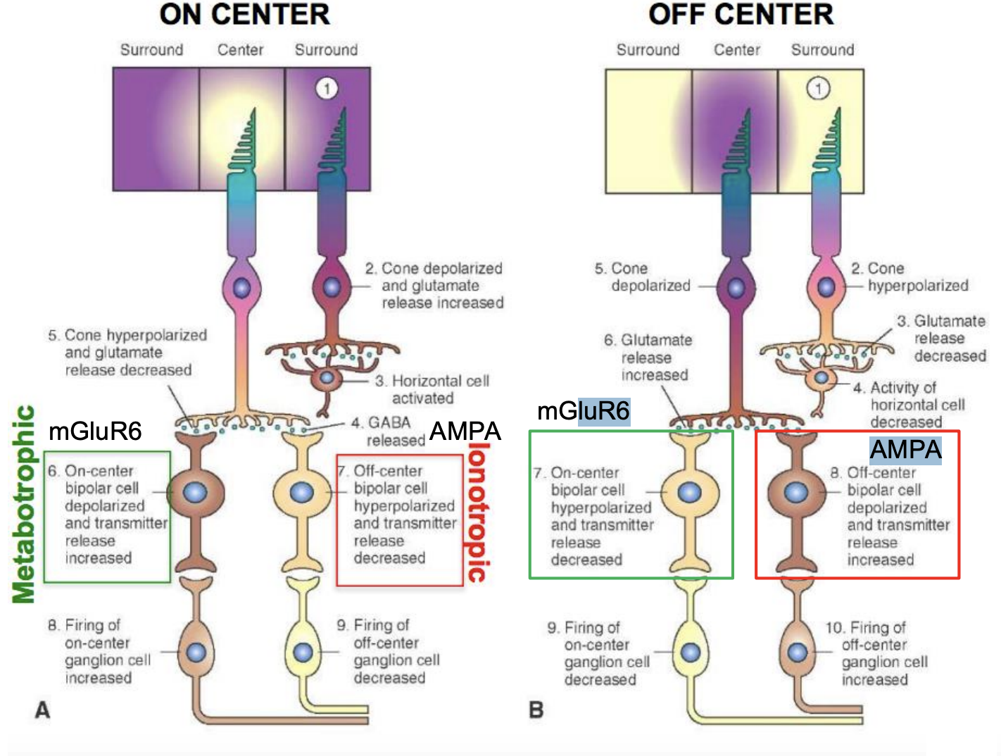
Two kinds of receptive fields for bipolar cells
draw the two parts and name them
Explain what on-center is and what off center cells is
Explain area of sensitivity and pathways that are affected (metabotropic vs ionotropic pathways and what receptor for each pathway - name them)
Bipolar cells have center-surround receptive fields
Every neuron in the visual system has a receptive field, also called its visual field — the region of the retina where light affects the cell’s activity, i.e. the set of photoreceptors which affect the cell.
Bipolar cells have center-surround fields, with a round center region and a doughnut-shaped surround.
On-center cells are excited by light in the center of their field, and inhibited by light in the surround. So these cells respond most when a light spot fills their center and the surround is dark.
Off-center cells are inhibited by light in the center, and excited by light in the surround. They respond best when a dark spot fills their center and the surround is light.
What type of light do bipolar cells react to?
explain why they aren’t activated in some cases
Explain how they activate when they react to ___
Firing method
React to contrast
When the lighting is uniform, whether bright or dim, neither type of bipolar cell responds, because the effects of the center and surround cancel, leaving the cell at its resting level of activity (only weakly active).
In other words, bipolar cells react strongly only when the lighting is not uniform, i.e. they respond to contrast.
They respond with graded membrane potentials; they do not fire action potentials.
retinal ganglion cells
type of firing of receptors
similarity to bipolar cells
Two things
Type of cells
What they detect
Fires action potential
But like bipolar cells, most retinal ganglion cells also have center surround receptive fields, and these fields can also be on-center or off-center.
Detect contrast
Ganglion cells in different parts of the retina have different-sized receptive fields
near the fovea vs in the peripheral
number of bipolar cells converging
Level of sensitivity
level of spatial resolution
A ganglion cell near the fovea gets input (via bipolars) from only a few photoreceptors, mostly cones. Farther out, each ganglion cell gets input from many receptors (up to 75,000), mostly rods.
So in the periphery, each ganglion cell is very sensitive to light but poor at reporting spatial detail because it blends information from a wide swathe of receptors.
And near the fovea, ganglion cells are less sensitive to light but have better spatial resolution because each one gets input from just a few densely packed cones.
Ganglion cells
types (in terms of classification)
M cells - full name - what. it detects
P cells - full name - what it detects
Melaonpsin - what does it have - what do they do to the brain
Percentage of ganglion cells that are M cells, P cells, Melanopsin cells
Large, magnocellular ganglion cells, or M cells, provide information that is used by the brain to infer the movement of objects. These cells are phasic. ~10% of retinal ganglion cells are M.
Small, parvocellular ganglion cells, or P cells, provide information that is used to infer form and fine detail, such as texture. ~70% of retinal ganglion cells are P.
~1% of retinal ganglion cells are melanopsin ganglion cells, which are photoreceptors, with their own visual pigment, melanopsin. They project to the suprachiasmatic nucleus, a center for circadian rhythms.
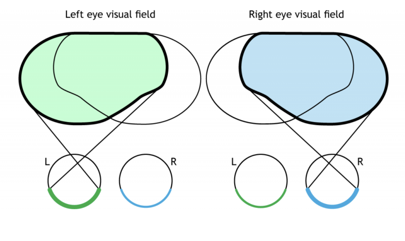
Full visual field involves…
explain each individual field
two eyes
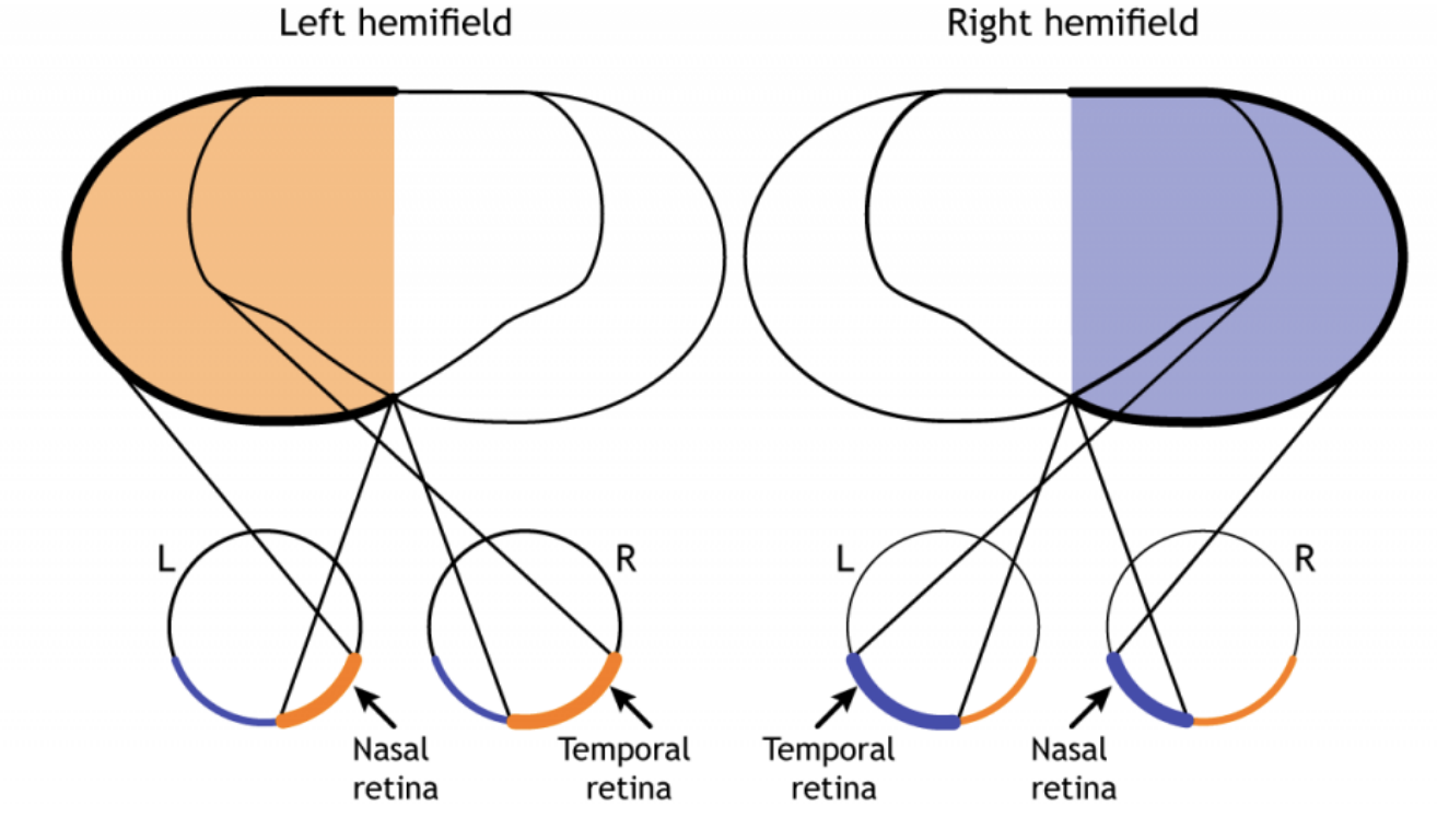
left hemifield involves which part of each eye
Right hemifield involves which part of each eye
hint: think of the located toward the nose vs away from the face
the section of each eye will be the same side
left hemifield - right side of each eye
left eye: nasal retina - more closer to the nose
right eye: temporal retina - away from the nose
Right hemifield - left side of each eye
Left eye: temporal retina - away from the nose
eight eye: nasal retina - closer to the nose
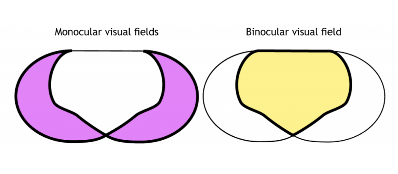
What field is the monocular visual field/central overlap
draw location in the field of the general eyes
what is it helpful for in vision
What field is the binocular visual field
draw location in the field of the general eyes
what is it helpful for in vision
Central overlap/binocular visual field is essential for depth perception and 3D spatial awareness
the monocular zones extend your peripheral awareness
Where doe the visual info from the retina reach what type of nerves
two names for it
Where is these kind of nerve located in the eye - what is this located called
Visual information leaves the retinas in the optic nerves
Each of the million ganglion cells in each retina sends its axon out the back of the eye through the blind spot.
These million fibers from each eye form its optic nerve, also known as cranial nerve II.
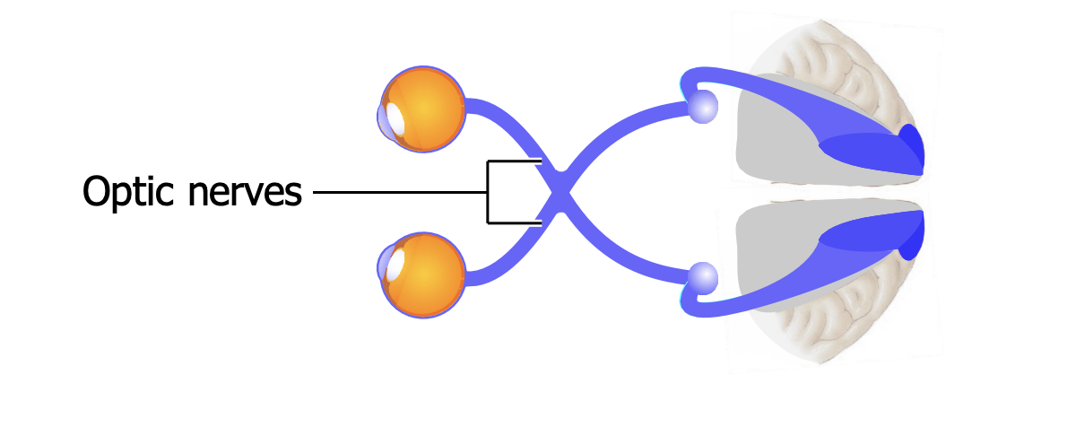
Where in the nerve does half of the nerve fibers split in two separate ways (for left and right hemifield information)
draw a diagram if needed
In what cerebral hemisphere do each group go to (draw if needed)
the optic chiasm
In the eye, the right side of the scene (the right visual hemifield) projects onto the left side of each retina, i.e. onto the nasal side of the right retina and the temporal side of the leftretina.
Because the nasal fibers cross, all the information from the right hemifield comes together in the left cerebral hemisphere, and vice versa.
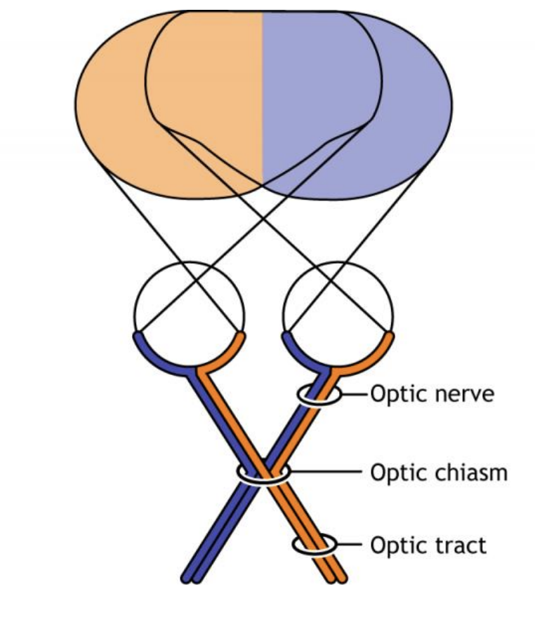
What are the optic tracts
When it splits where does it end up in the thalamus - name
After the thalamus where does it go to in the brain and how does it reach there (what is the type of signalling does it do to reach there?)
Difference in number of neurons for each destination (2 destinations)
Through the type of signalling, what two lobes can it reach and pass through?
For each what is the type of visual field processing is it?
The nerve bundles emerging from the chiasm are called the optic tracts. They end in the 2 lateral geniculate nuclei (LGN) in the thalamus, which project via the optic radiations to primary visual cortex,V1.
Optic radiations are going through either the parietal lobe carrying information from the lower visual field vs. one looping through the temporal lobe — called Meyer’s loop — carrying information from the upper visual field
Together, the 2 LGN have ~2 million neurons — the same as the number of ganglion cells in the 2 retinas — whereas V1 has far more neurons, ~300 million.
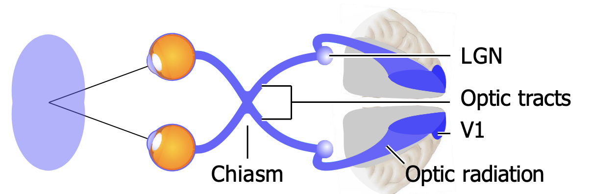
In what lobes of the brain is the primary visual cortex located?
What area (name) is most of the visual cortex concentrated in this lobe?
in the occipital lobe (back of the brain)
The calcarine sulcus is associated with the visual cortex. It is where the primary visual cortex (V1) is concentrated.
Organizing the visual areas in the brain in connection with what part of the eye?
What is this called?
Where is this arrangement organization found in which areas of the brain
Give an example. Light hitting in the center vs peripheral
visual areas connected with retina areas
That is, neurons close to each other in the brain get information from close-together parts of the retina.
Called retinotopically organized
This arrangement is found in the lateral geniculate nuclei, V1, and many higher visual processing areas.
E.g. when light hits the central retina, shown in red, then the region shown red in V1 is active; light in more-peripheral retina activates more-peripheral parts of V1.
The retinotopic map
which part of the eye gets more are in the brain for processing and why?
The fovea, which covers only a small area of the retina (0.01% of the retina’s total surface area), projects to large areas in V1 (and in other cortical regions and LGN),
i.e. a large proportion of visual cells in the brain receive and process data from foveal photoreceptors.
The fovea gets a lot of space because it has many photoreceptors, bipolars, and ganglion cells, and so carries a lot of information.
Visual field deficits
explain each type
Anopia
Scotoma
Monocular
Binocular
Hemianopia
Quandrantanopia
Homonymous
Bitemporal
Contralateral
Anopia = General term for vision loss
Scotoma = Focal area of lost vision
Monocular = Affecting one eye
Binocular = Affecting both eyes
Hemianopia = Loss of half the visual field
Quadrantanopia = Loss of one-quarter of the visual field
Homonymous = Same side lost in both eyes
Bitemporal = Outer halves of both eyes lost
Contralateral = Lesion is on the opposite side of the lost visual field