Week3/4- Topic A (1) Cardiovascular system- Heart and mediastinum
1/64
There's no tags or description
Looks like no tags are added yet.
Name | Mastery | Learn | Test | Matching | Spaced | Call with Kai |
|---|
No analytics yet
Send a link to your students to track their progress
65 Terms
what makes up the cardiovascular system
The cardiovascular system is made up of the heart, blood vessels, and blood.
what is the heart
The heart is a self-adjusting double pump that propels blood around two circulatory systems in series, the pulmonary and systemic systems.
It consists of four chambers: two atria and two ventricles.
what muscle is the heart formed from
specialised, cardiac muscle- myocardium.
Contraction of the cardiac muscle pushes blood out of the chambers.
Relaxation of a chamber allows blood to flow or be pushed into it but the heart does not actively draw blood in.
what is a heartbeat
Each cycle of contraction and relaxation is known as a heartbeat.
systole-Contraction of the ventricles
diastole- relaxation of the ventricles
describe the flow of pulmonary circulation
The pulmonary circulation is pumped at a low pressure.
Not much force is required to send blood the short distance through the lungs from the right to the left heart
high pressure in pulmonary capillaries would force fluid out of the blood, into the lung tissue and we would drown.
where does blood flow in pulmonary circulation
Blood pumped to the lungs from the right ventricle has a very low oxygen concentration.
Blood leaves the right ventricle through the pulmonary trunk which divides to give right and left pulmonary arteries.
Oxygenation of this blood occurs in the lungs before it returns to the heart through a series of pulmonary veins which enter the left atrium.
what is the systemic circulatory system
The systemic circulation feeds into and out of all the organs of the body except the lungs.
The left ventricle is the origin of the systemic circulation and the right atrium is its termination.
what are the features of the systemic circulatory system
operates at a high pressure (compared to the pulmonary).
Considerable force is required to adequately perfuse all the tissues in the human body and the blood has a much larger distance to travel from left to right heart than right to left.
The supply to the organs is rich in oxygen, the blood returning to the heart is low in oxygen.
what is the anatomy of the thorax
The thorax sits superiorly in the trunk of the body.
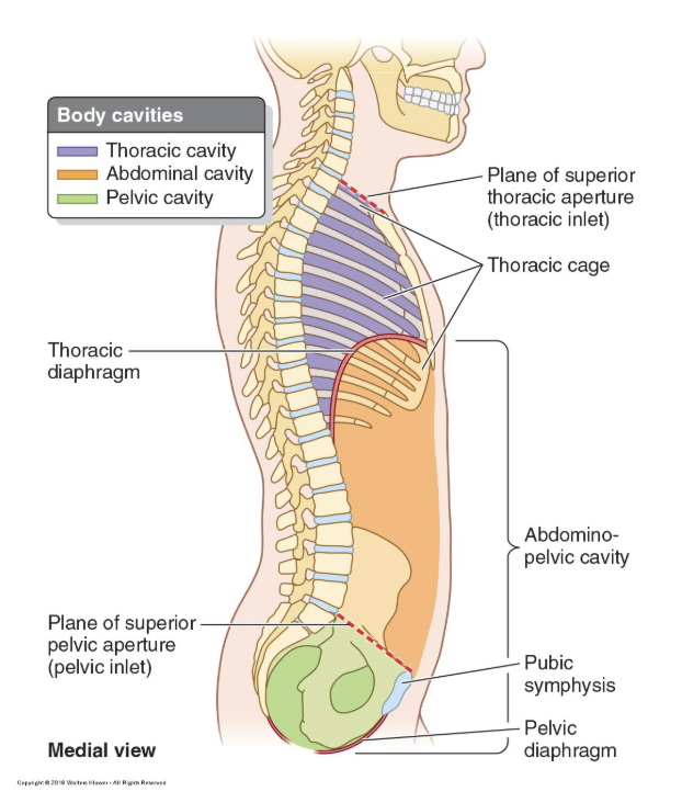
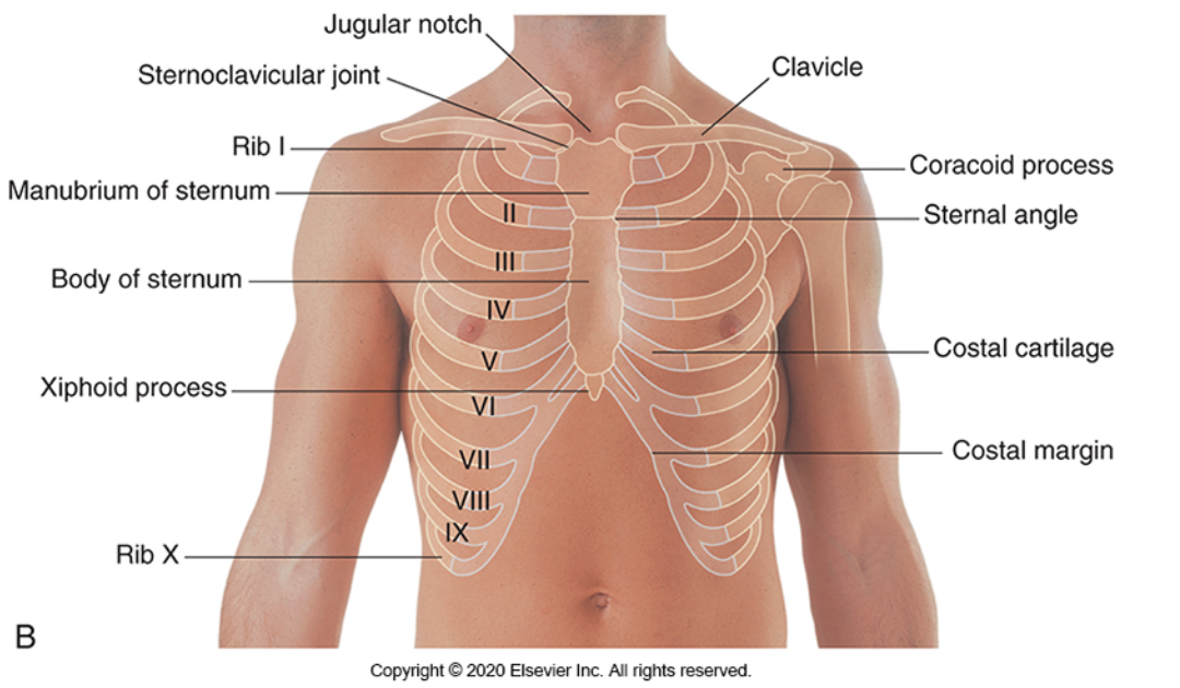
what is the thoracic cage
The thoracic cage protects the contents of the thorax.
It is made up of the ribs, costal cartilage, sternum (anteriorly), and thoracic vertebrae (posteriorly).
These structures, along with thoracic muscles, help form the boundaries of the thorax.
what are the regions of the thoracic cavity
mediastinum- sits centrally
left pleural cavity
right pleural cavity
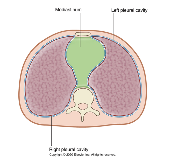
what is the mediastinum
the central compartment of the thoracic cavity- situated between the lungs
extends from the superior thoracic aperture (superiorly) to the diaphragm (inferiorly) and from the sternum and costal cartilages (anteriorly) to the bodies of the thoracic vertebrae (posteriorly).
what does the mediastinum contain
all the thoracic structures except the lungs and is a highly mobile region in the living because it consists primarily of hollow visceral structures which are joined by loose connective tissue.
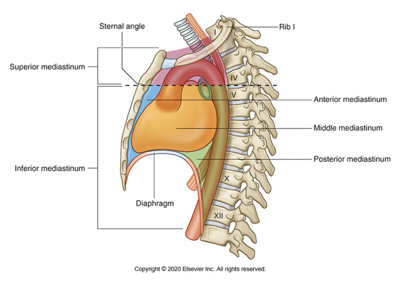
what is the sternal angle
the joint between the manubrium (top part) and the body (middle part) of the sternum
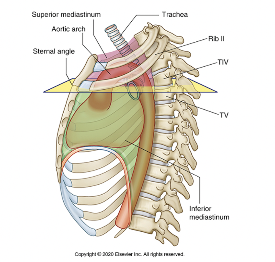
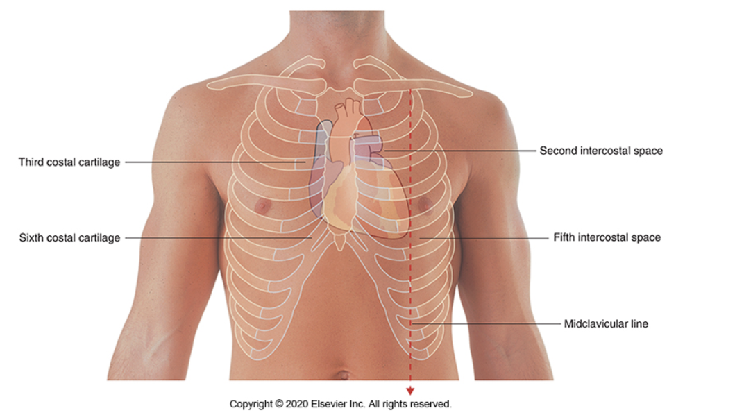
what is the thoracic plane
A horizontal plane can be drawn from the sternal angle to the level between T4 and T5, as shown on the diagram below.
This is imaginary line is known as the thoracic plane.
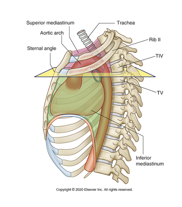
what does the thoracic plane split the mediastinum into
splits the mediastinum into two main compartments.
the superior mediastinum
the inferior mediastinum.
The inferior mediastinum is then further subdivided into:
anterior mediastinum
middle mediastinum
posterior mediastinum.
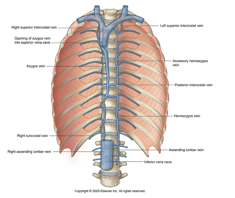
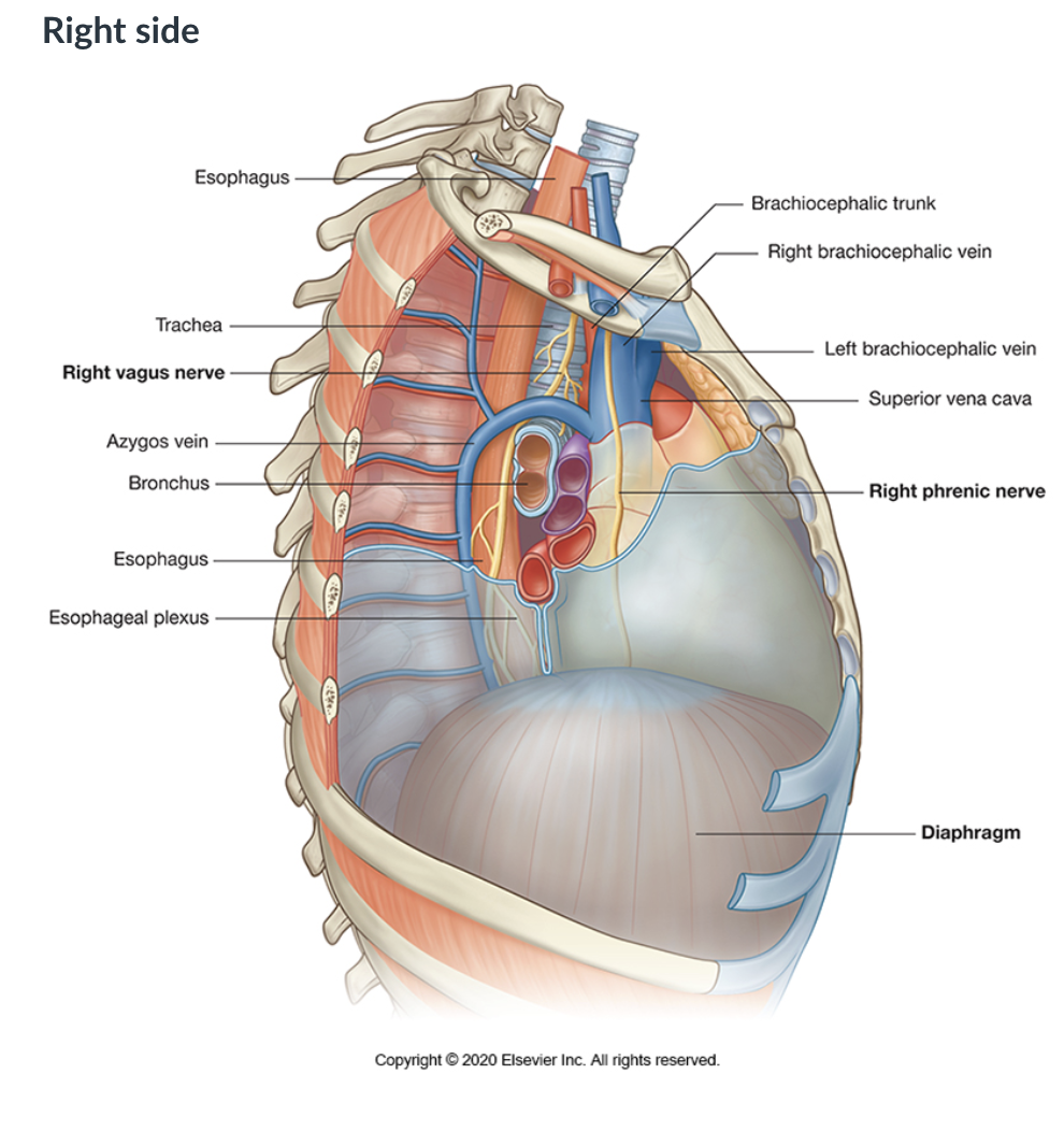
what is the Azygos system of veins
A H-shaped configuration of the azygos, hemiazygos, and accessory hemiazygos veins
This system drains the posterior thoracic wall.
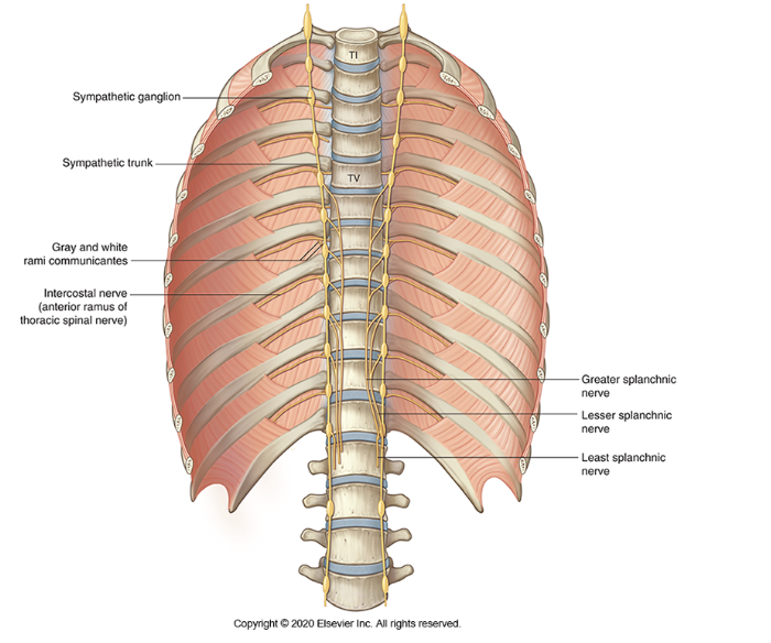
what are the Sympathetic chains
part of the sympathetic nervous system
The sympathetic chain (also known as the sympathetic trunk) is external to the spinal column, adjacent to the vertebral bodies
It is a paired structure (one on each side of the body).
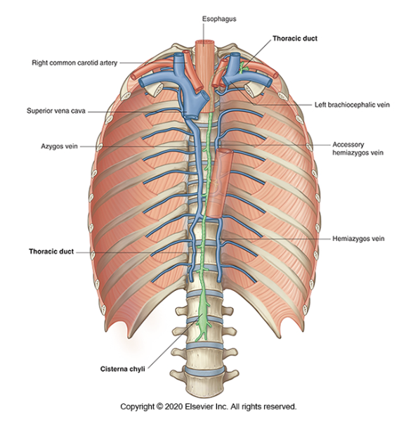
what is the Thoracic duct
the main lymphatic vessel for the return of chyle/lymph to the systemic venous system.
It drains lymph from both lower limbs, abdomen (except the convex area of the liver), left hemithorax, left upper limb and left side of face and neck.
what is the thymus
a T-cell producing lymphoid organ that plays a role in the development of the immune system particularly, maturation of T-cells.
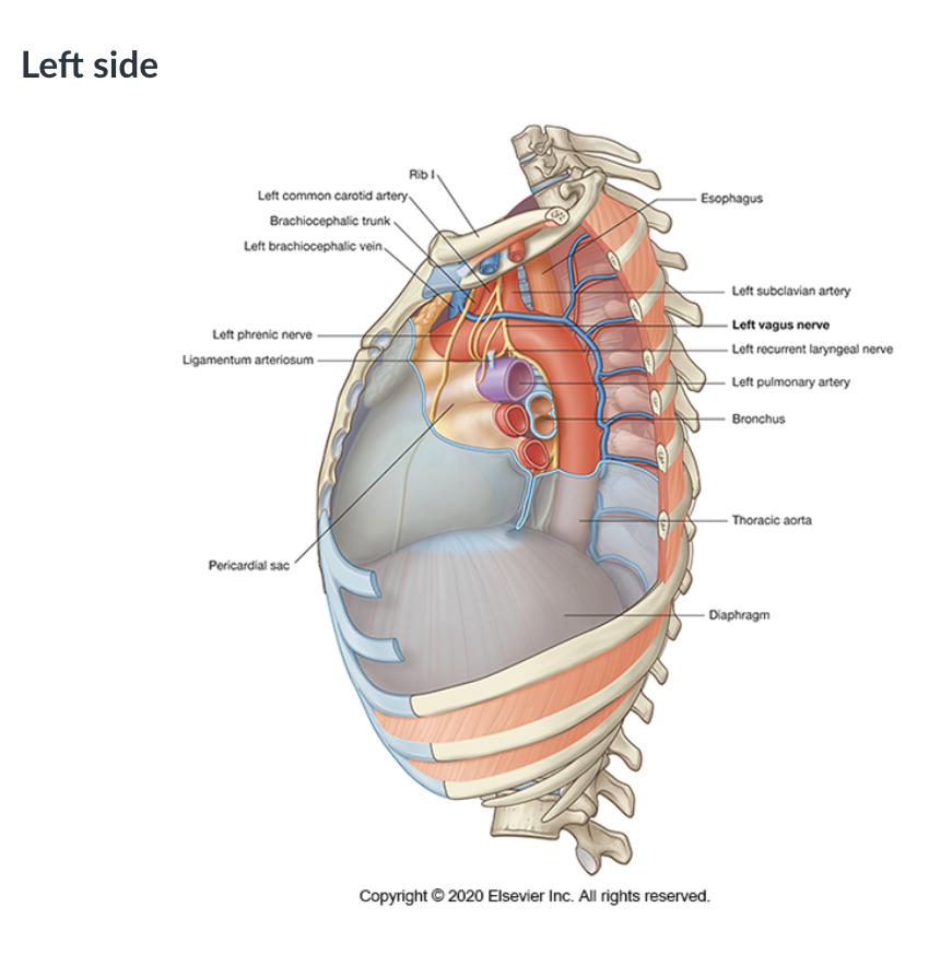
what is the phrenic nerve
A paired nerve (one of the left and one on the right) that supplies the diaphragm. It comes from the nerve roots C3, C4, C5.
what is the vagus nerve
The tenth cranial nerve (CNX). It is paired (there is one of the left and one on the right).
It provides the bulk of the parasympathetic input to the gastrointestinal system and to the heart.
It is a complex mixed sensory, motor and parasympathetic nerve.
In a transverse (cross-sectional) view of the superior mediastinum (viewed as if looking toward the patient’s head), identify the key structures
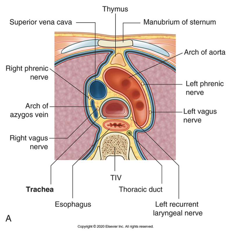
what does the left side of the superior mediastinum look like
what does the right side of the superior mediastinum look like
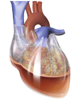
what is the pericardium, what are the 2 layers
a membrane that covers the heart. It is positioned within the middle mediastinum of the thorax.
It consists of two layers:
An outer fibrous layer
An inner thin serous layer that reflects from the inside of the fibrous sac onto the surface of the heart

what is the fibrous pericardium
Outer layer
Continuous with great vessels adventitia (aorta and pulmonary trunk)
Blended inferiorly with the central tendon of the diaphragm.
Rigid structure
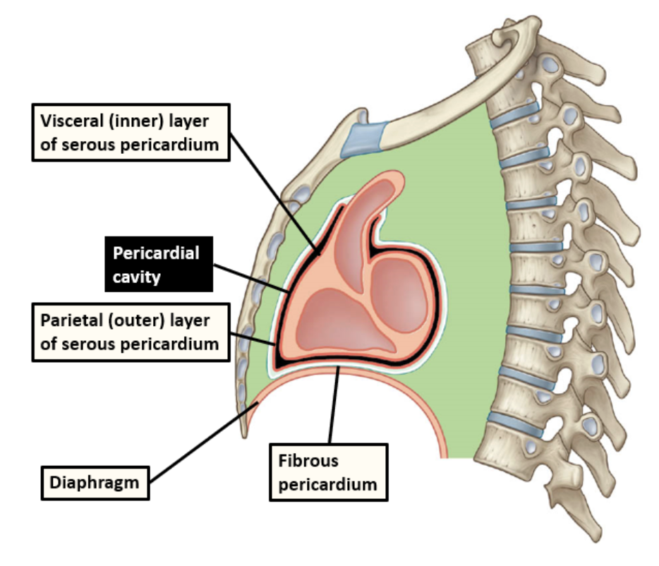
what is the Serous pericardium
Contained within the fibrous pericardial sac
Analogous to pleural membrane (Respiratory system)
Double layer
Between the double layer is a lubricating fluid which reduces the friction caused when the heart contracts

what are the 2 layers of the Serous pericardium
Visceral layer (epicardium) - the inner layer of the serous pericardium. The epicardium is the name given to the external layer of the heart but is actually just the visceral layer of the serous pericardium.
Parietal layer - the outer layer of the serous pericardium which lines the fibrous pericardium

what are the 4 functions of the pericardium
protection from infection
fixes the heart in the mediastinum and limits its motion
lubrication
prevents rapid overfilling of the heart
how does the pericardium provide protection from infection
The fibrous layer serves as a physical barrier between the muscular body of the heart and adjacent organs prone to infection, such as the lungs.
how does the pericardium fix the heart in the mediastinum and limit its motion
The pericardium is attached to the diaphragm, the sternum, and the tunica adventitia (outer layer) of the great vessels which fixes the heart in place.
how does the pericardium provide lubrication
The serous pericardium and the small amount of serous fluid within the pericardial cavity reduces friction between the heart and the surrounding structures.
how does the pericardium prevent rapid overfilling of the heart
The fibrous layer of the pericardium prevents the heart from increasing in size too rapidly, thus placing a physical limit on the potential size of the organ.
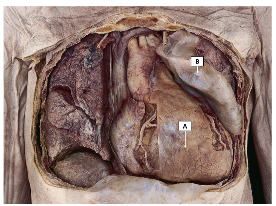
what are structures A and B in the image
A = Visceral layer of the serous pericardium
B = Parietal layer of the serous pericardium adhered to the internal surface of the fibrous pericardium
what is a cardiac tamponade
a potentially fatal situation when blood or fluid accumulates in the pericardium.
This compresses the heart, preventing the ventricles from expanding fully and impeding it’s blood supply.
why might a cardiac tamponade occur
most commonly associated with pericarditis caused by either bacterial or viral infections.
may also be attributed to trauma where damage to the heart or its vessels causes the pericardium to fill with blood.
Other causes include heart surgery, a dissecting aortic aneurysm, end stage lung cancer or acute myocardial infarction (MI).
how is cardiac tamponade treated
Treatment of cardiac tamponade requires immediate hospitalisation where a pericardiocentesis is usually performed.
During this fluid is drained from the pericardium in order to reduce the pressure.
The underlying cause of the tamponade may then be addressed, for example by repairing heart tissue or giving antibiotics to fight an infection.
what is Pericarditis
inflammation of the pericardium
what is Dissecting aortic aneurysm
an aortic dissection is a serious condition in which a tear occurs in the inner layer of the body's main artery (aorta).
Blood rushes through the tear, causing the inner and middle layers of the aorta to split (dissect)
what is Acute myocardial infarction
a life-threatening condition that occurs when blood flow to the heart muscle is abruptly cut off, also known as a heart attack
what is Pericardiocentesis
a procedure done to remove fluid that has built up in the sac around the heart (pericardium).
It's done using a needle and small catheter to drain excess fluid
what great vessels are in the right side of the heart
The Superior Vena Cava
The Inferior Vena Cava
The Pulmonary Trunk

function of The Superior Vena Cava
This brings de-oxygenated blood to the right atrium from the systemic circulation superior to the heart, i.e. the upper limbs, thorax, head and neck.
function of The Inferior Vena Cava
This brings deoxygenated blood to the right atrium from the systemic circulation inferior to the heart, i.e. the abdomen. pelvis and lower limbs.
function of The Pulmonary Trunk
This exits the right ventricle, taking deoxygenated blood to the lungs.
Immediately superior to the heart it bifurcates into the right and left pulmonary arteries which run into their respective lungs.
what great vessels are in the left side of the heart
the pulmonary veins
the aorta
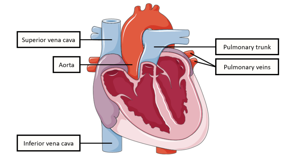
function of The Pulmonary Veins
These enter the left atrium on the posterior aspect of the heart, carrying oxygenated blood from the pulmonary circulation.
There are 4 pulmonary veins:
Left superior and left inferior pulmonary veins
Right superior and right inferior pulmonary veins.
function of The Aorta
Known as the ascending aorta as it leaves the heart, this vessel carries high pressure, oxygenated blood to the body in the systemic circulation
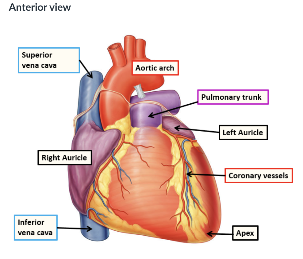
what does the anterior view of the external surface of the heart look like when the pericardium has been removed
what does the posterior view of the external surface of the heart look like when the pericardium has been removed
what is the Anterior (sternocostal) surface of the heart
This is formed mostly from the right ventricle, this surface is related anteriorly to the sternum and ribs.
what is the Inferior (diaphragmatic) surface of the heart
This surface is formed mostly by the left and partly from the right ventricle. It is related inferiorly to the centre of the diaphragm.
what is Left (pulmonary) surface the of the heart
This surface is formed mostly by the left ventricle. It is related laterally with the left lung and occupies a depression in this lung know as the cardiac impression.
what is the base of the heart
The base is situated on the posterior aspect, directed towards the vertebrae T6-9.
t is formed mostly from the left and partly from the right atrium and extends from the bifurcation of the pulmonary trunk superiorly to the atrioventricular groove (depression between the atrium and ventricle on each side of the heart) inferiorly.
what is the base of the heart
The apex lies posterior to the 5th intercostal space in the midclavicular line. It is directed antero-inferiorly and to the left.
what are the 4 borders of the heart
visible in both posterior and anterior views
right
superior
left
inferior
describe the 4 borders of the heart
Right border
formed by the right atrium. extends from the superior to inferior vena cava.Inferior border
roughly horizontal and mostly formed by the right ventricle, with a small contribution by the left ventricle.Left border
mostly formed from the left ventricle with the superior portion being formed by the auricular appendage of the left atrium.Superior border
formed by both atria. The aorta and pulmonary trunk arise from this border and the superior vena cava enters the heart at the right side of the superior border.
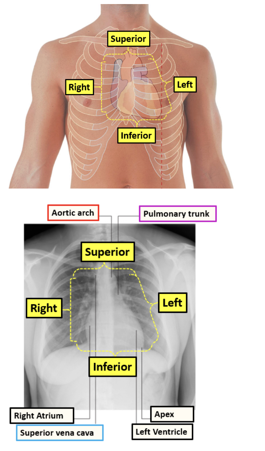
how can underlying structures be located
surface landmarks are used. When locating the heart, we can use the bony landmarks of the thoracic cage; for example, counting ribs and intercostal spaces (the spaces between the ribs).
what is the sternal angle, why is it an important landmark
the joint between the manubrium of the sternum and the sternal body
as there are many underlying structures at this level. For example, the arch of the aorta, the bifurcation of the trachea, the second rib, the pulmonary trunk, and nerves.
What is the superior border of the heart in surface anatomy?
Extends from the 3rd costal cartilage (right side of sternum)
to the 2nd intercostal space (left side of sternum)
Where is the right margin of the heart located?
Runs from the right 3rd costal cartilage
to near the right 6th costal cartilage

Where is the left margin of the heart located?
Descends from the left 2nd intercostal space
to the apex at the 5th intercostal space, midclavicular line

Where is the inferior (lower) border of the heart located?
Runs from the sternal end of the right 6th costal cartilage
to the apex in the 5th intercostal space (midclavicular line)

Where is the apex of the heart located?
Left 5th intercostal space
At the midclavicular line
