Head and Eyes - H&P
1/126
There's no tags or description
Looks like no tags are added yet.
Name | Mastery | Learn | Test | Matching | Spaced | Call with Kai |
|---|
No analytics yet
Send a link to your students to track their progress
127 Terms
the skull is composed of what bones
temporal
parietal
frontal
occipital
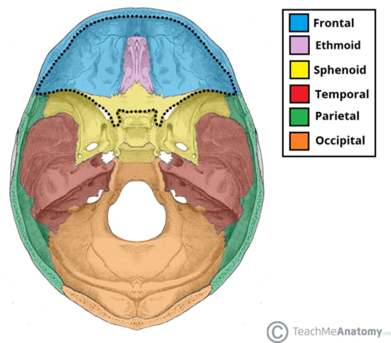
base of skull
occipital
Vertex of head
the top of the head, the crown; the highest point; the top or apex of something
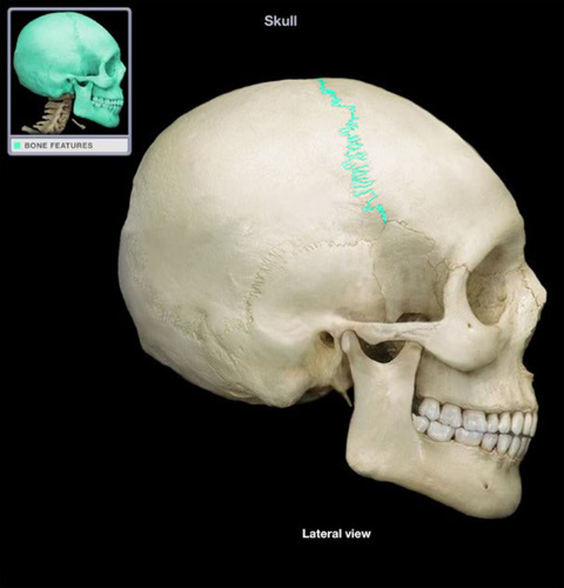
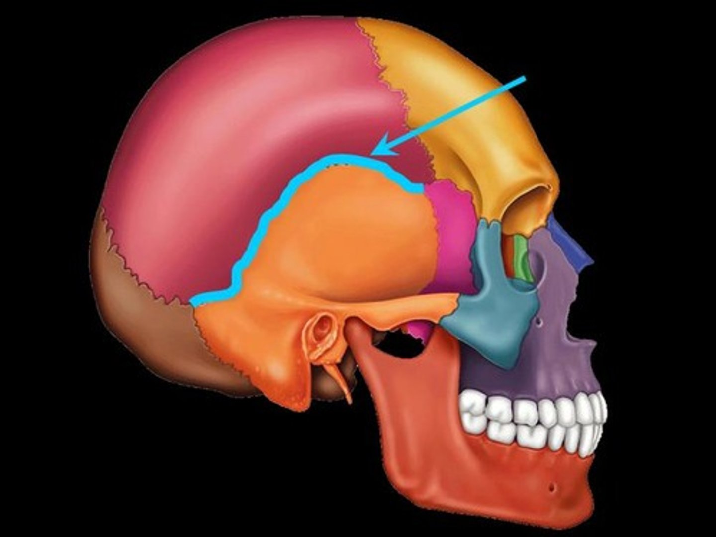
coronal suture
the suture between the parietal and frontal bones of the skull
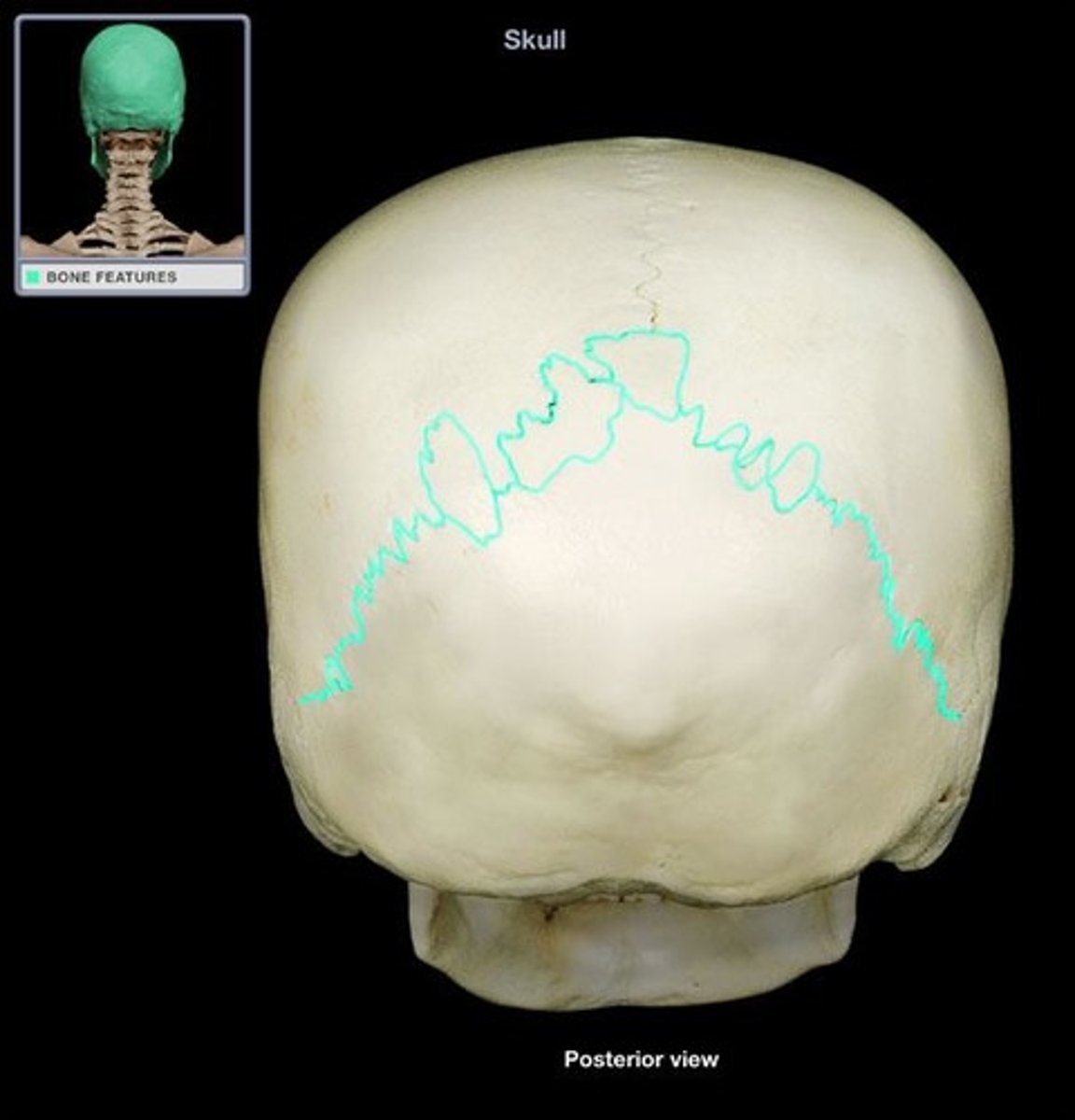
lamboidal suture (Skull)
between occipital and parietal bones
zygomatic arch of temporal bone
cheek bone
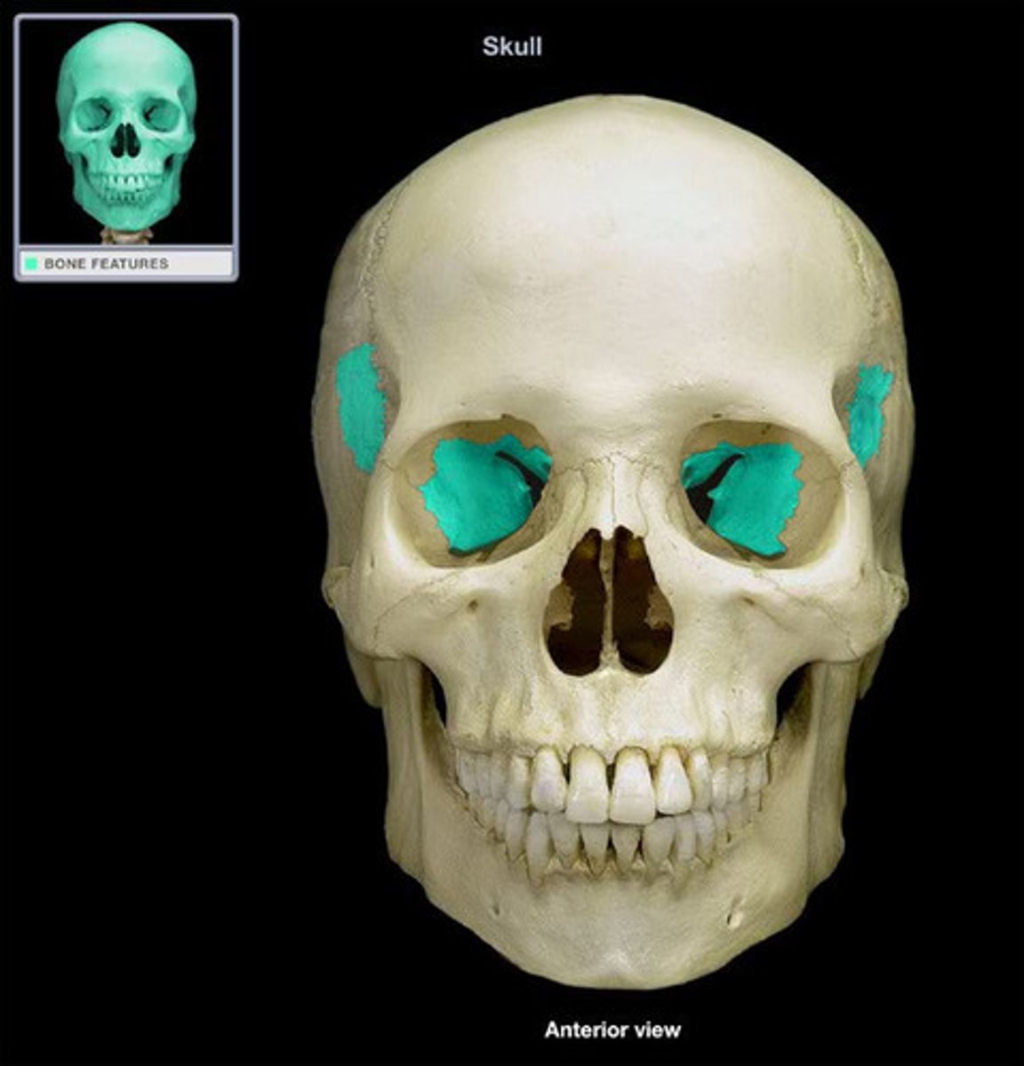
sphenoid bone
forms part of the base of the skull and parts of the floor and sides of the orbit
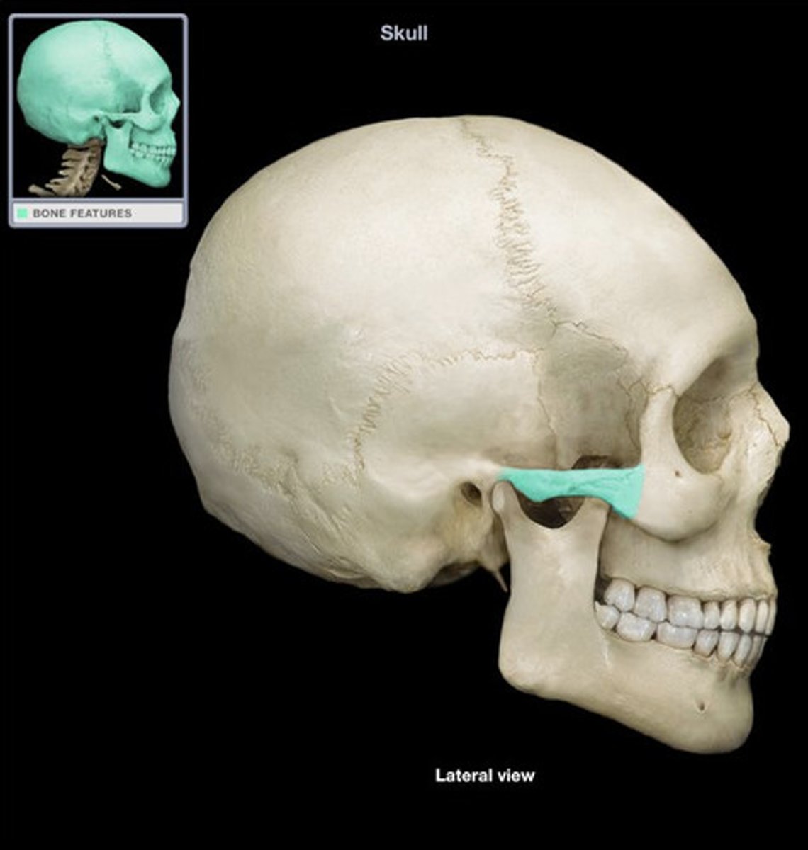
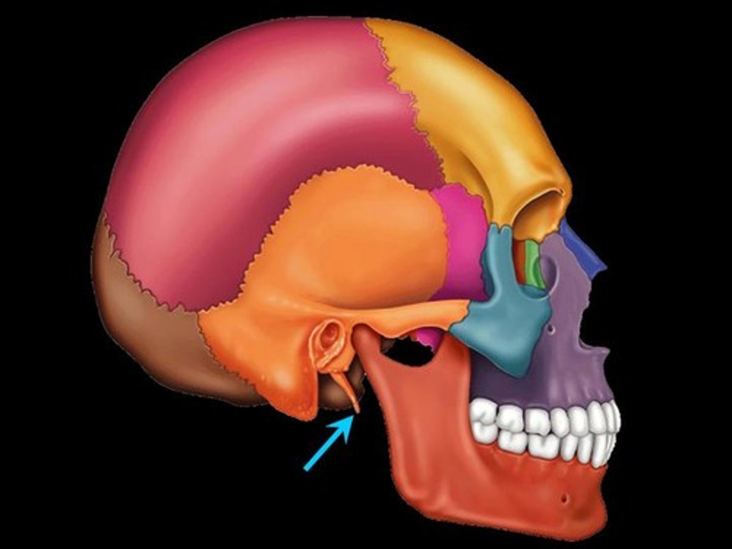
styloid process skull
pole-like process extending downward from the temporal bone on each side of the skull
skull sutures
coronal, sagittal, squamous, lambdoid
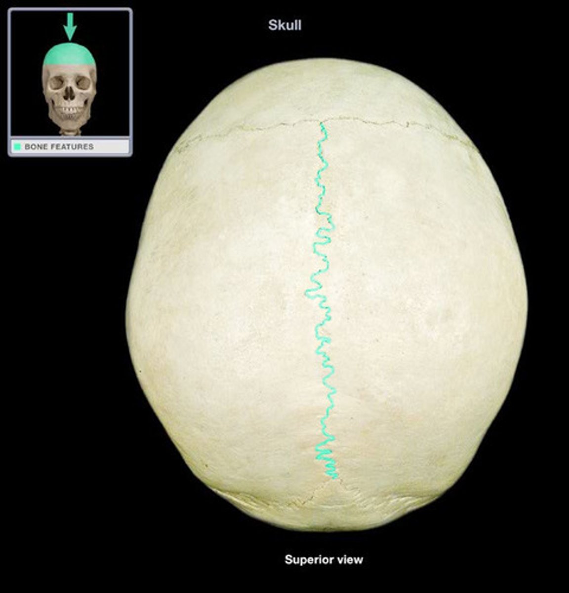
sagittal suture of the skull
between parietal bones
squamous skull suture
between the parietal and temporal
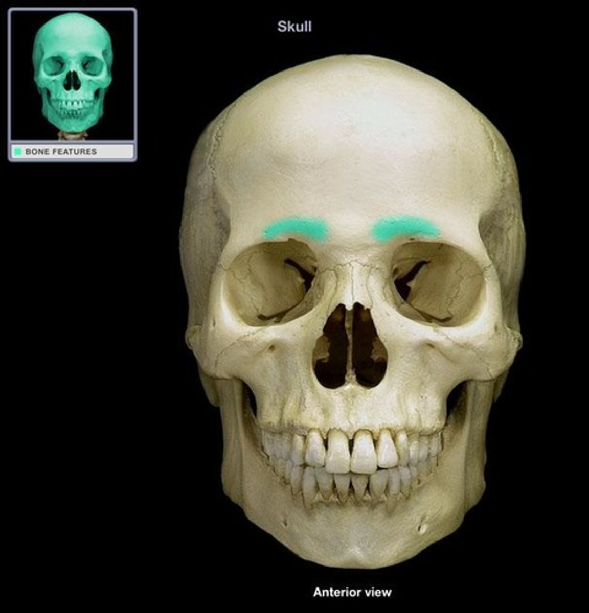
supraorbital ridge
bony projection in the eyebrow area of the frontal bone
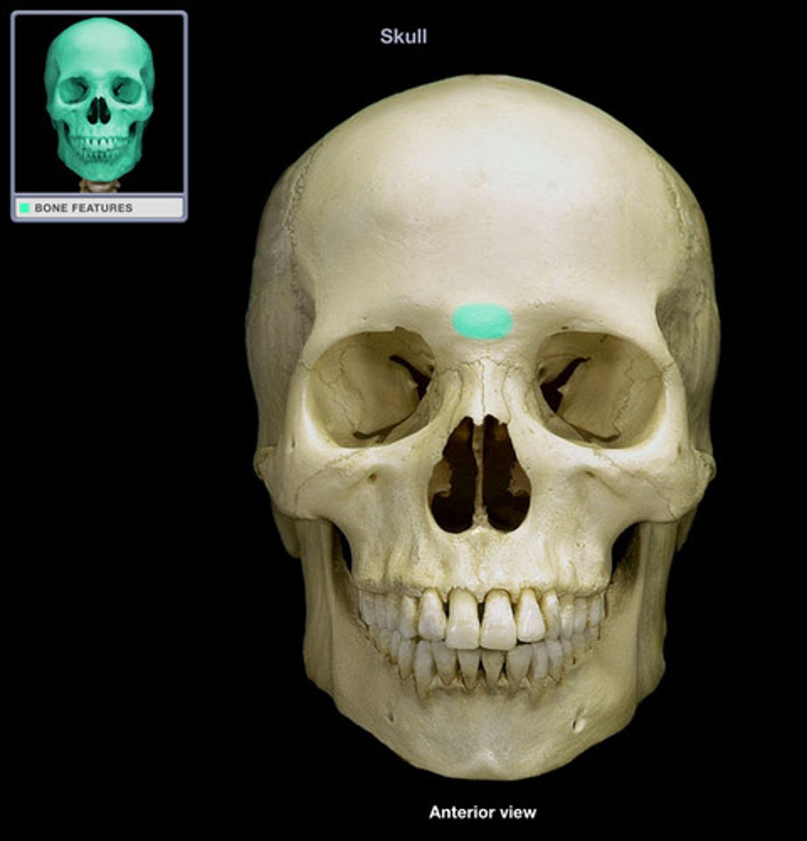
glabella of frontal bone
Smooth area between the eyes
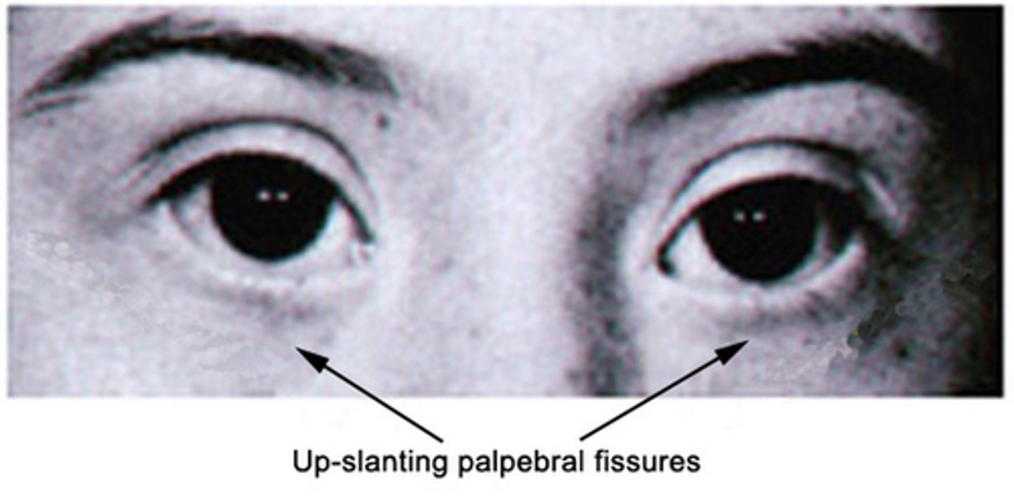
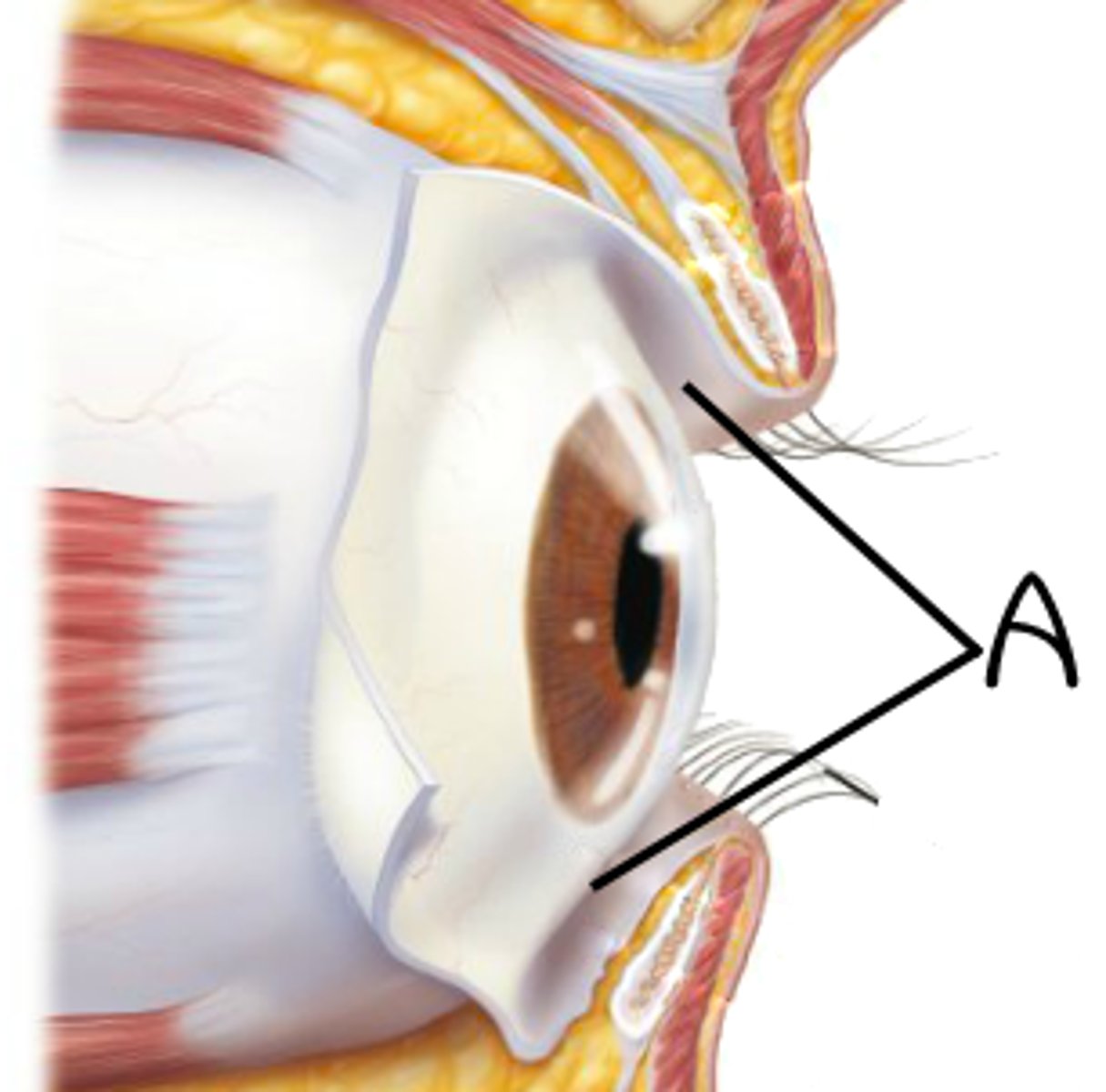
palpebral fissures
spaces between the eyelids, should be equal in size
- indicates down syndrome
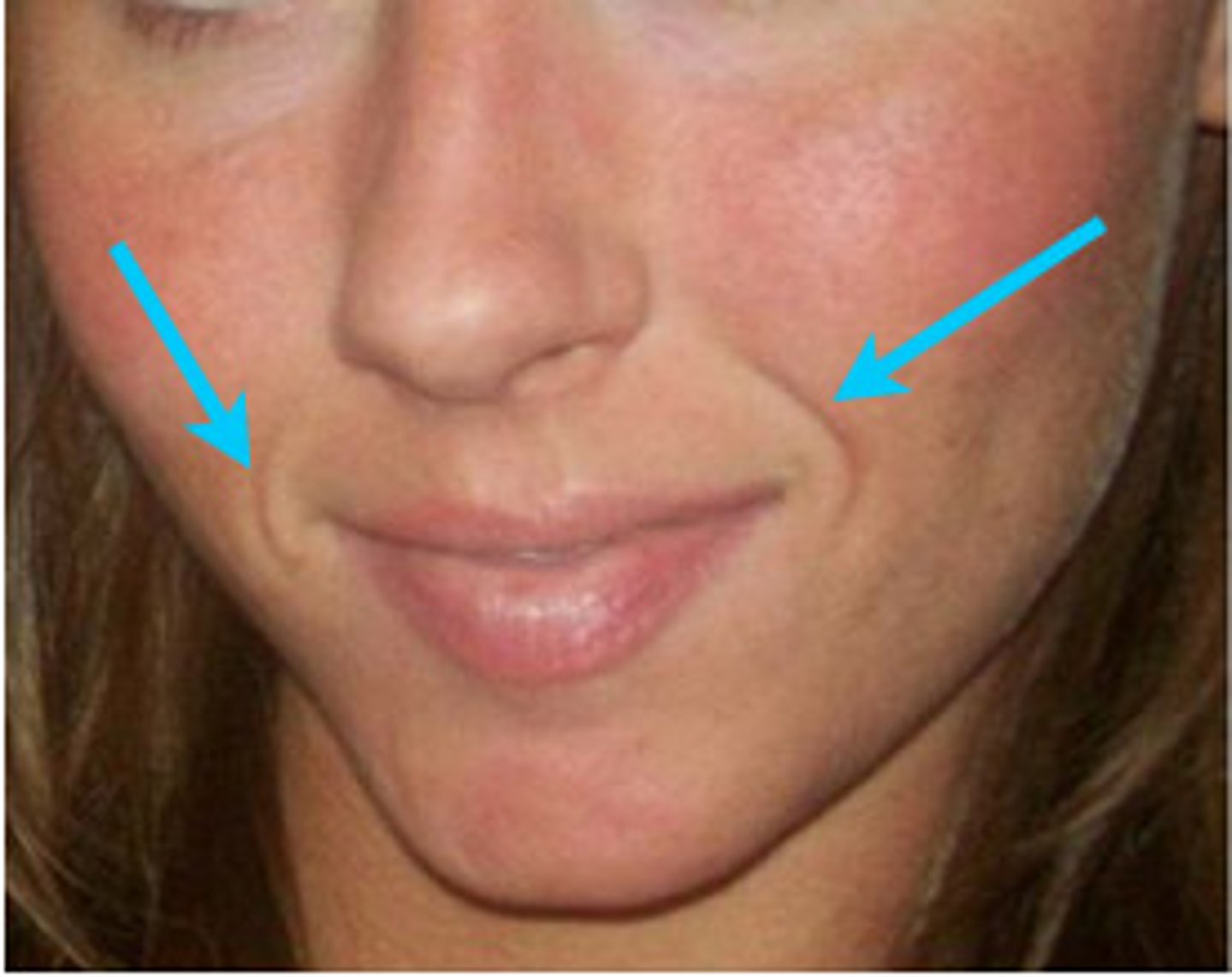
nasolabial fold
skin crevice between the nose and the corner of the mouth
The frontalis and occipitalis muscles together are called the
epicranius
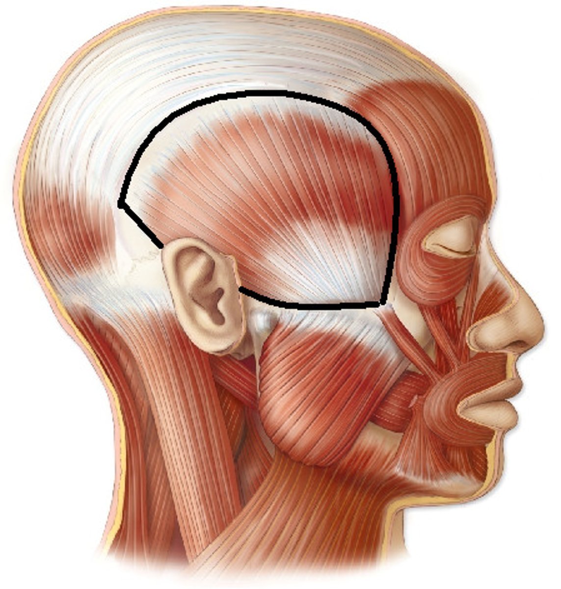
Temporalis
elevates and retracts mandible
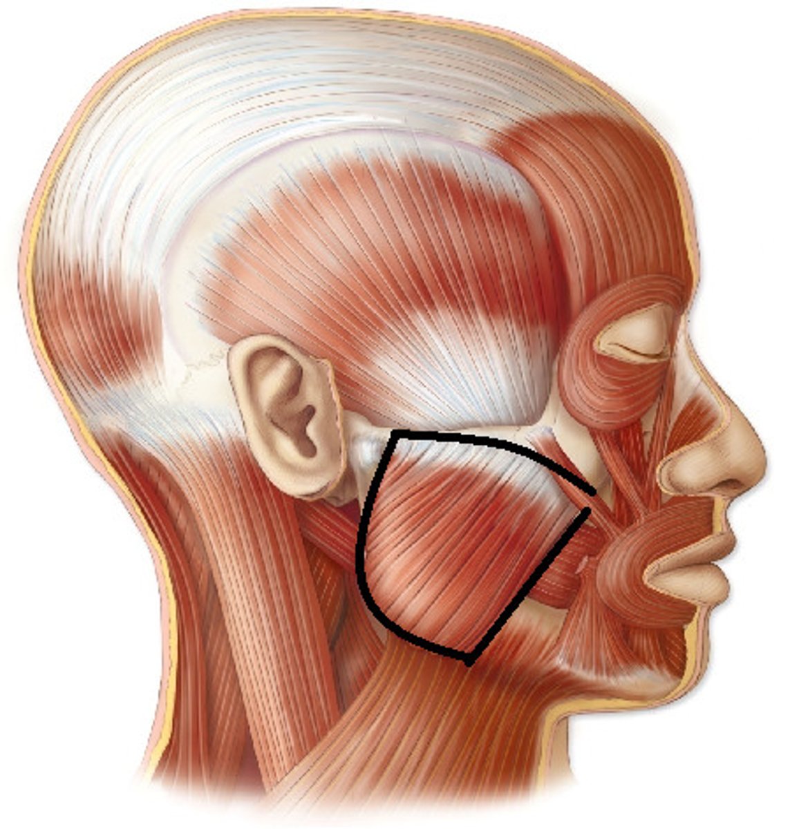
Masseter
elevates mandible and closes jaw
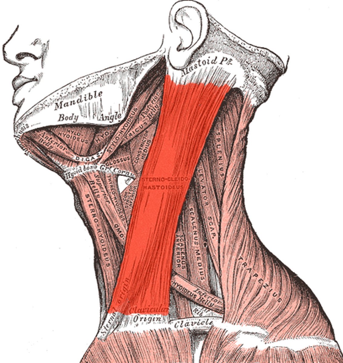
Sternocliedomastoid
flex neck, rotates neck
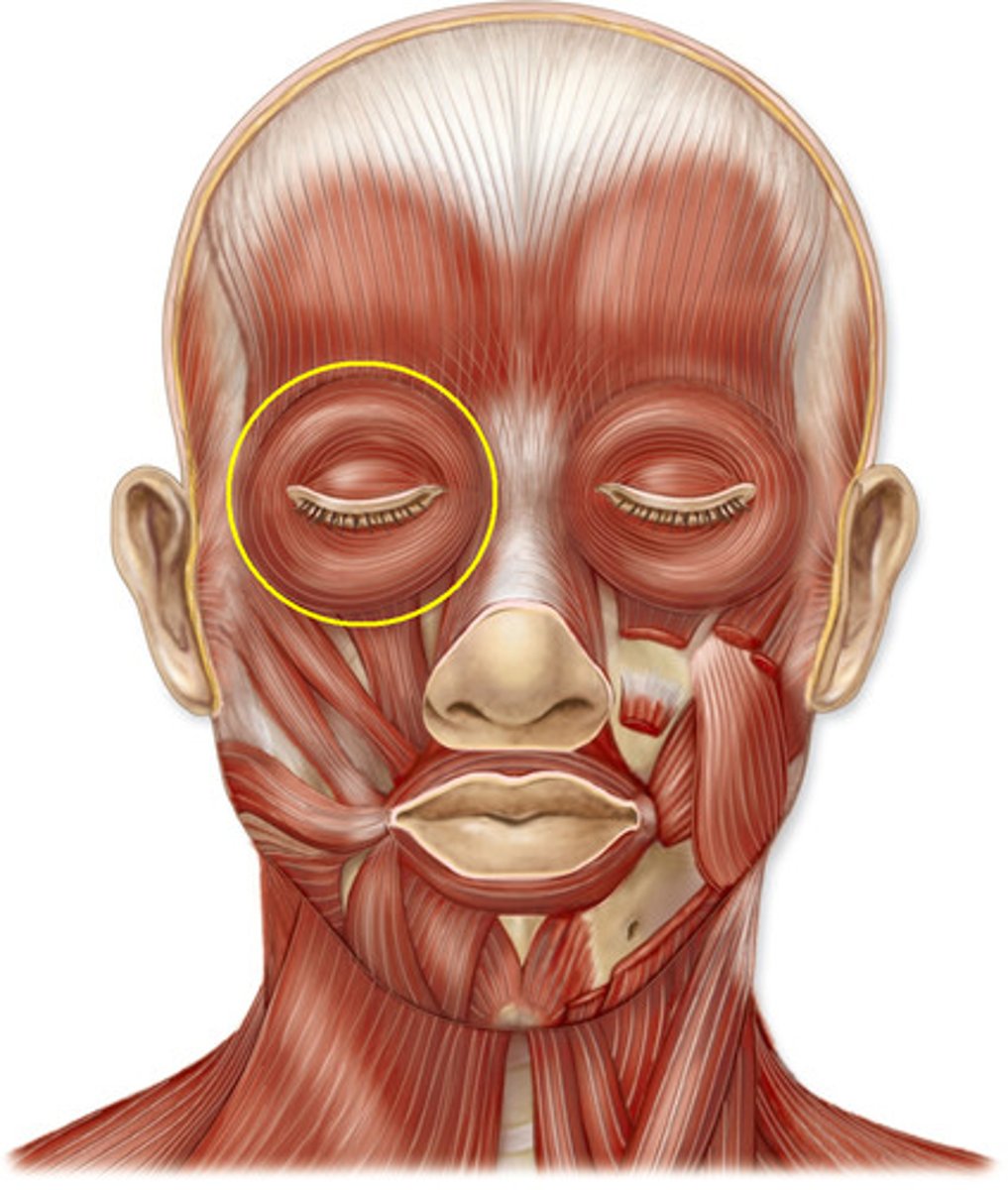
orbicularis oculi
Closes eyelids; used in blinking, winking, and squinting
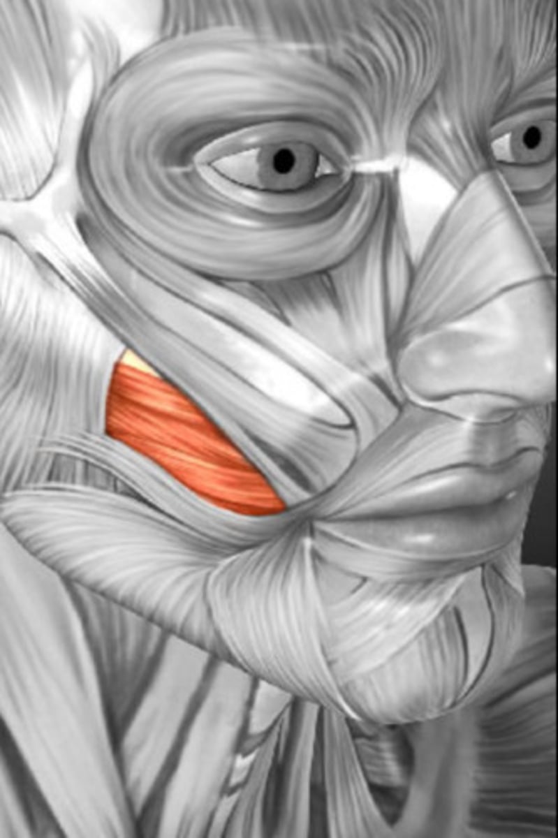
Zygomatic muscle (Zygomaticus)
smiling muscle, pulls corners of the mouth upward
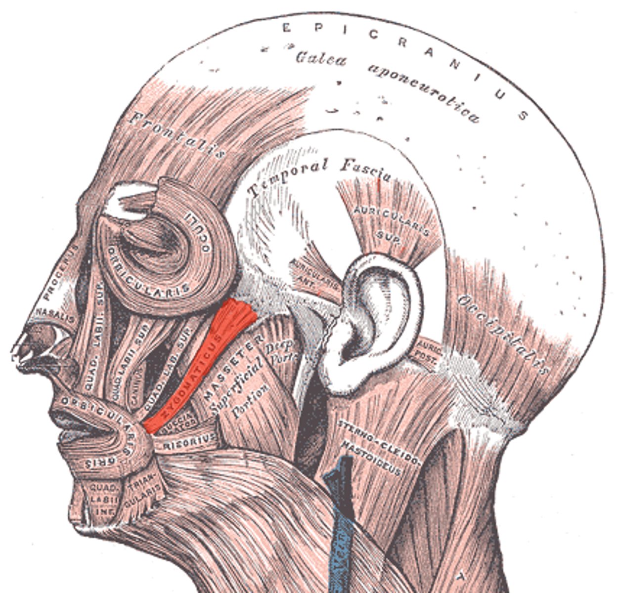
buccinator muscle
Thin, flat muscle of the cheek between the upper and lower jaw that compresses the cheeks and expels air between the lips.
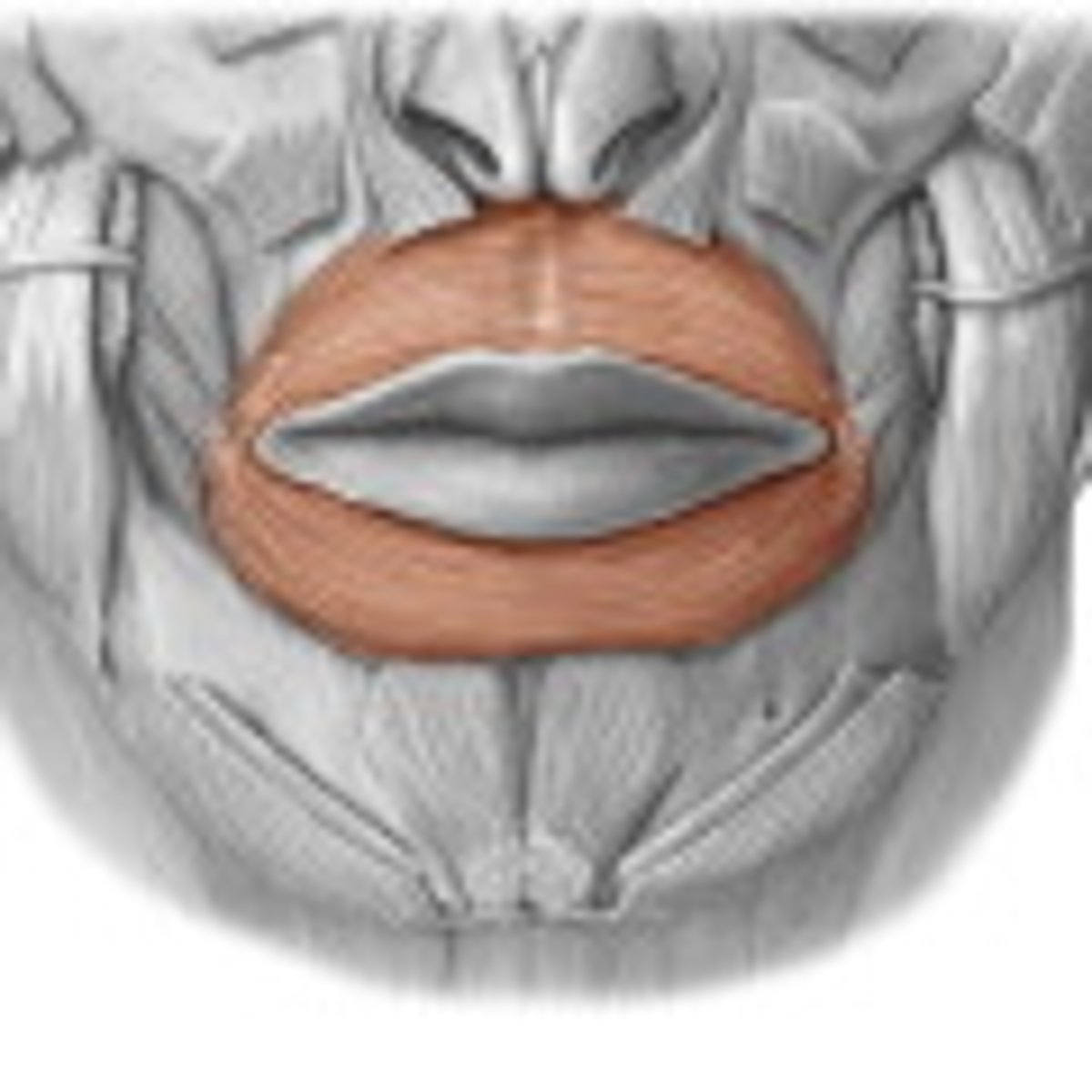
orbicularis oris
closes and protrudes lips
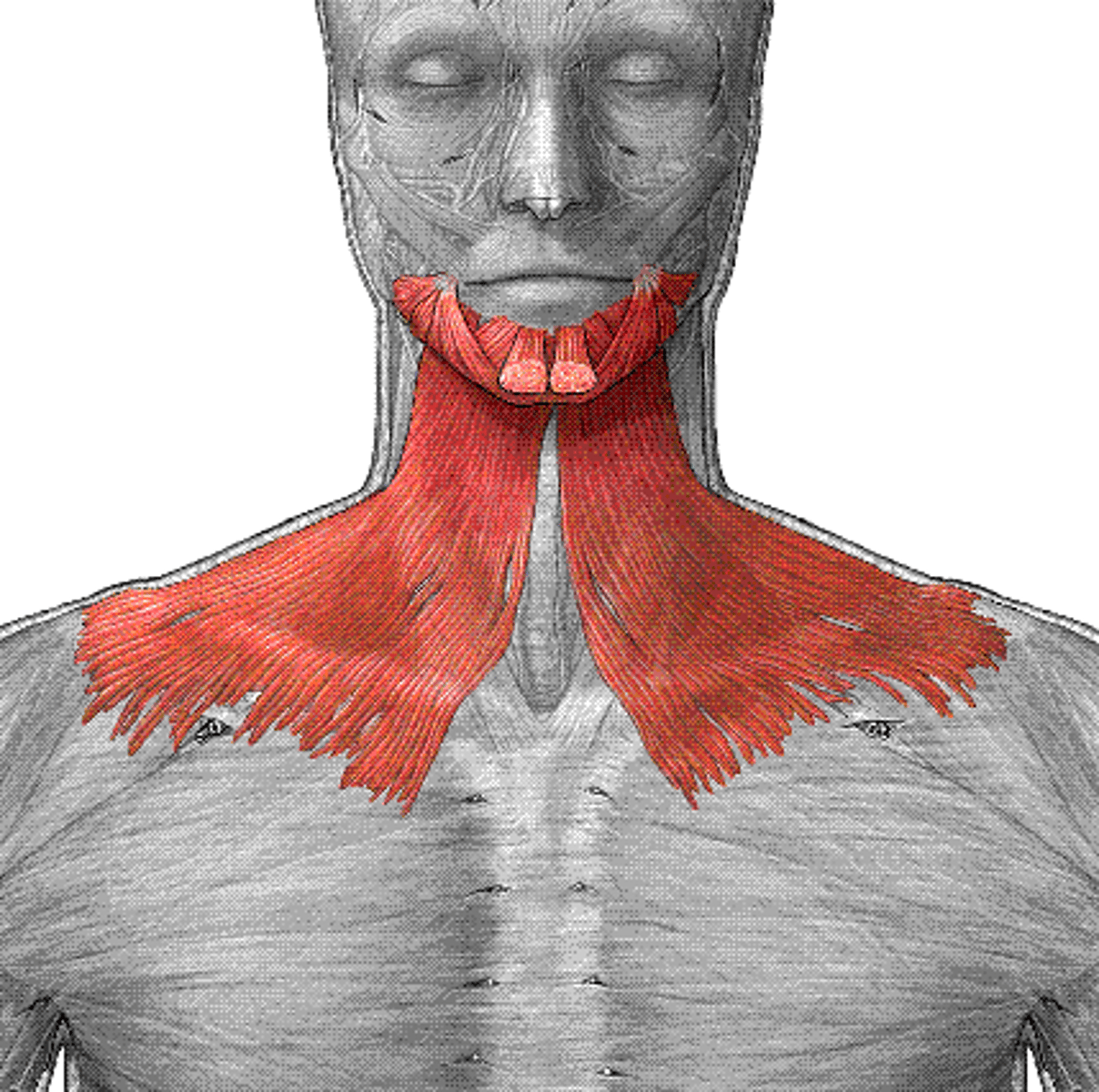
Platysma
tenses skin of neck, depresses mandible
Parotid (Stensen's) duct drains where?
drains to top of mouth
Where does the submandibular (wharton) duct open?
drains into bottom of the mouth under the tongue
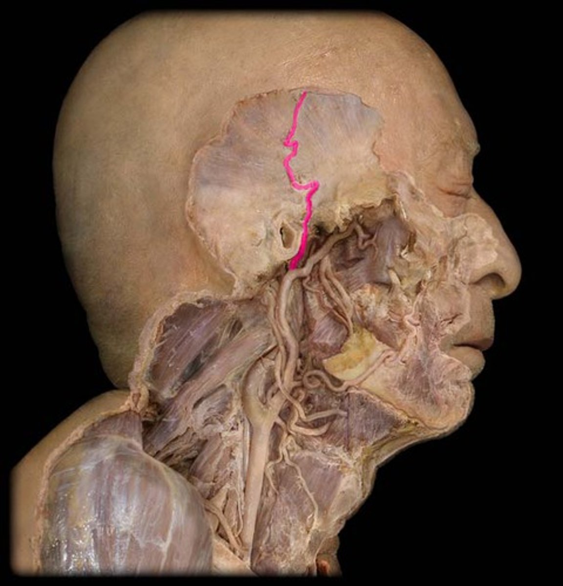
superficial temporal artery
A continuation of the external carotid nerve artery; supplies blood to the muscles of the front, side, and top of the head.
pathological facies
facial expressions that are indicative of an underlying pathology such as cushing's or down syndrome
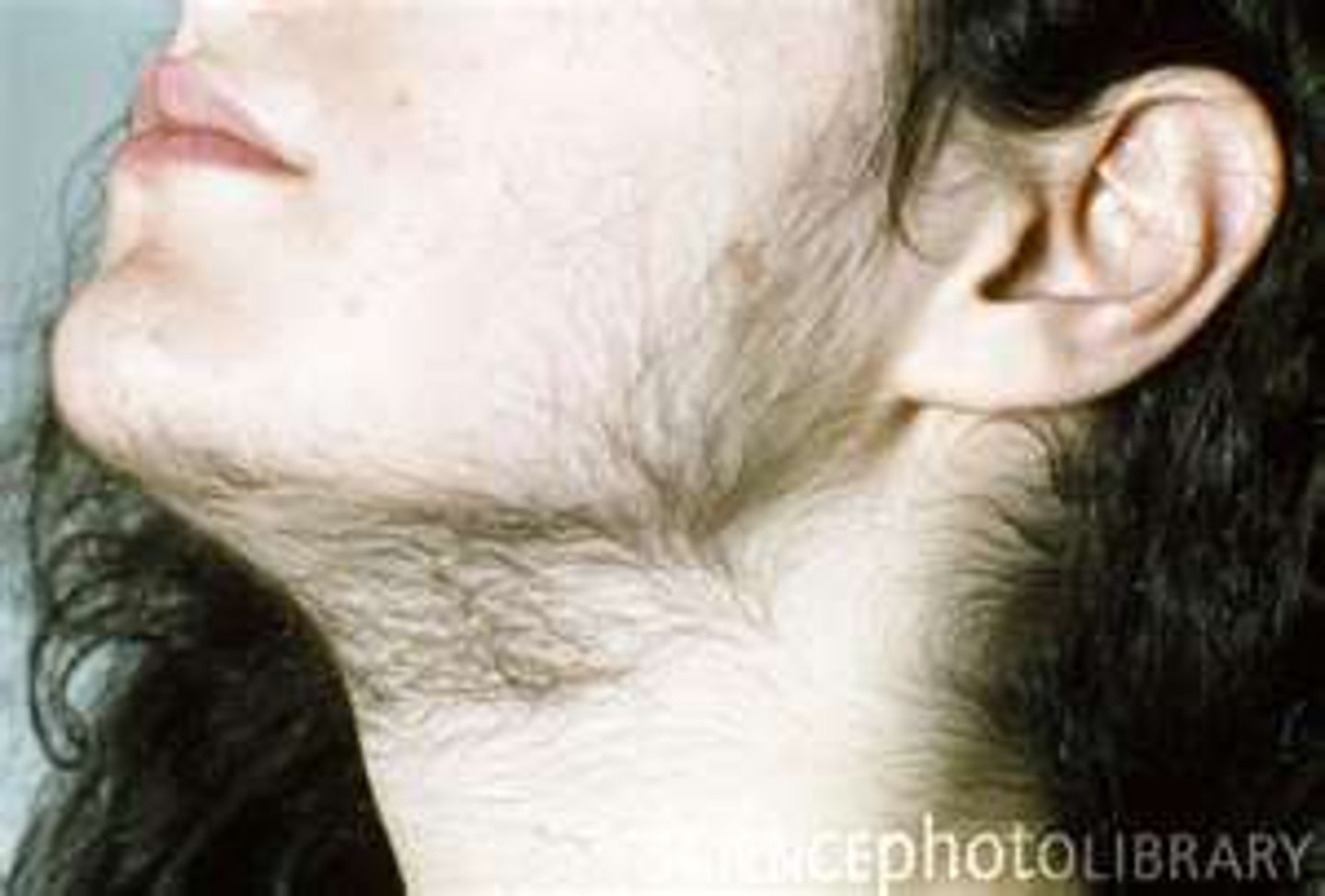
hirsutism
excessive hair growth over the body
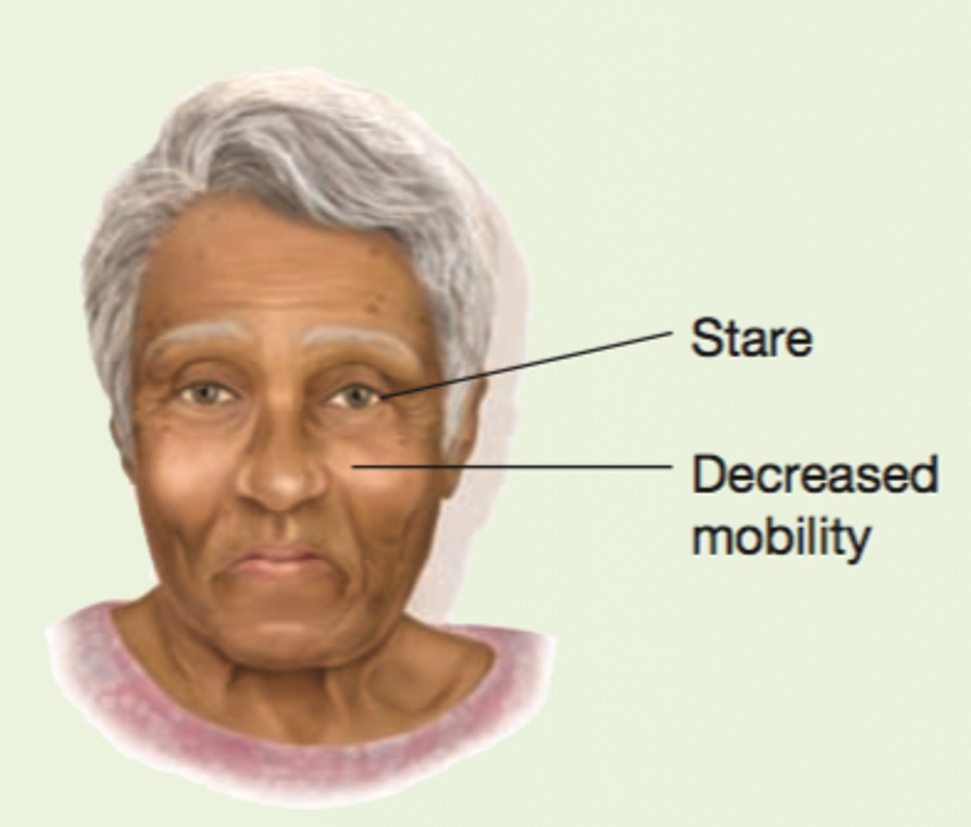
Parkinson's Facies
decreased facial mobility blunts expression.
decreased blinking and a characteristic stare present.
patient seems to peer upward towards observer.
facial skin becomes oily and drooling may occur.
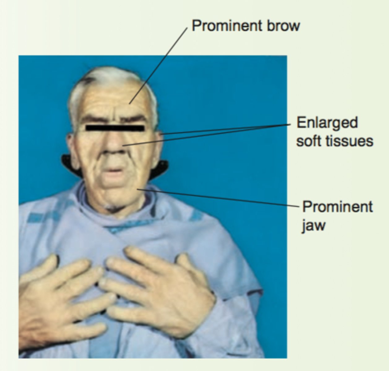
Acromegaly Facies
enlargement of both bone and soft tissues. head is elongated with bony prominence over forehead, nose, and lower jaw. nose, lips, and ears also enlarge.
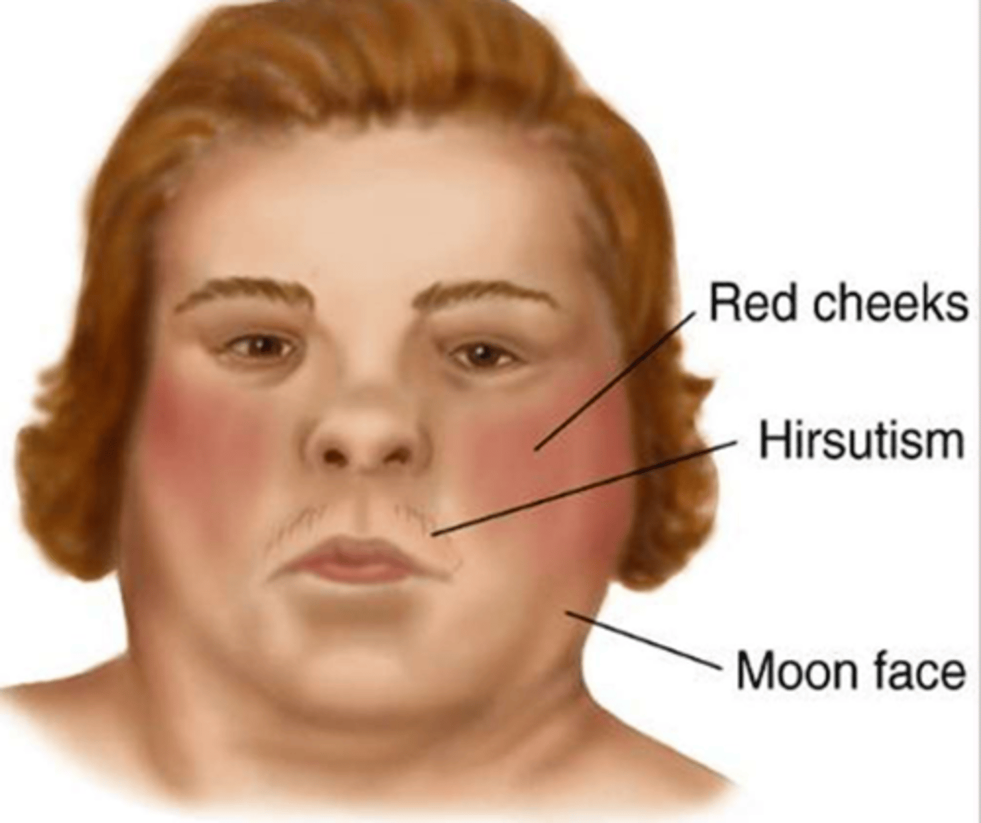
Cushing's syndrome facies
moon face
red cheeks
hirsutism
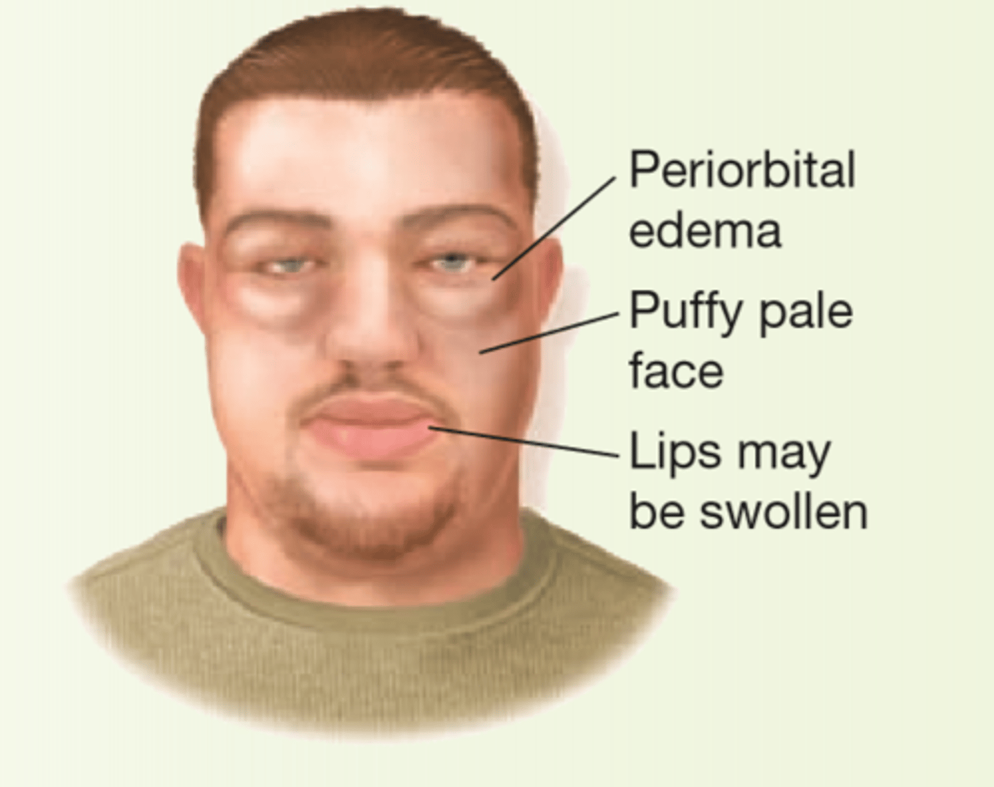
nephrotic syndrome facies
group of clinical signs and symptoms caused by excessive protein loss in urine
- periorbital edema
- puffy pale face
- lips was swell
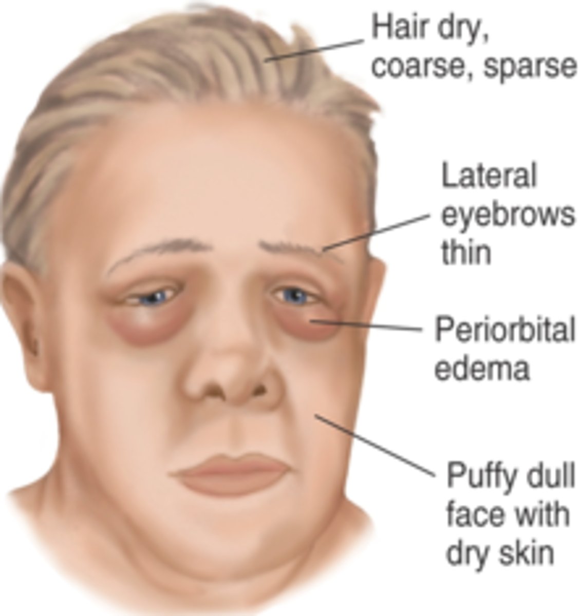
Myxedema facies
hair dry, sparse
lateral eyebrows thin
periorbital edema
puffy dull face
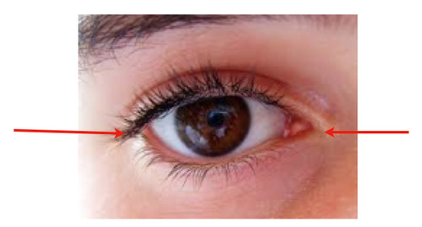
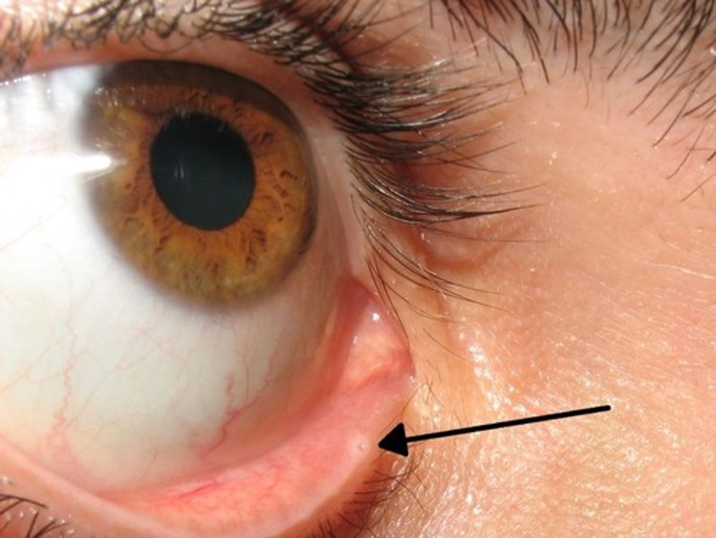
medial and lateral canthus
corners of the eye
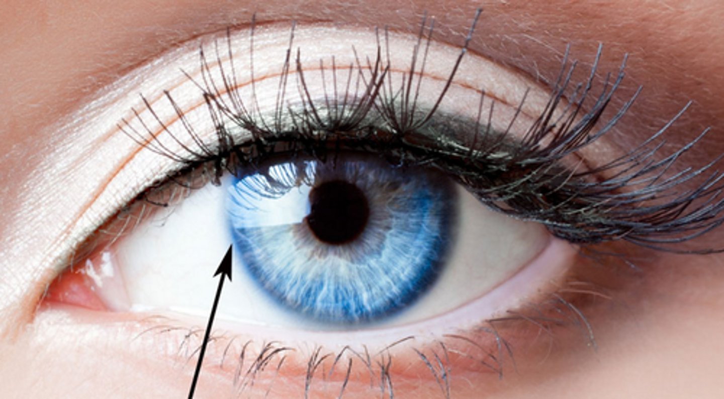
limbus
border between cornea and sclera
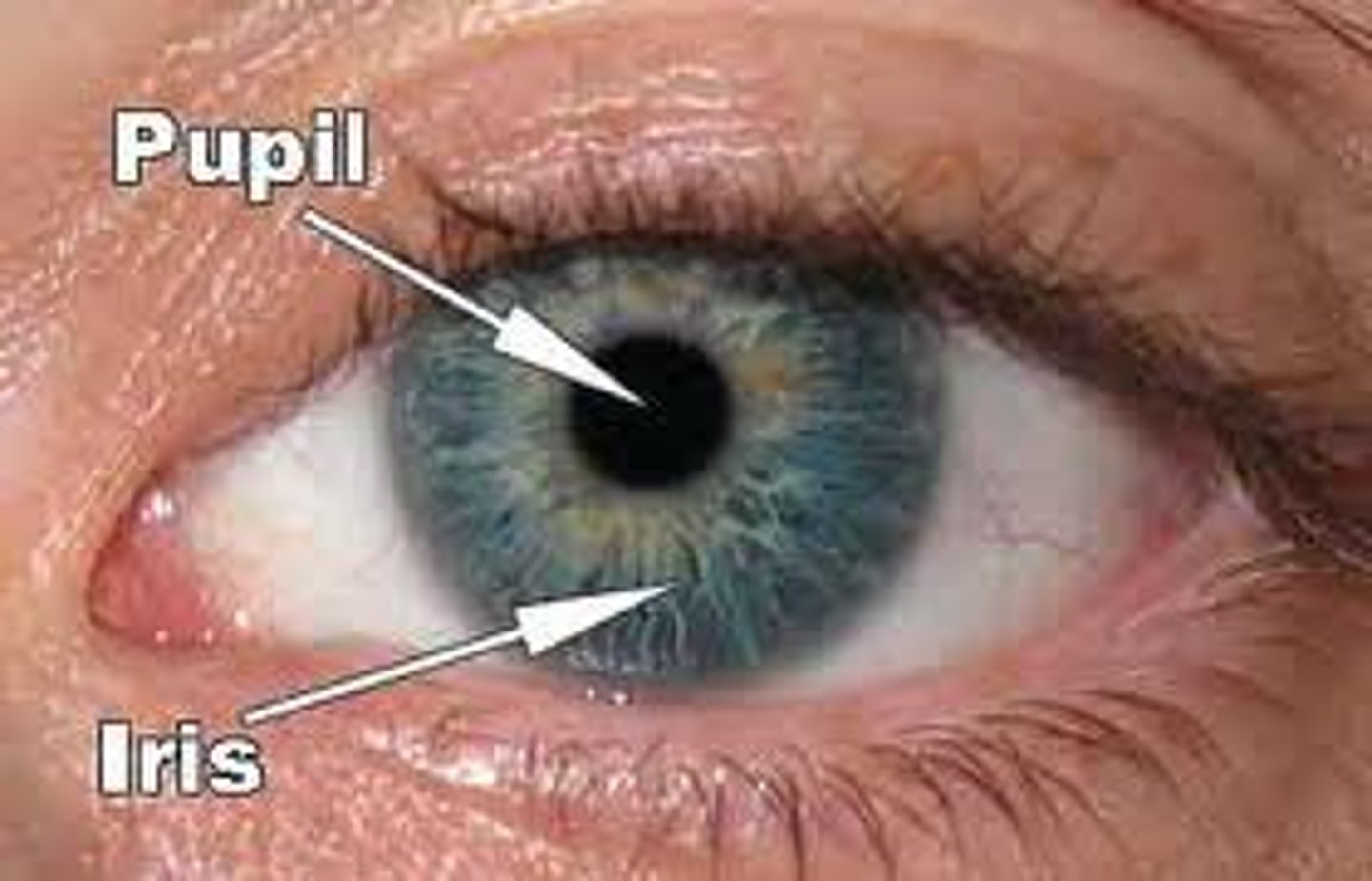
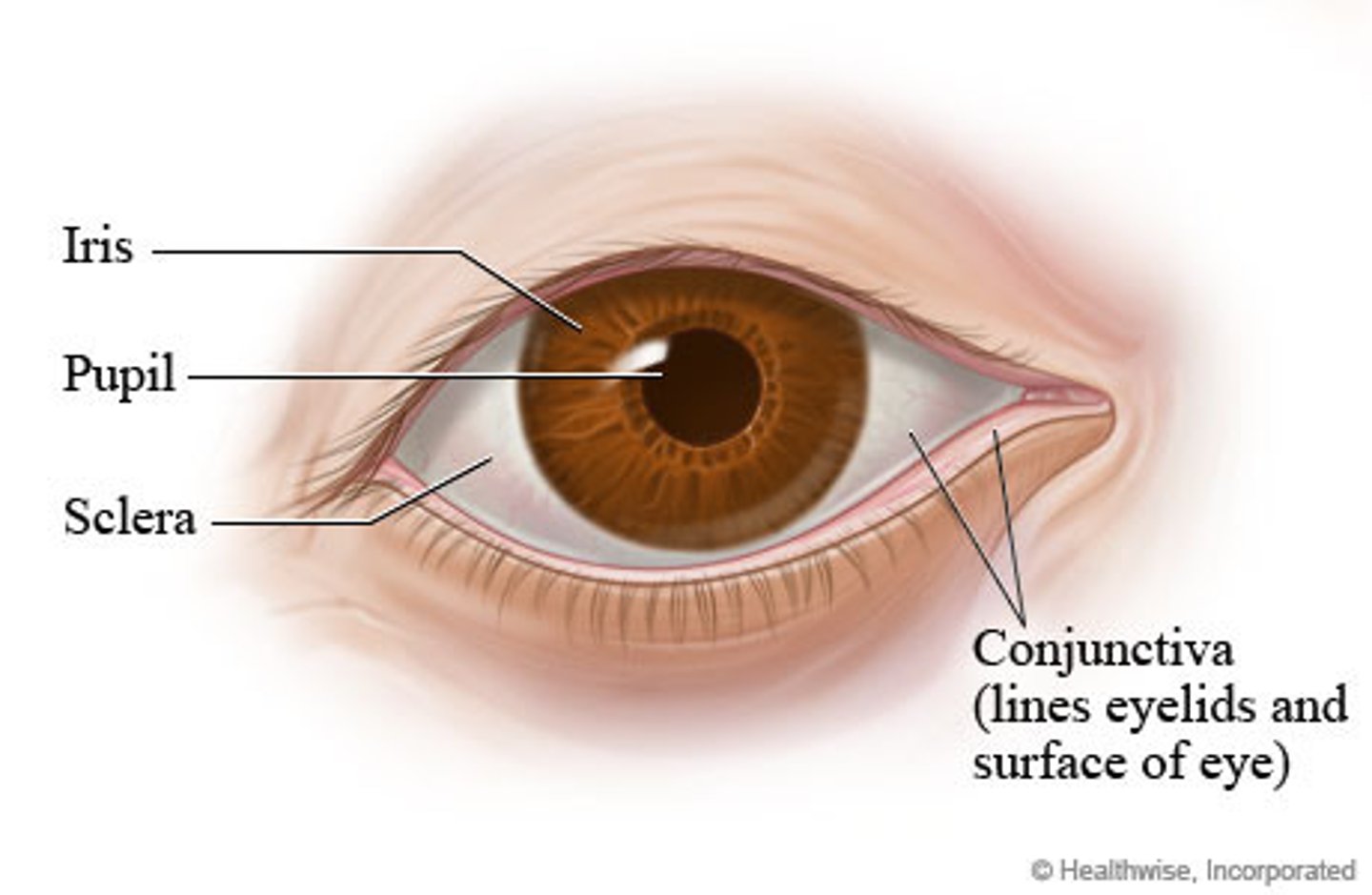
Iris
Colored part of the eye

pupil
the adjustable opening in the center of the eye through which light enters
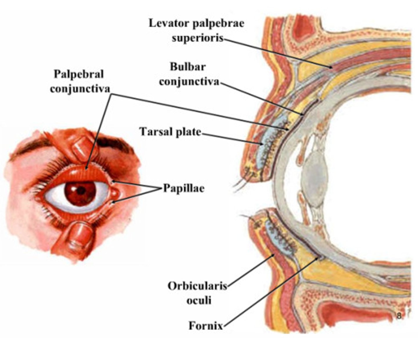
The tarsal plate of the eyelid ________.
is connected to the levator palpebrae
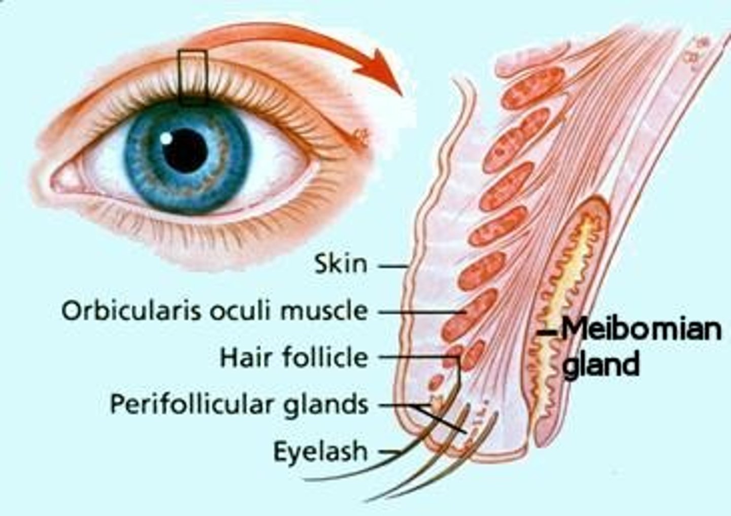
meibomian glands
oil glands found in the upper and lower edges of the eyelids that help lubricate the eye
conjunctiva
Delicate membrane lining the eyelids and covering the eyeball
palpebral conjunctiva
membrane that lines underside of eyelids (vascular and red)
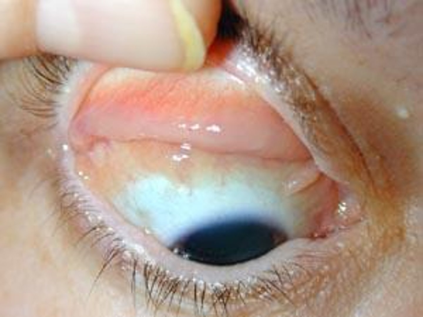
bulbar conjunctiva
overlays the eyeball, with the white sclera showing through (clear)
lacrimal gland
gland located in the upper outer region above the eyeball that secretes tears

nasolacrimal duct
passageway for tears from the lacrimal sac into the nose
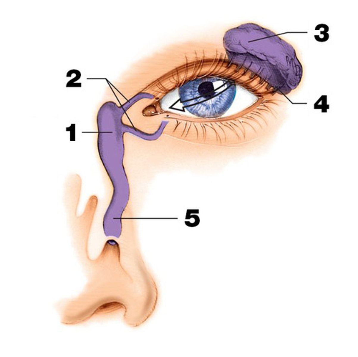
Canaliculi eye
portion of the lacrimal sac
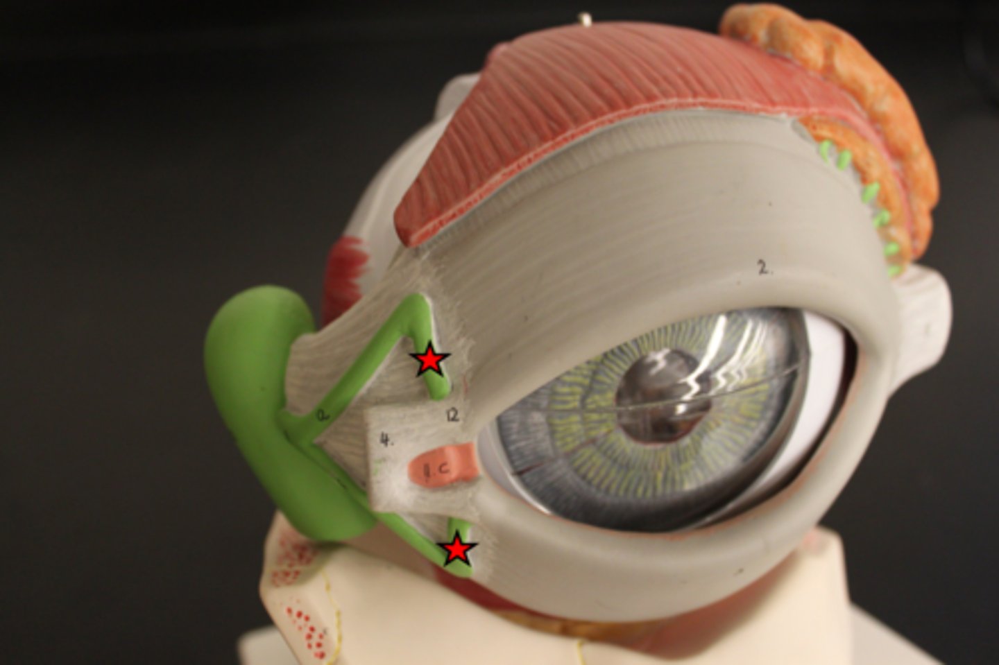
puncta
Tiny openings of the tear ducts.
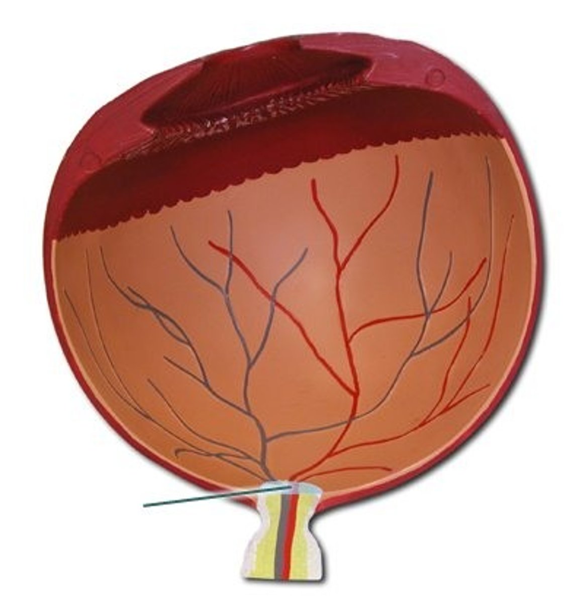
what are the 3 layers of the eye?
external - sclera and cornea
middle or vascular layer - iris, ciliary body and choroid = uveal tract
internal layer - retina
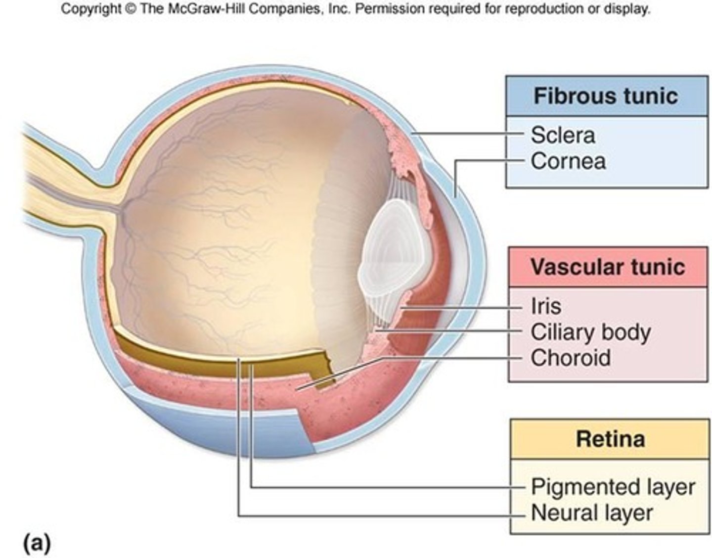
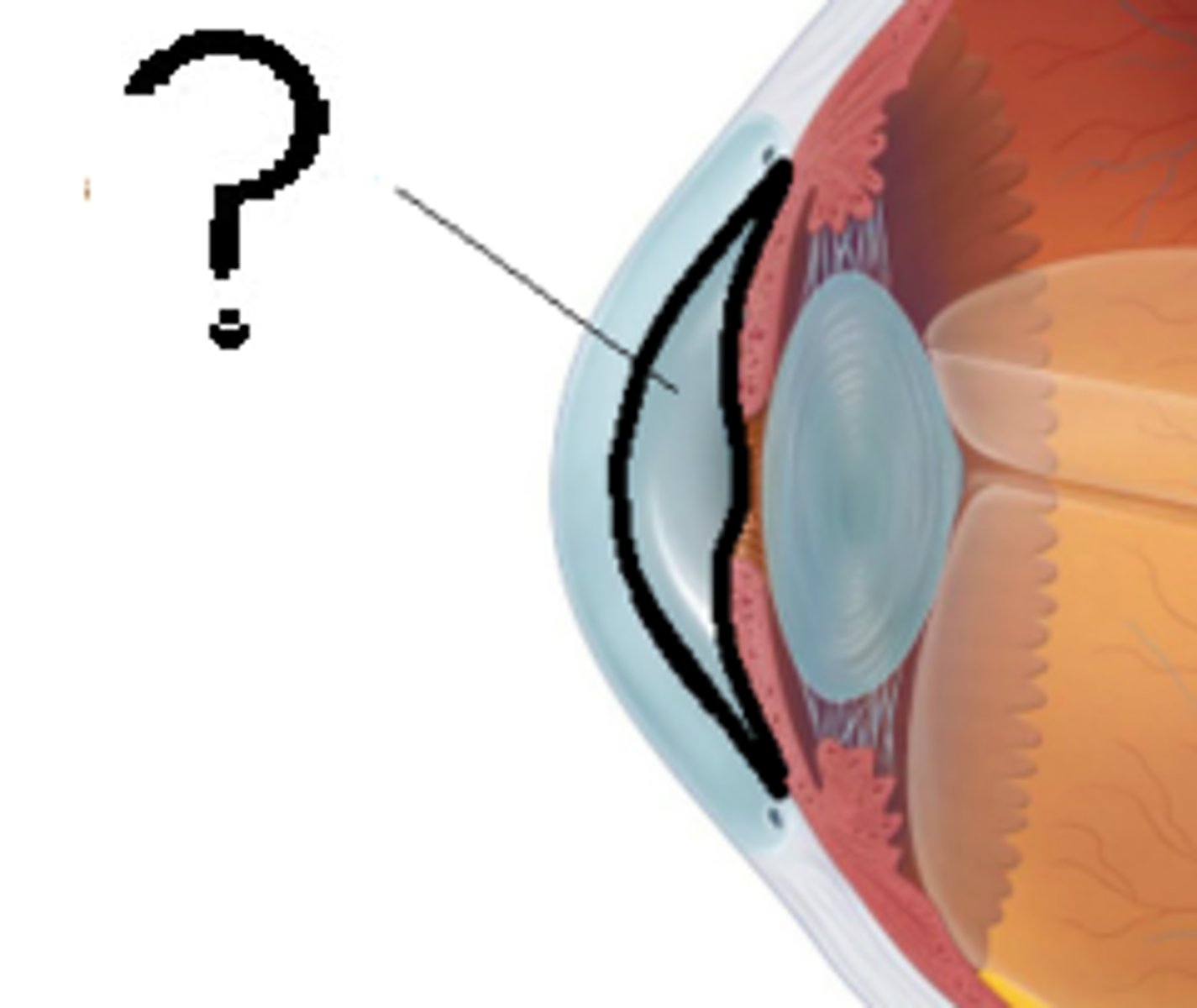
ciliary body
ring of tissue behind the peripheral iris that is composed of ciliary muscle and ciliary processes - attached to the lens and moves it
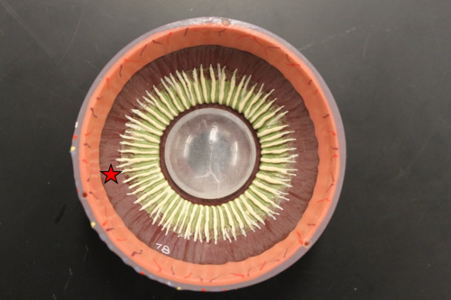
lens
the transparent structure behind the pupil that changes shape to help focus images on the retina
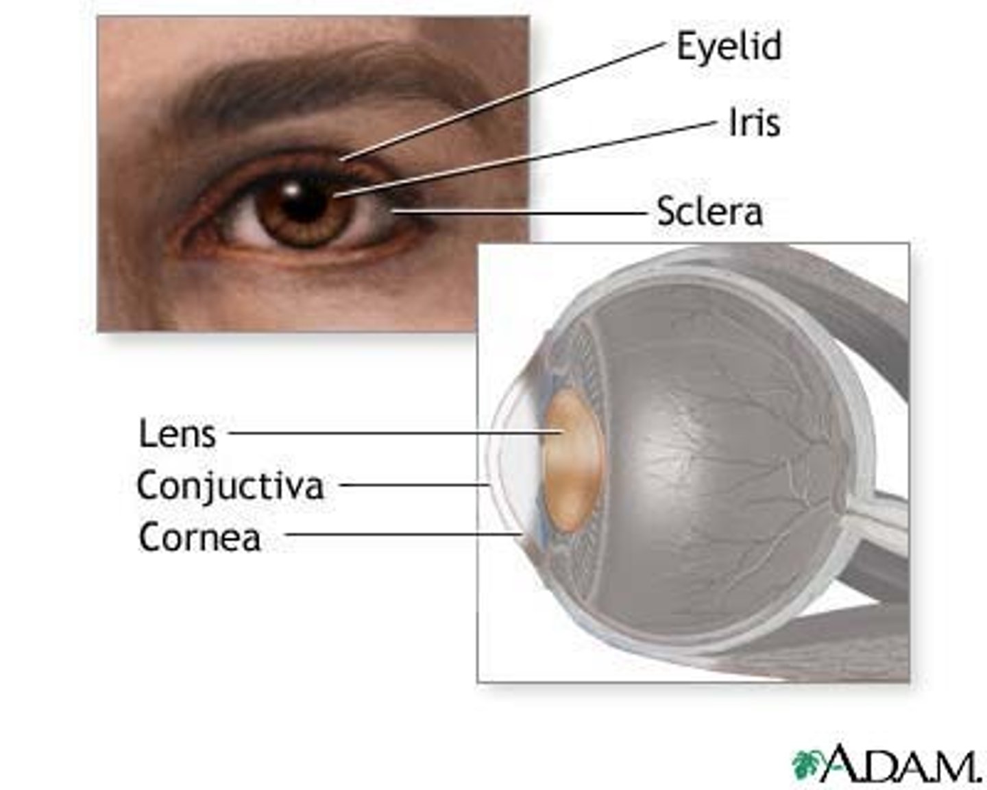
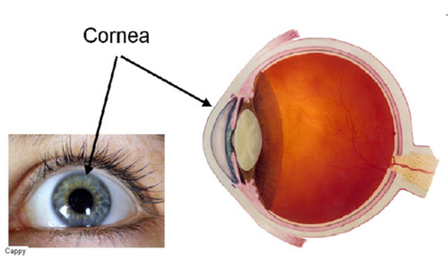
Cornea
The clear tissue that covers the front of the eye
macula lutea
a yellowish central area of the retina that is rich in cones and that mediates clear detailed vision
choroid
middle, vascular layer of the eye, between the retina and the sclera
Retina
the light-sensitive inner surface of the eye, containing the receptor rods and cones plus layers of neurons that begin the processing of visual information
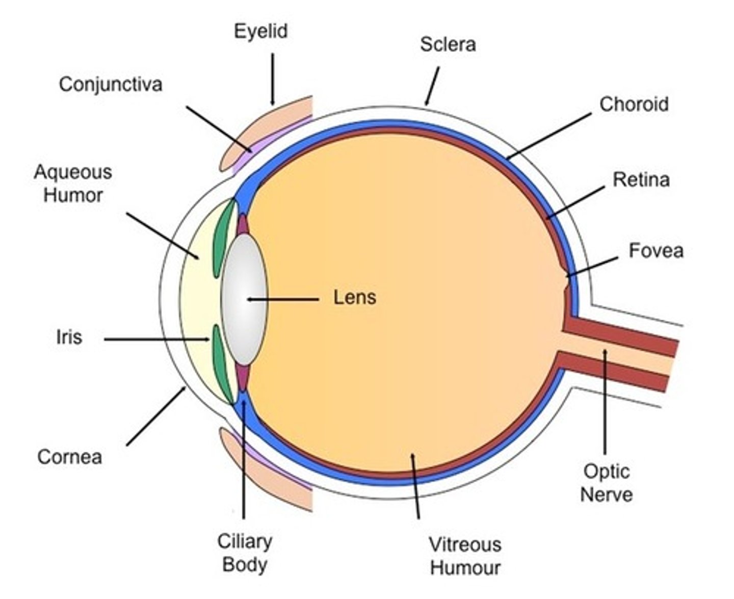
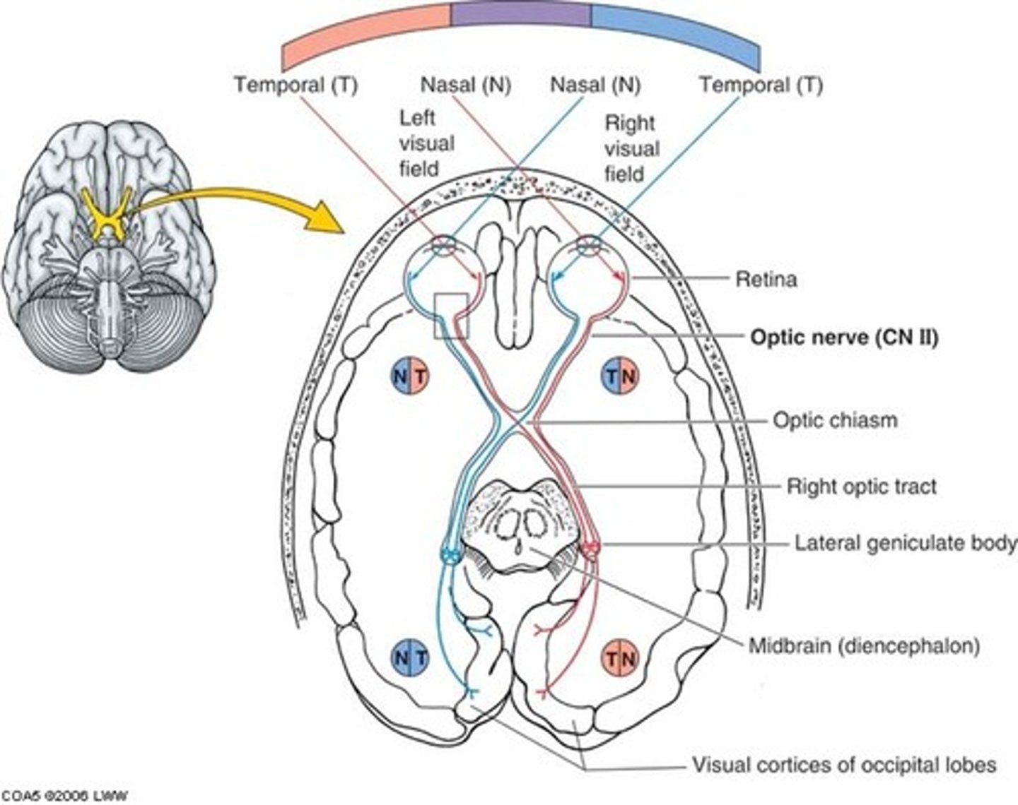
optic nerve
the nerve that carries neural impulses from the eye to the brain
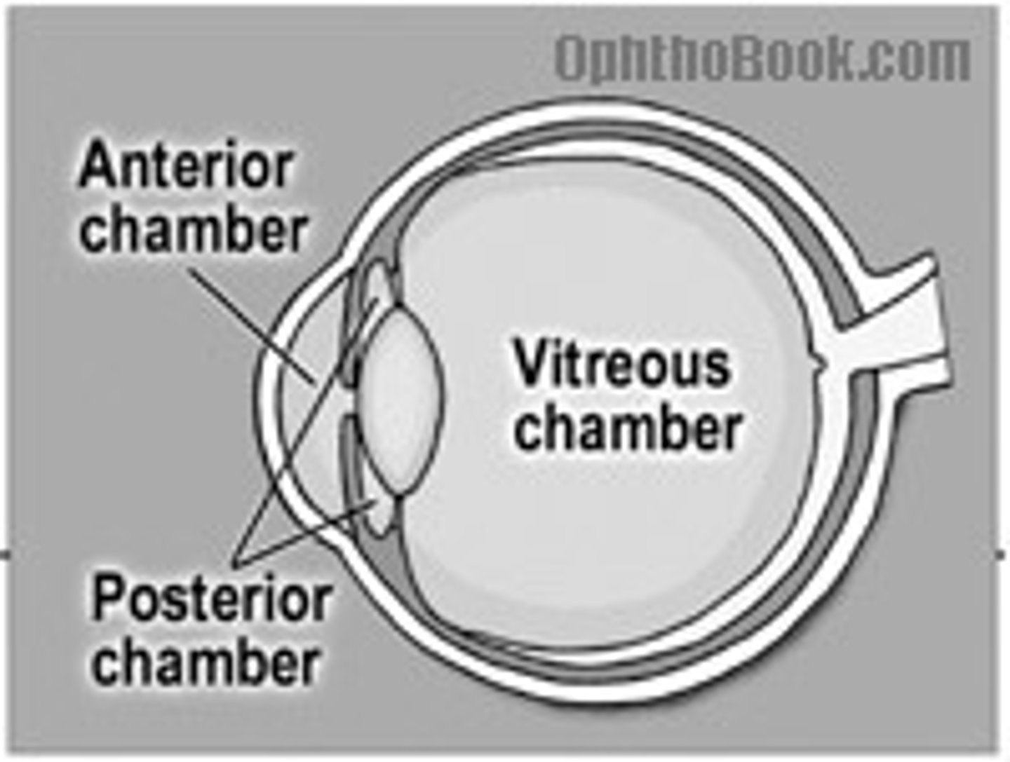
aqueous humor
fluid in the eye, found between the cornea and the lens (anterior chamber)
Canal of Schlemm
duct in the anterior chamber that carries filtered aqueous humor to the veins and bloodstream
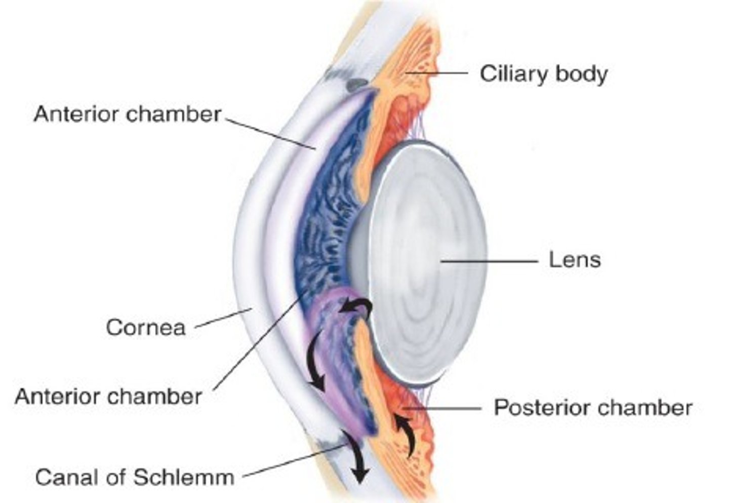
posterior chamber of eye
between iris and lens, filled with aqueous humor
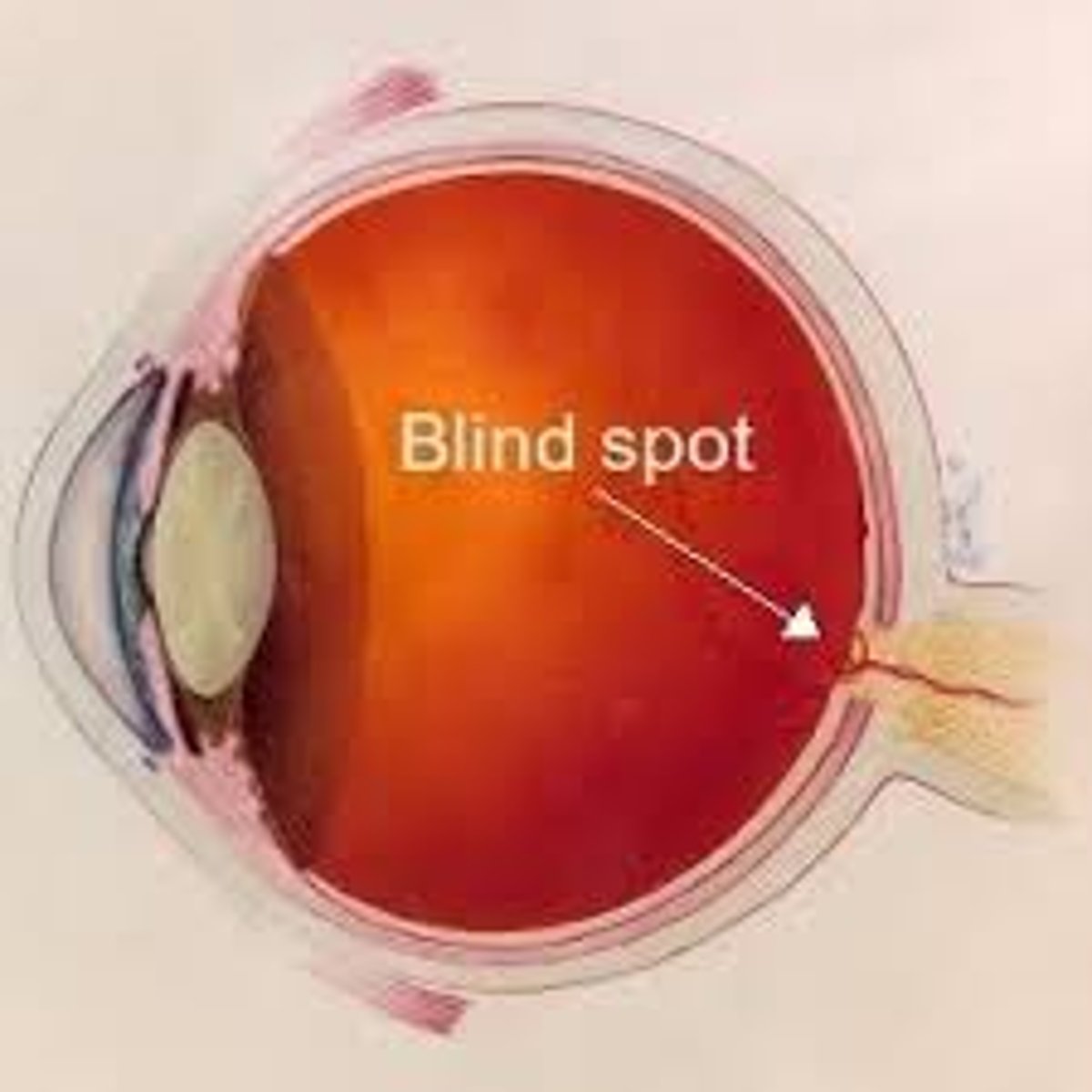
What is the blind spot?
the point at which the optic nerve leaves the eye, creating a "blind" spot because no receptor cells are located there
Is the sclera vascular or avascular?
avascular
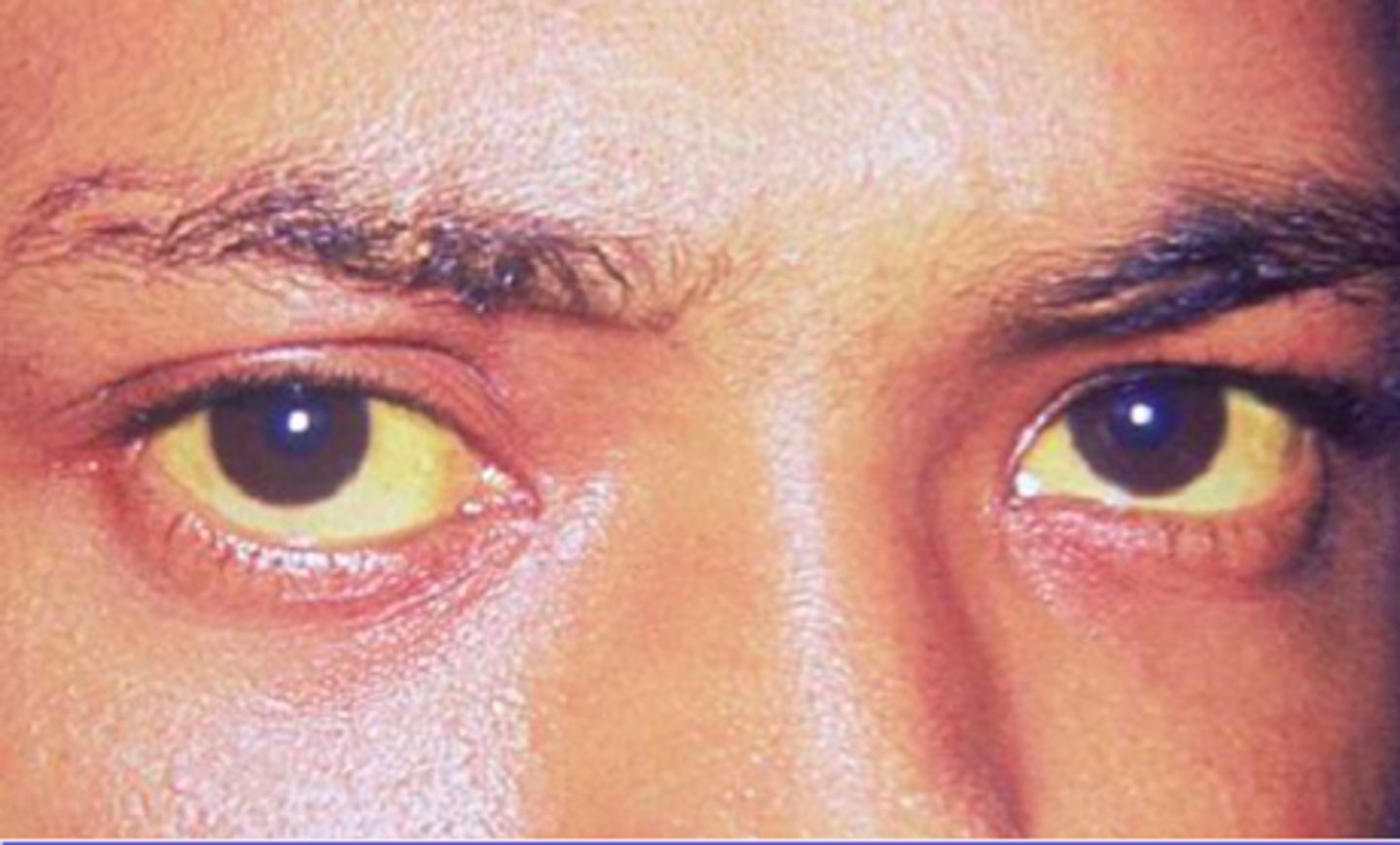
Scleral icterus
yellowing of the sclera due to jaundice
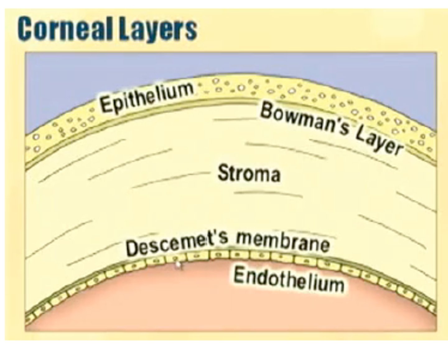
Is the cornea vascular or avascular?
avascular (has no blood vessels) - continuous with sclera
Bowman's membrane of cornea
tough, thin membrane of tightly woven collagen - prevents corneal scarring
what structure preforms the most refraction of incoming light?
cornea
uveal tract
the pigmented, vascular layer of the eye (iris, choroid), ciliary muscle, lens
What does the choroid do?
provides blood supply to retina and absorbs scattered light
iris controls
amount of light passing through the lens
circulation of aqueous humor
posterior chamber, anterior chamber, exits canal of schlemm, entering venous circulation
is the retina nervous tissue?
yes
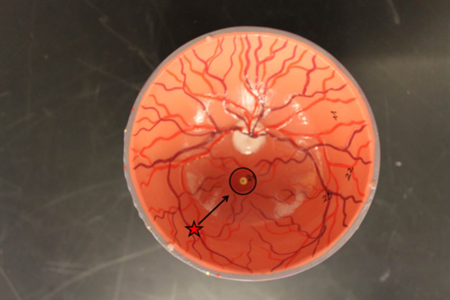
optic disc
is the origin of the optic nerve
optic cup
the depression in the center of the optic disc; ratio should be 1:2
Fovea
the central focal point in the retina, around which the eye's cones cluster (best spot of sight)
are arteries or veins more prevalent in the eye
A:V ratio should be 2:3
Snellen chart
used to measure visual acuity - should be 20 ft away
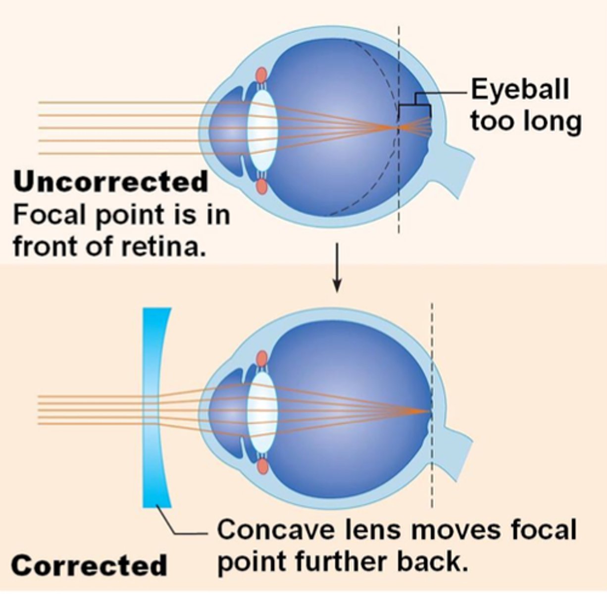
Myopia (nearsightedness)
occurs when the image is focused in front of the retina
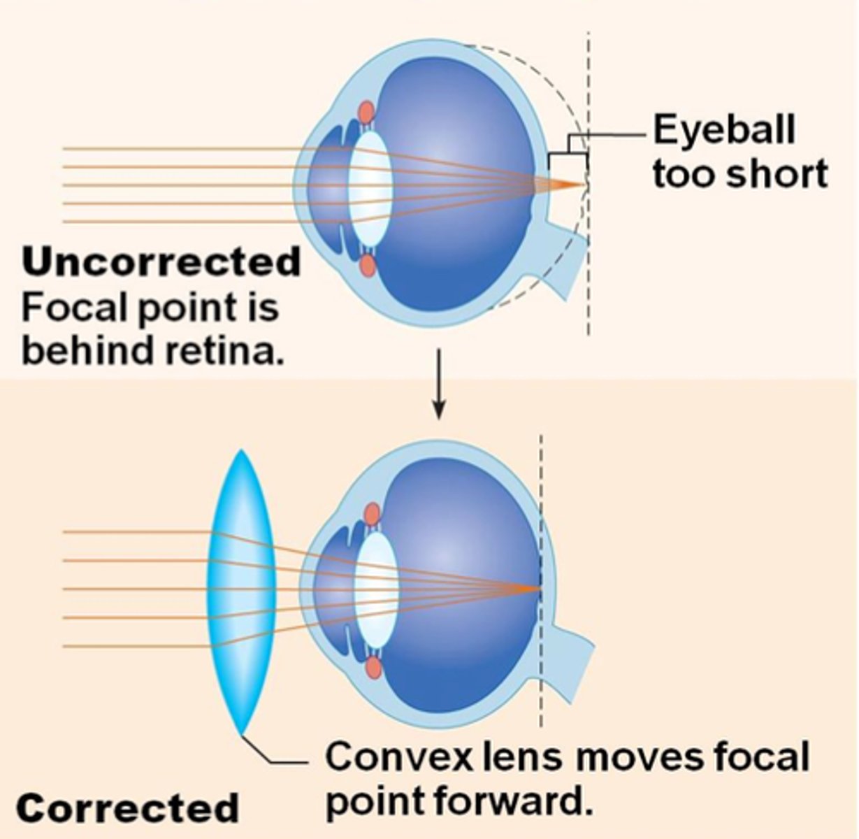
Hyperopia (farsightedness)
occurs when the image is focused behind the retina
OD, OS, OU
right eye,
left eye,
both eyes
what line should you read on snellen chart?
smallest line of print where the patient can read at least half
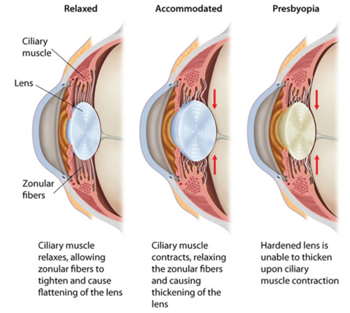
presbyopia
farsightedness caused by loss of elasticity of the lens of the eye, occurring typically in middle and old age.
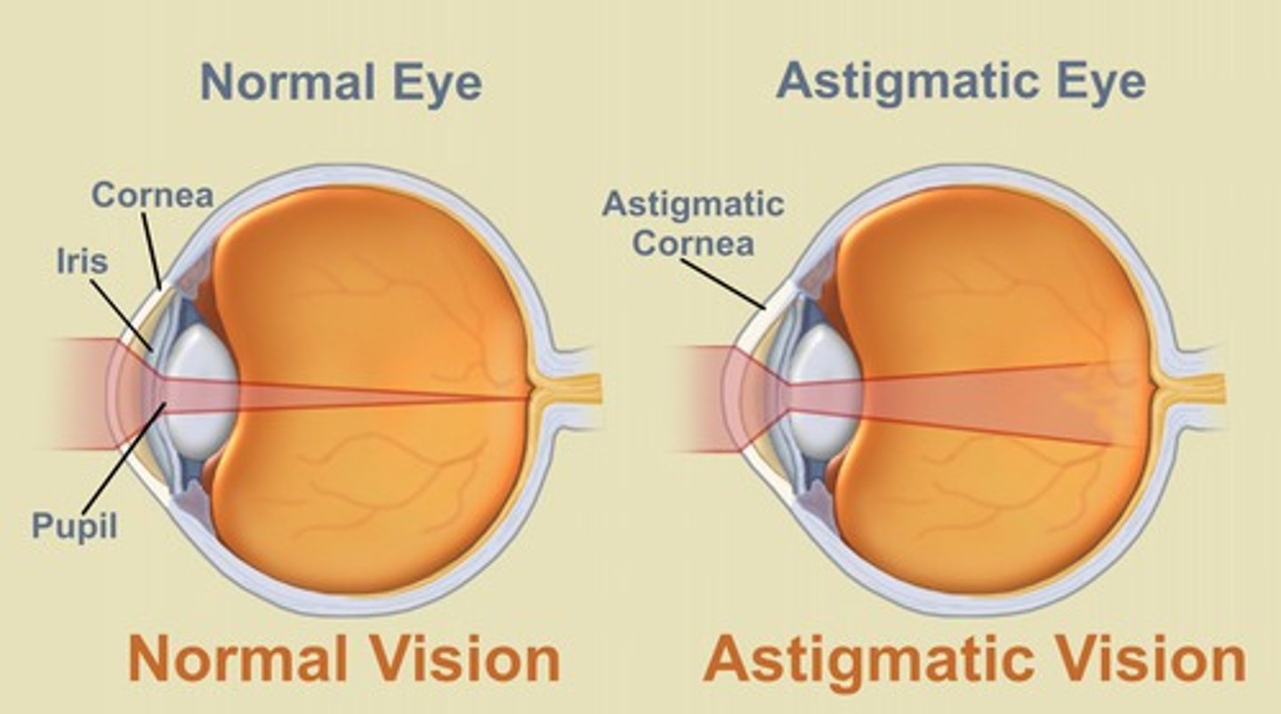
astigmatism
defective curvature of the cornea or lens of the eye - causes distortion
visual field
the whole area that you can see without moving your head or eyes
testing visual fields by confrontation
- take fingers from behind patient's shoulders, should see fingers at 90 degree
- take fingers from behind head, should see fingers at 50 degrees just after eye brows
- take fingers from behind torso, should see fingers at 70 degrees
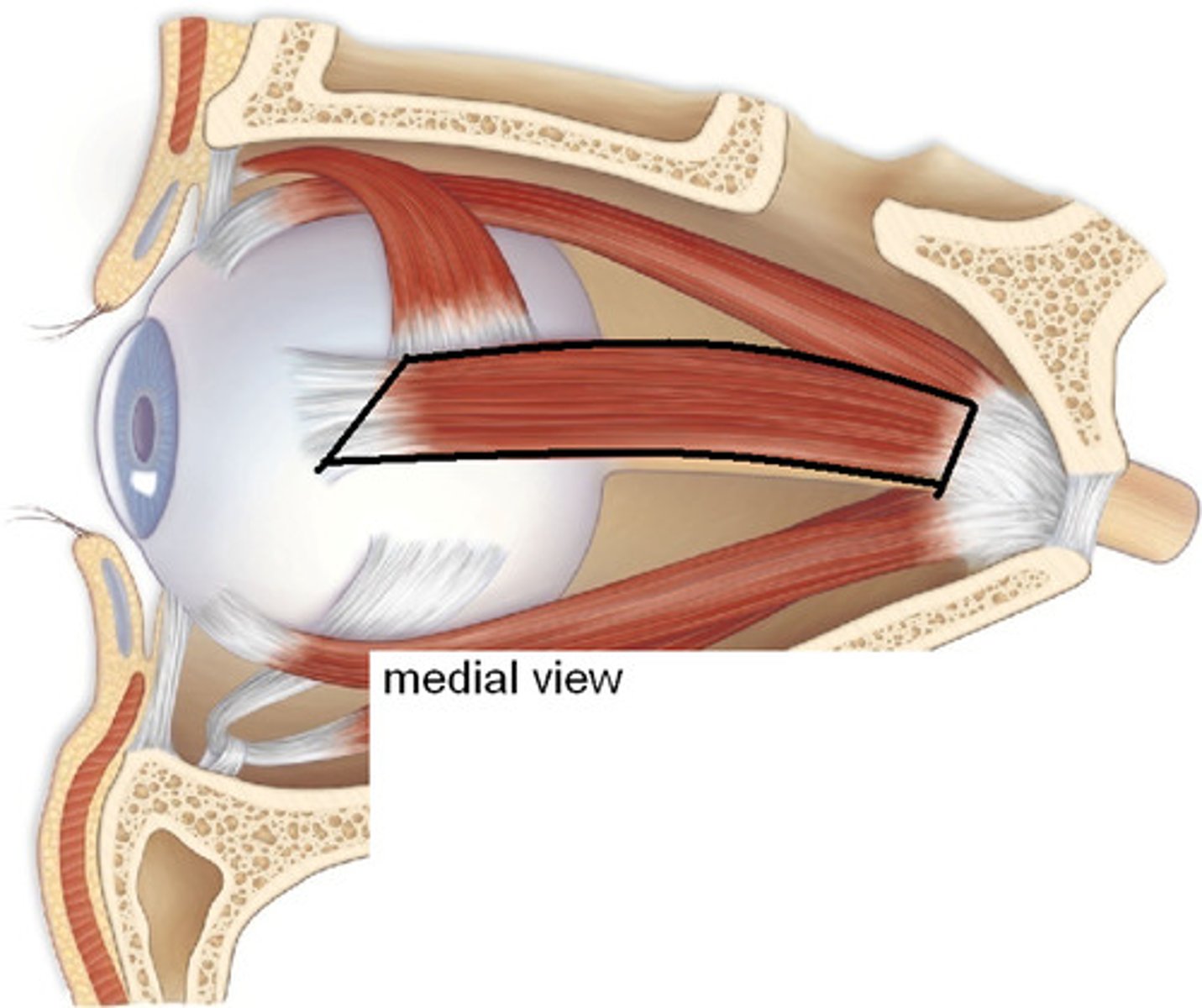
LR6 SO4 all the rest are 3
lateral rectus (6 - abducens)
superior rectus ( 4 - trochlear)
superior oblique (4 - trochlear)
inferior oblique (3 - oculomotor)
medial rectus (3 - oculomotor)
inferior rectus (3 - oculomotor)
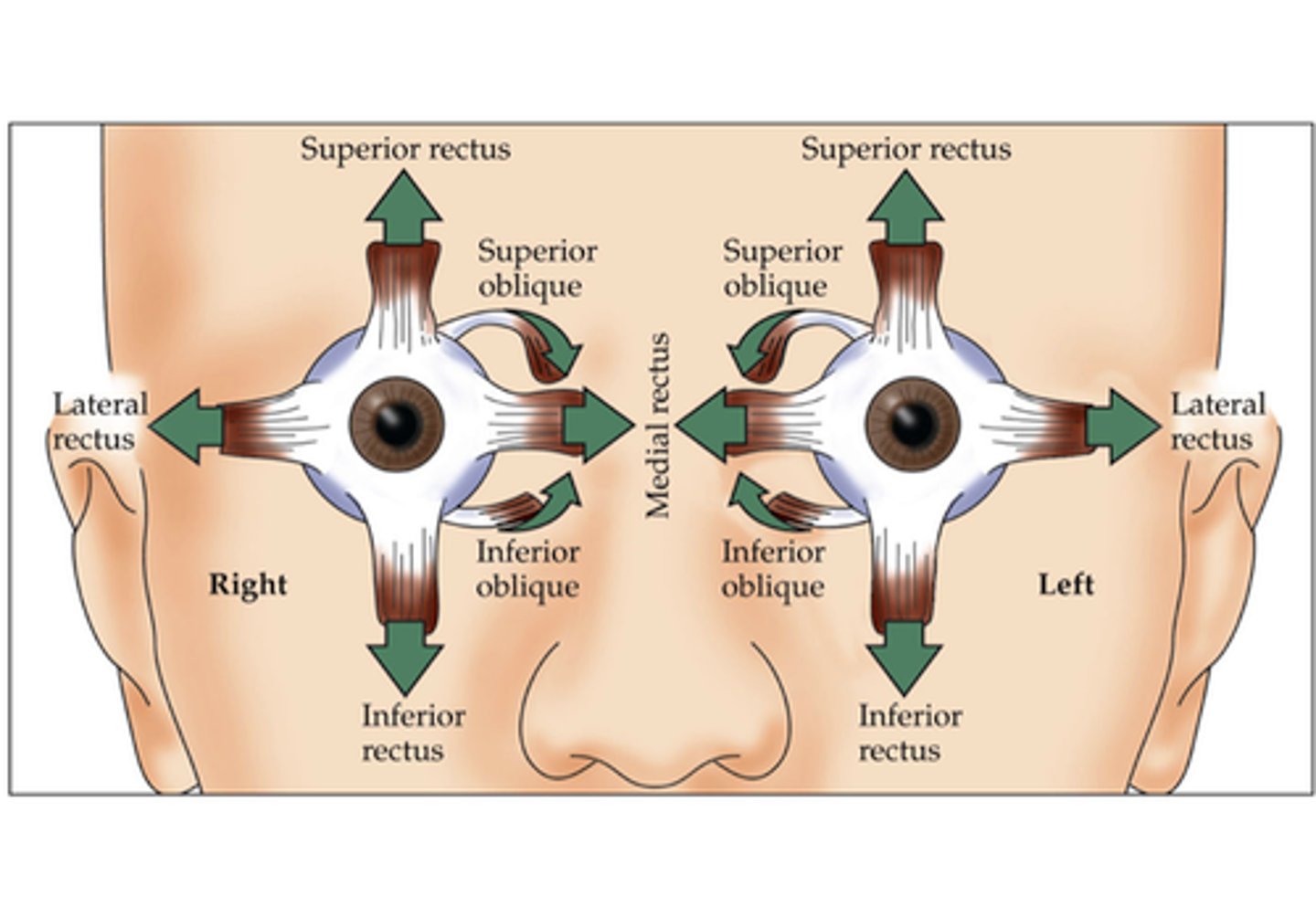
action of superior rectus
elevates eye

action of inferior rectus
depresses eye
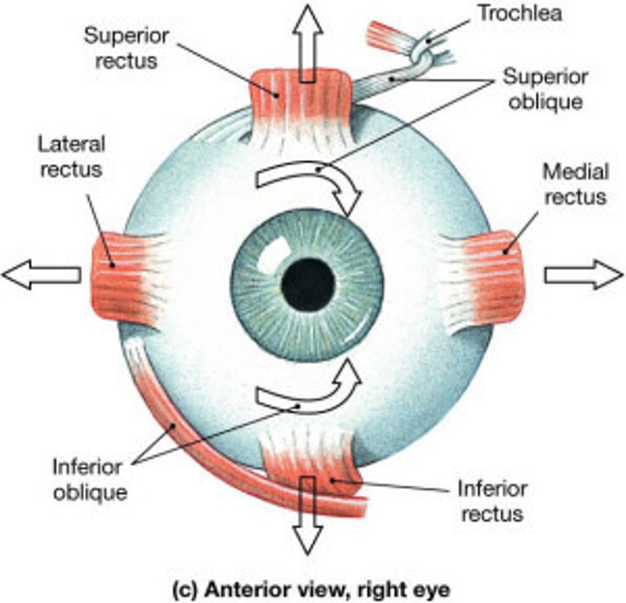
medial rectus
Turns the eyes inward towards the nose (add)
lateral rectus
moves eye laterally (VI abducens)
medial rectus innervation
Oculomotor nerve (CN III)
inferior rectus innervation
Oculomotor nerve (CN III)
superior rectus innervation
Oculomotor nerve (CN III)
superior oblique action
rotates eye towards nose and depresses eye

inferior oblique action
elevates eye and turns it laterally

superior oblique innervation
Trochlear nerve (CN IV)
inferior oblique innervation
Oculomotor nerve (CN III)
nystagmus
Involuntary rapid eye movements
convergence of eyes
eyes orient their visual axis toward object
what do pupils do during convergence?
pupils constrict to focus on a single point
accomodation of lens
lenses change shape to focus on objects near or far caused by contraction of the ciliary muscles
PERRLA stands for
pupils equal, round, reactive to light and accomodation
What does LLL mean?
Lids
Lashes
Lacrimals