Fetal Spine & Extremities
1/19
There's no tags or description
Looks like no tags are added yet.
Name | Mastery | Learn | Test | Matching | Spaced | Call with Kai |
|---|
No analytics yet
Send a link to your students to track their progress
20 Terms
what are the 5 sections of the spine?
cervical
thoracic
lumbar
sacral
coccyx
what are the 3 equidistant ossification centers of the vertebrae?
one anterior ossification center:
vertebral body
Two posterior ossification centers
pedicles and lamina
spinal defects are indicated by?
asymmetry of the ossification centers
splaying of the posterior centers
a break in the overlying skin
most common area of spinal defects?
lumbar region
which scan plane is most diagnostic?
transverse plane
skin and subcutaneous tissues along the posterior fetal back are best visualized in this plane…
LONG
describe US evaluation of the spine in long
two curvilinear lines extending from the cervical spine to the sacrum
referred to as the railway sign
required images are C, T, L, S spine
US evaluation of the spine coronally
spacing between lines is wider than in sag
cannot visualize the entire spine in one image due to fetal bedning/arching
required images are C,T,L,S spine
purpose of fetal extremity exams
to demonstrate and document
# of extremities
digits
contour of extremities
echoegenicity and shadowing
upper extremity
upper arm
humerus
forearm
radius
ulna
hand
digits
lower extremity
upper leg
femur
lower leg
tibia
fibula
foot
digits
describe US evaluation of humerus
long humerus found in sagittal plane-lateral from midline
shaft and acoustic shadow noted
cartilaginous head and distal elbow
US evaluation of radius and ulna
trace humerus to elbow to image radius and ulna
sag plane splays long axis of each bone
ulna is longer and located medially
radius is shorter than ulna and located laterally on thumb
Us evaluation of hands
count the digits
is the hand clenched all the time? try to specifically isolate the pinky finger
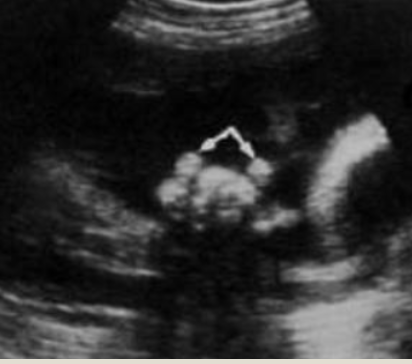
clenched fist
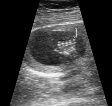
polydactyl
US evaluation of femur
distal epiphysis seen at the knee by 35 weeks
US evaluation of tibia and fibula
tibia lie medially and is larger than fibula
fibula lies laterally
US evaluation of feet
club foot suspected with abnormal flexion/inversion
toes should not be visualized with tib/fib