Models- Heart Lab, The Heart Model A&P Lab Practical II, The Heart, Heart Anatomy, A&P 2 Practical One
1/150
There's no tags or description
Looks like no tags are added yet.
Name | Mastery | Learn | Test | Matching | Spaced | Call with Kai |
|---|
No analytics yet
Send a link to your students to track their progress
151 Terms
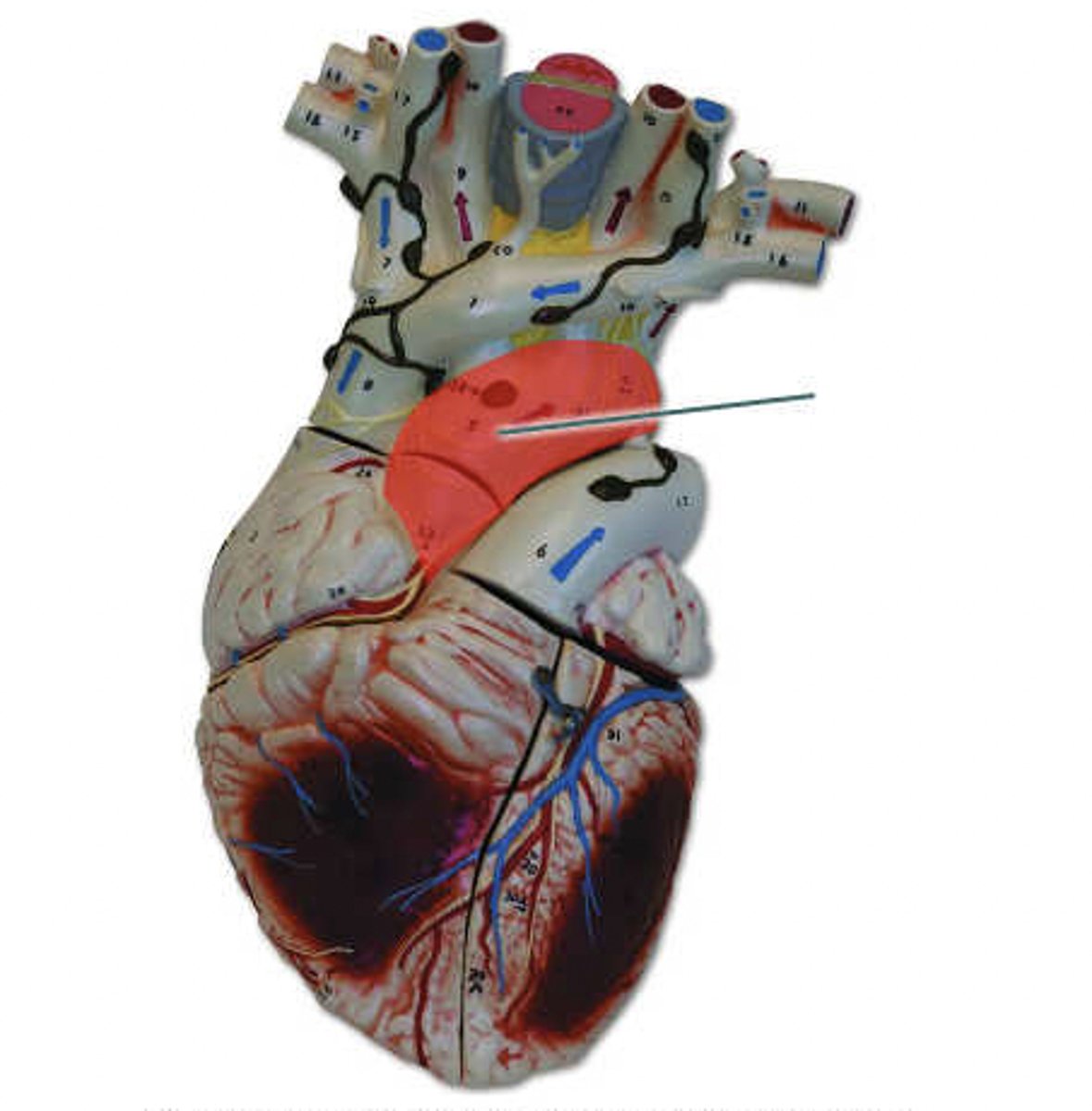
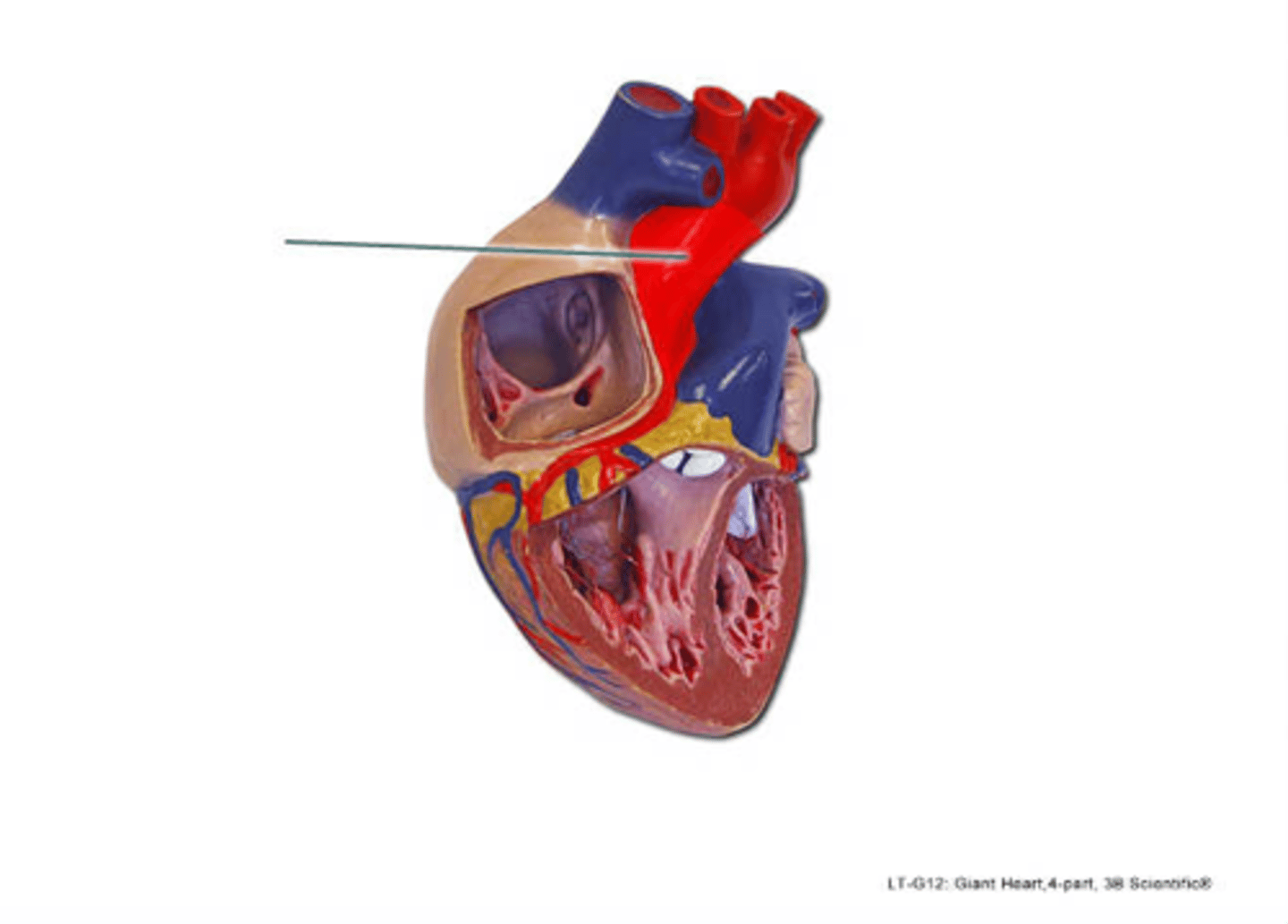
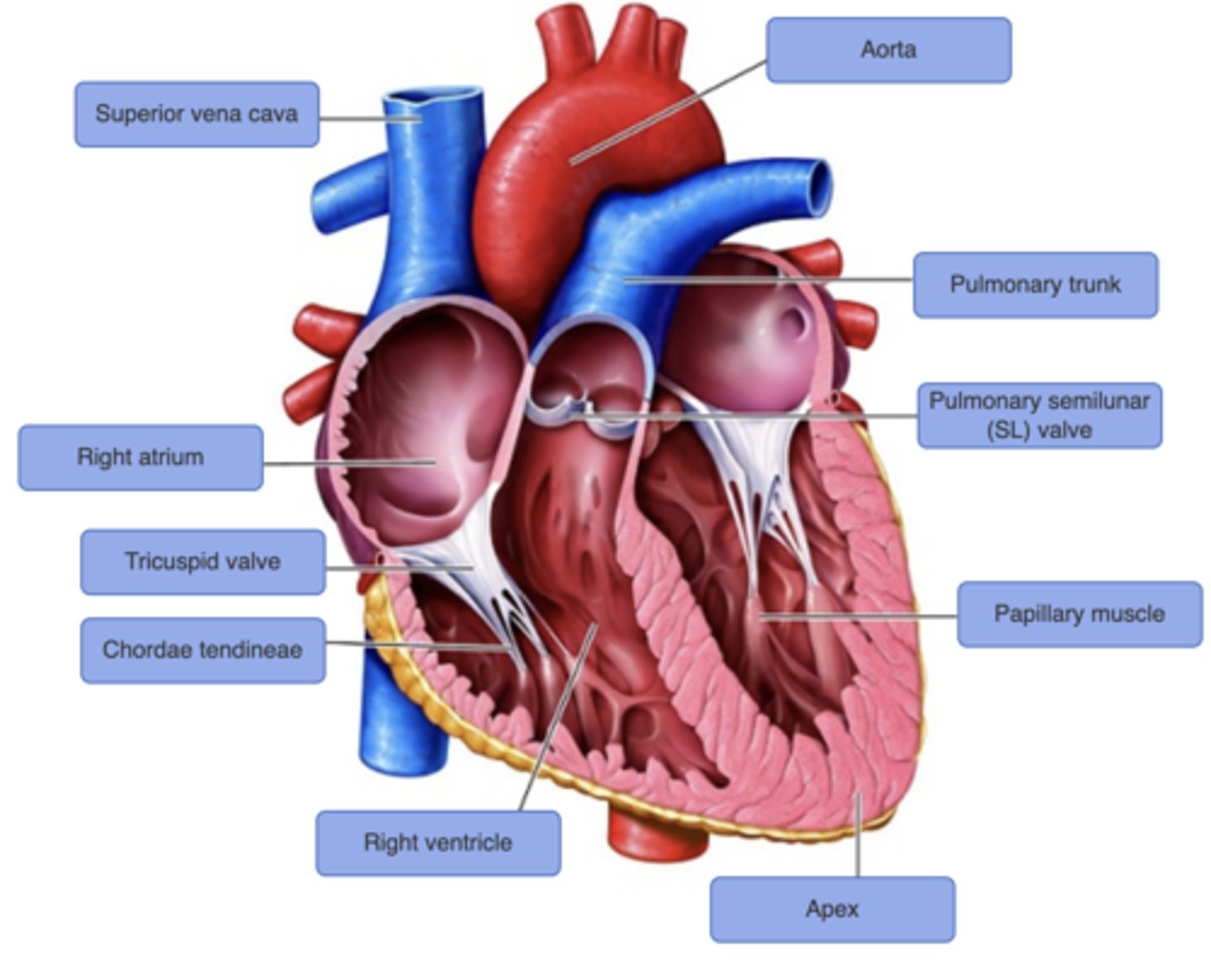
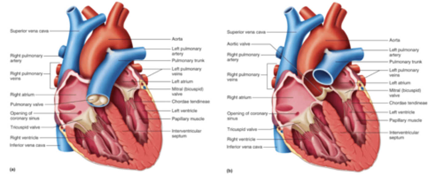
Aorta
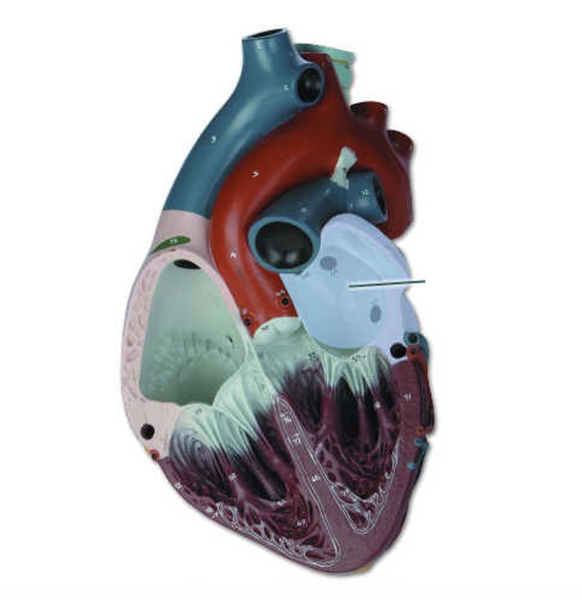
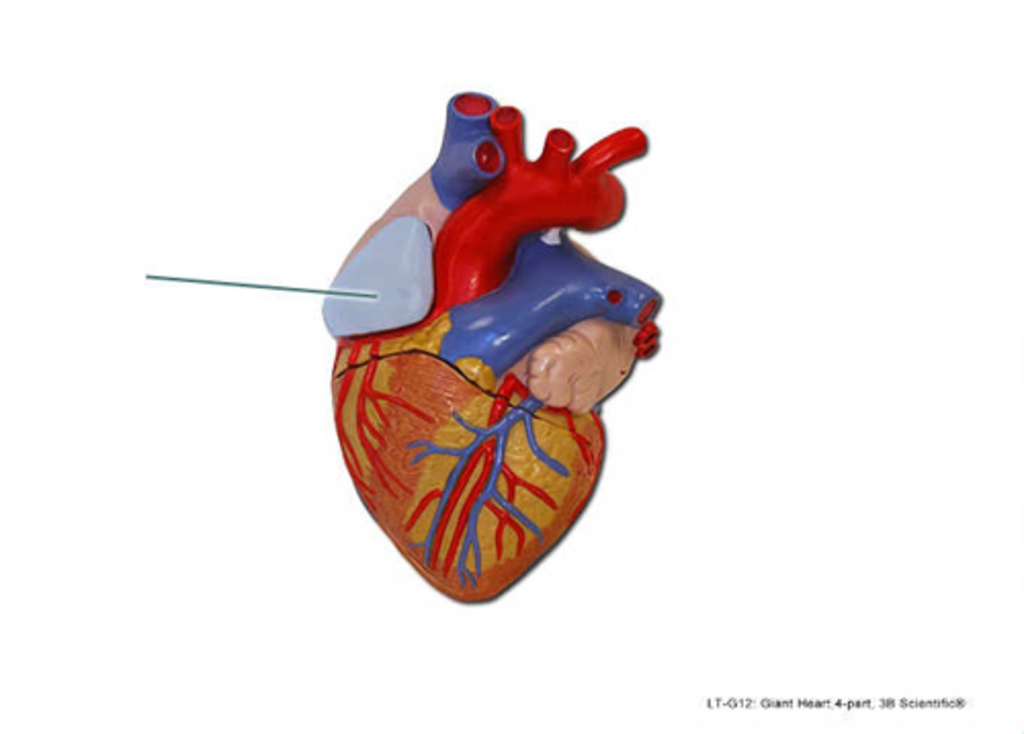
Identify the highlighted structure.
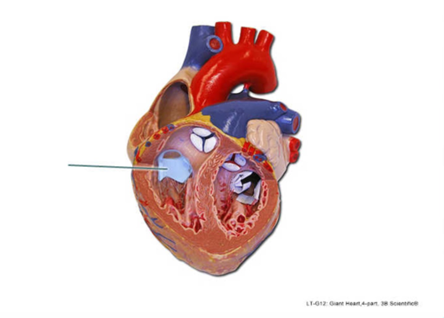
aortic semilunar valve
Identify the highlighted structure.
right auricle
Identify the highlighted structure.
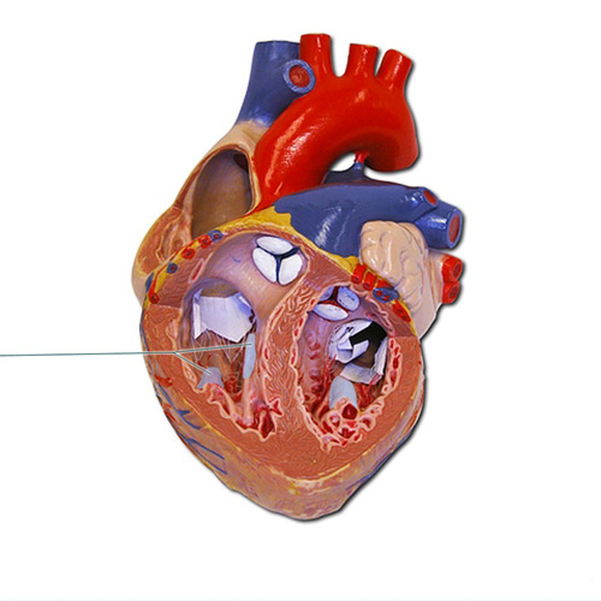
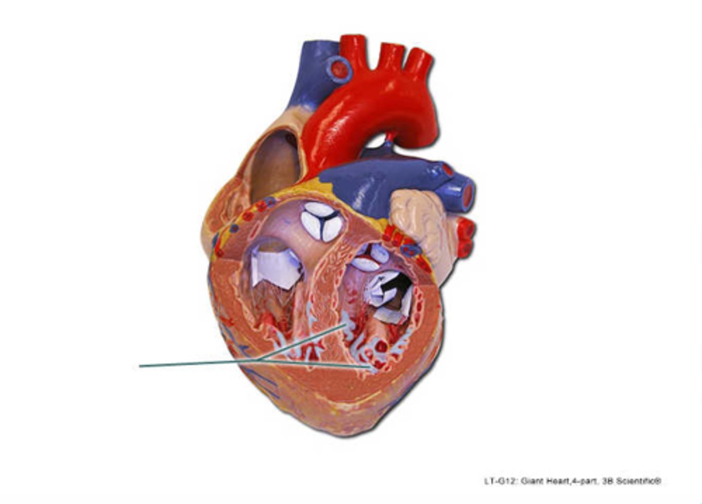
chordae tendineae
Identify the highlighted structure.
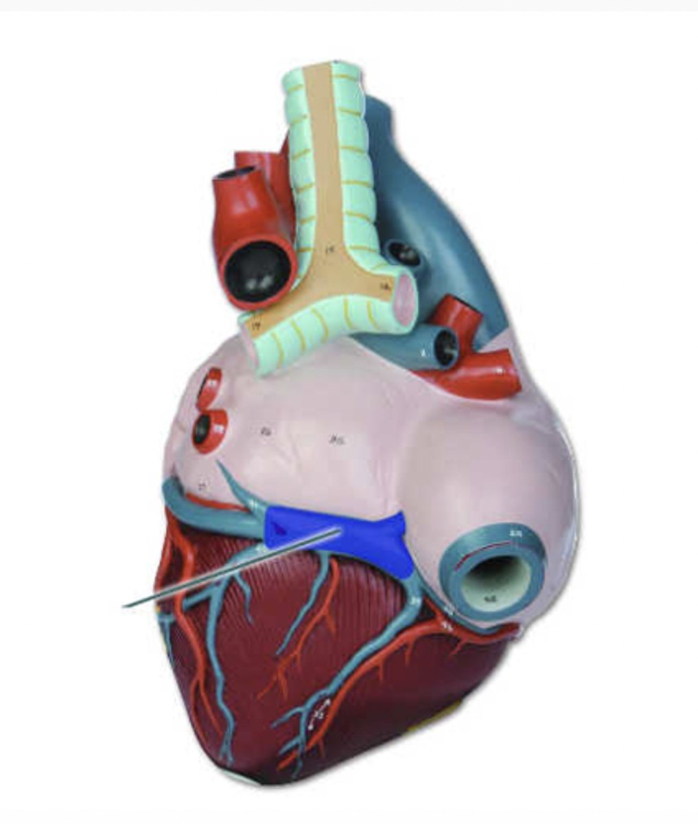
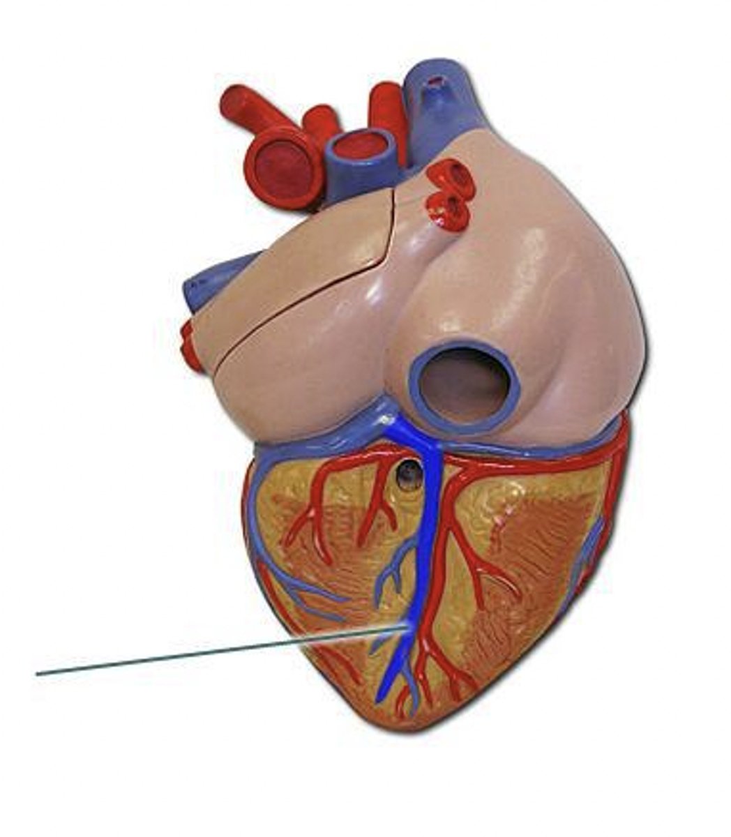
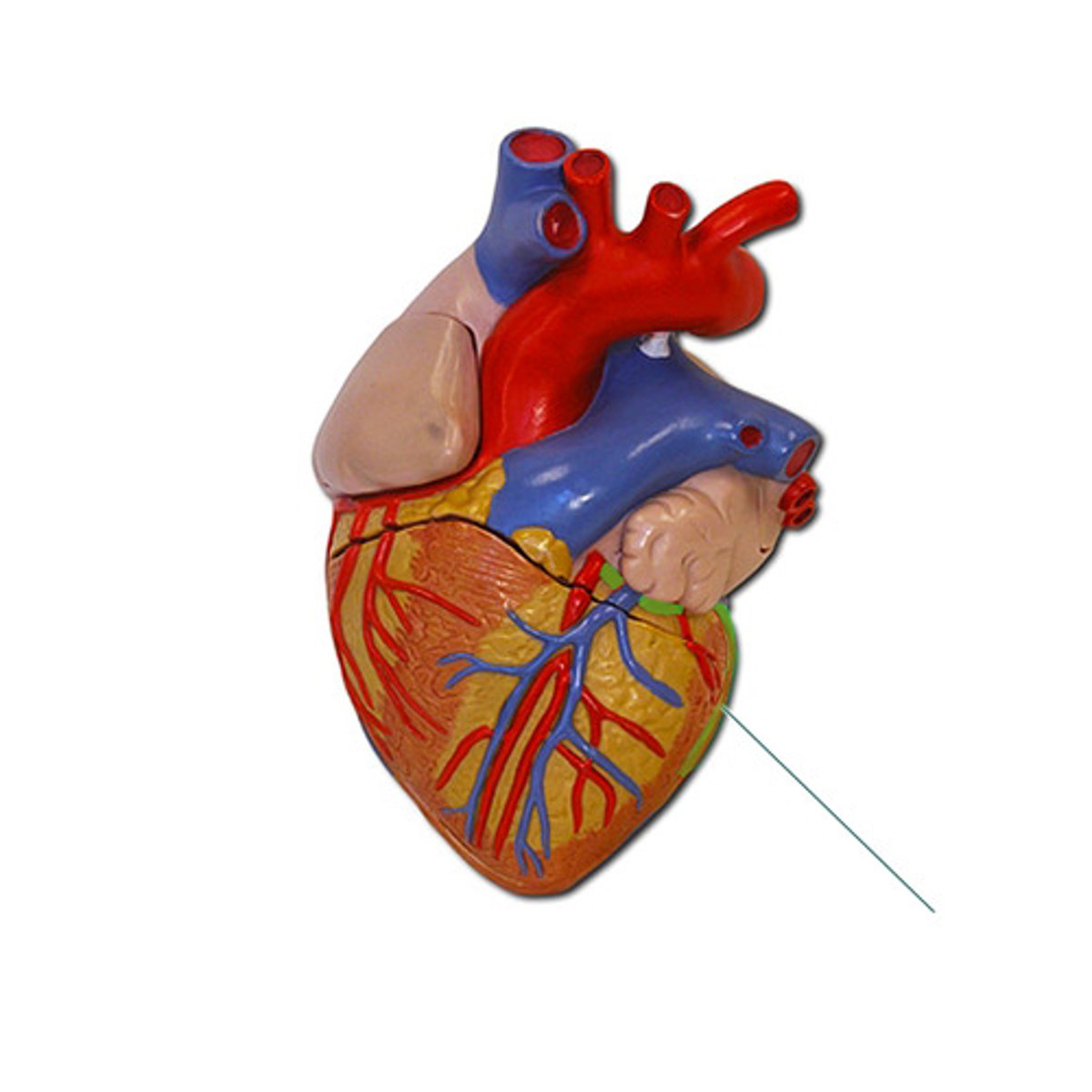
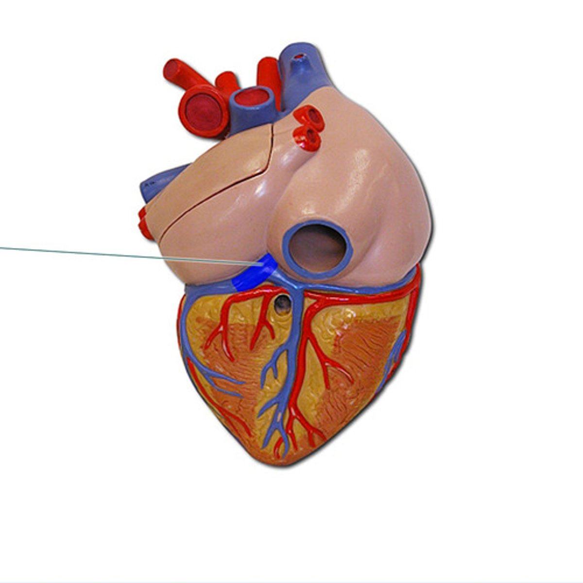
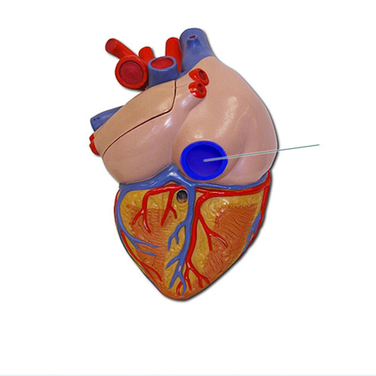
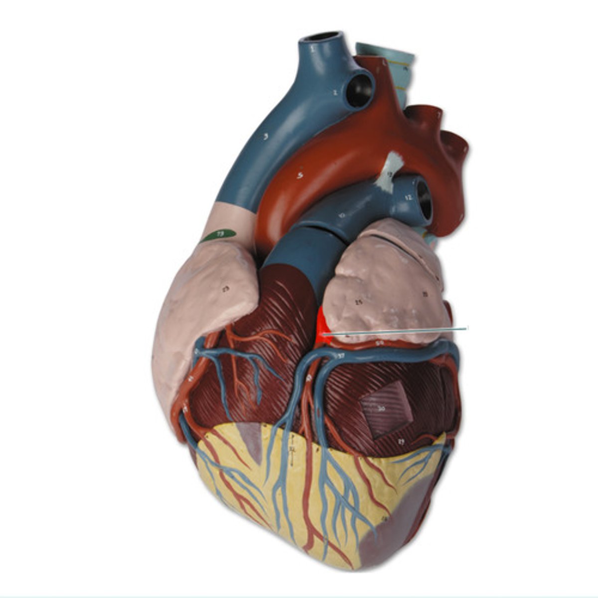
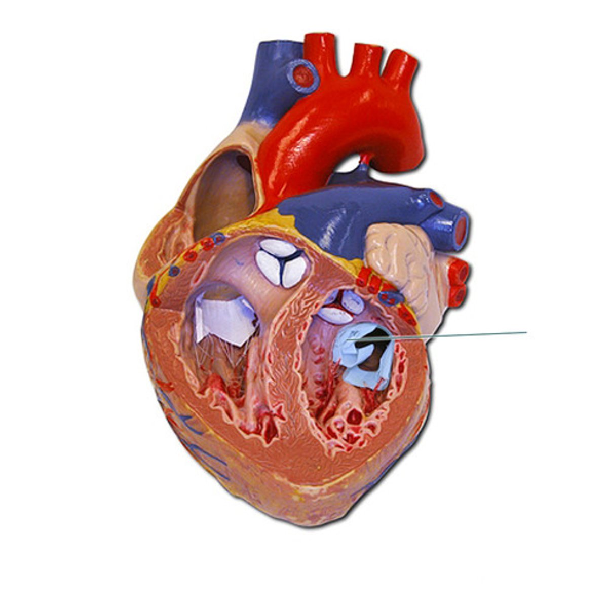
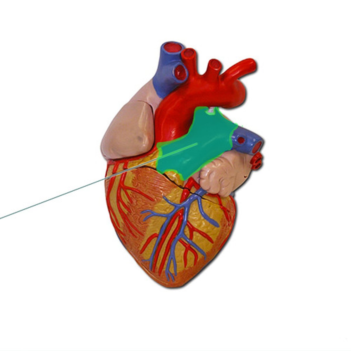
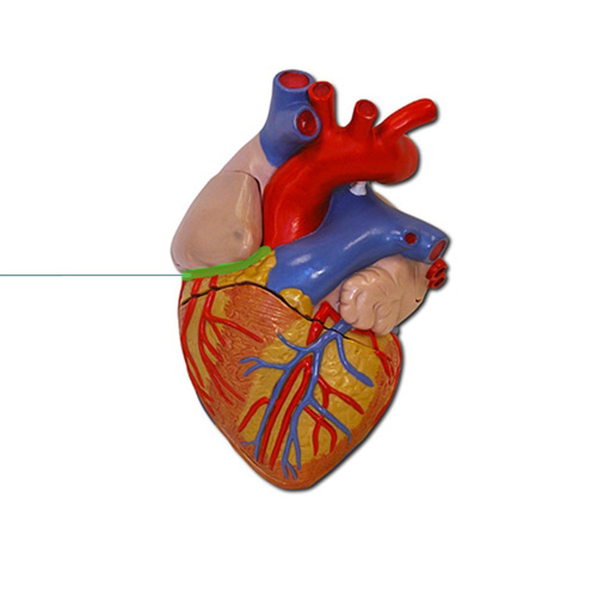
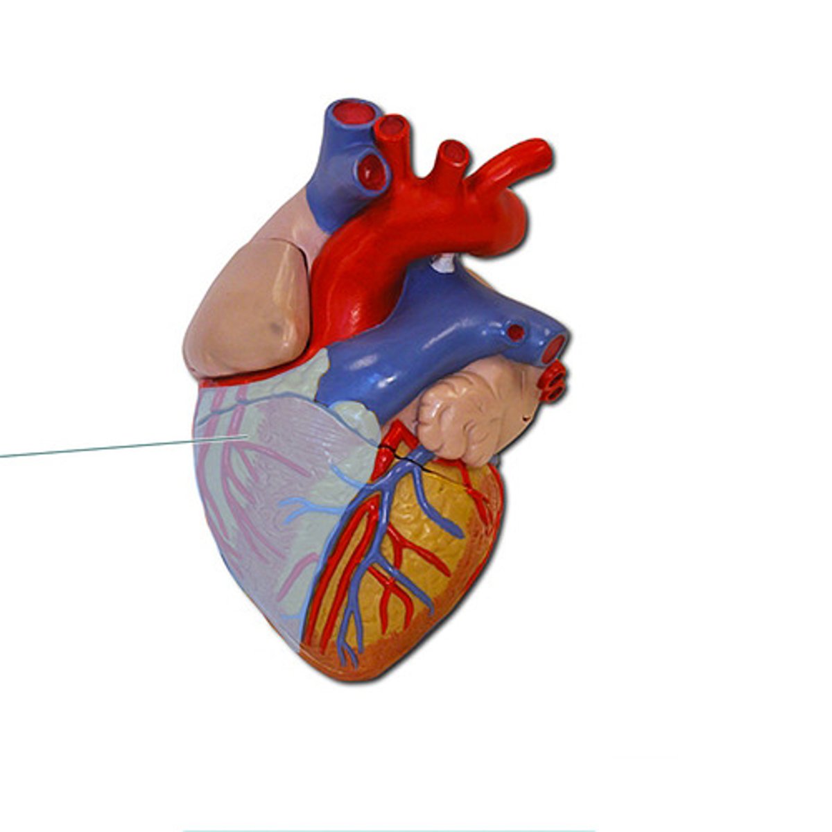
coronary sinus
Identify the highlighted structure.
Inferior vena cava, right atrium
The highlighted vessel (name it) returns blood from the body to which chamber of the heart?
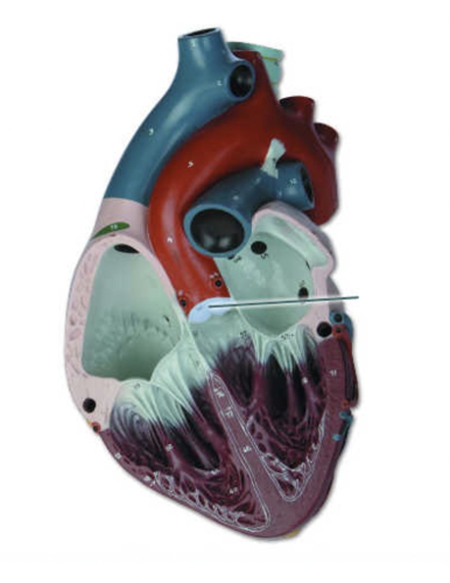
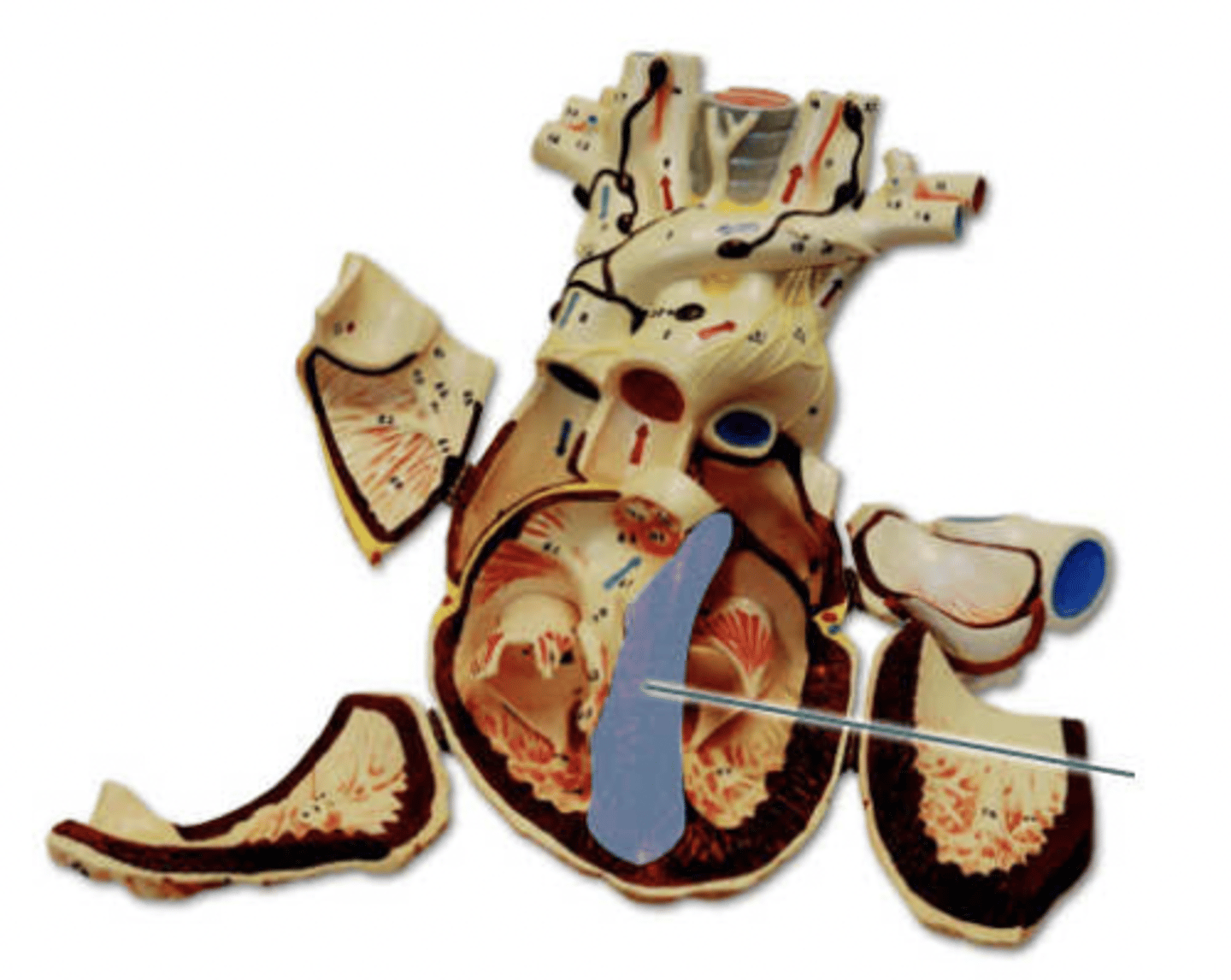
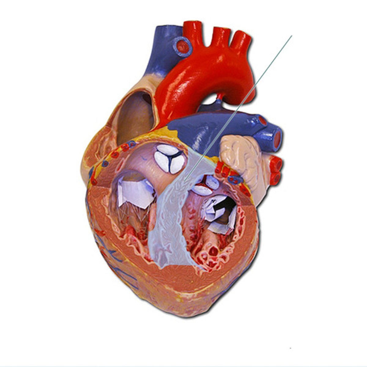
interatrial septum
Identify the highlighted structure.
interventricular septum
Identify the highlighted structure.
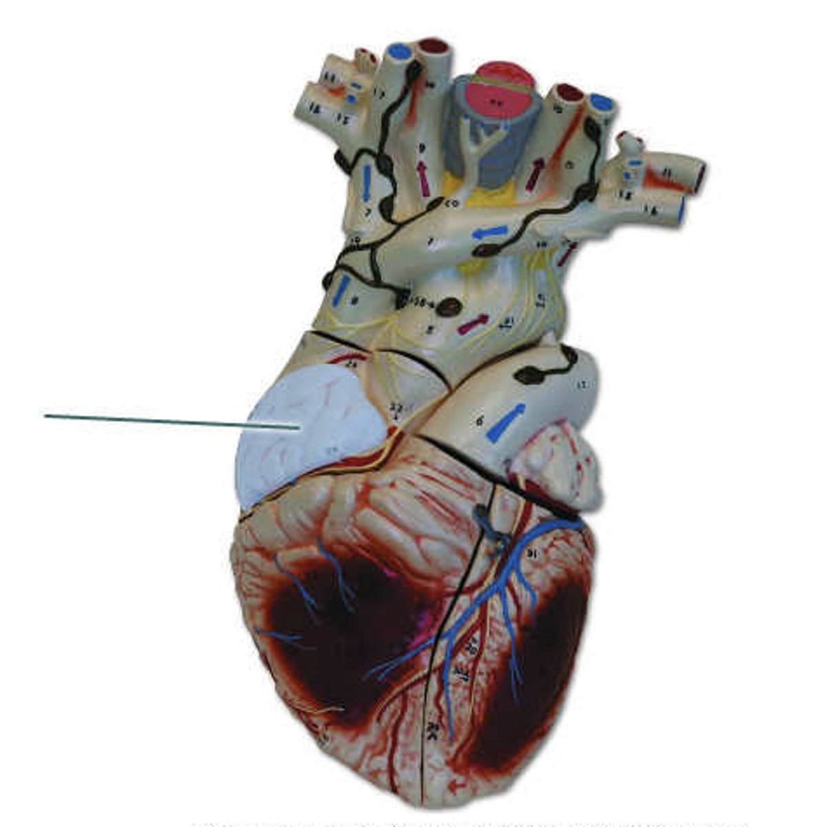
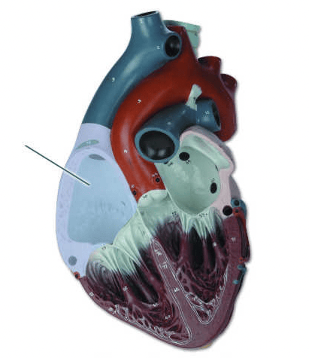
left atrium
Identify the highlighted structure.
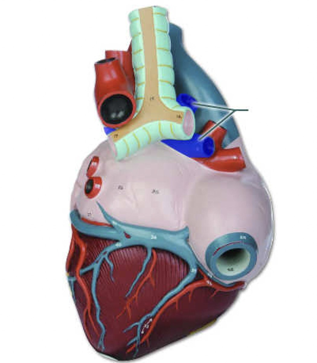
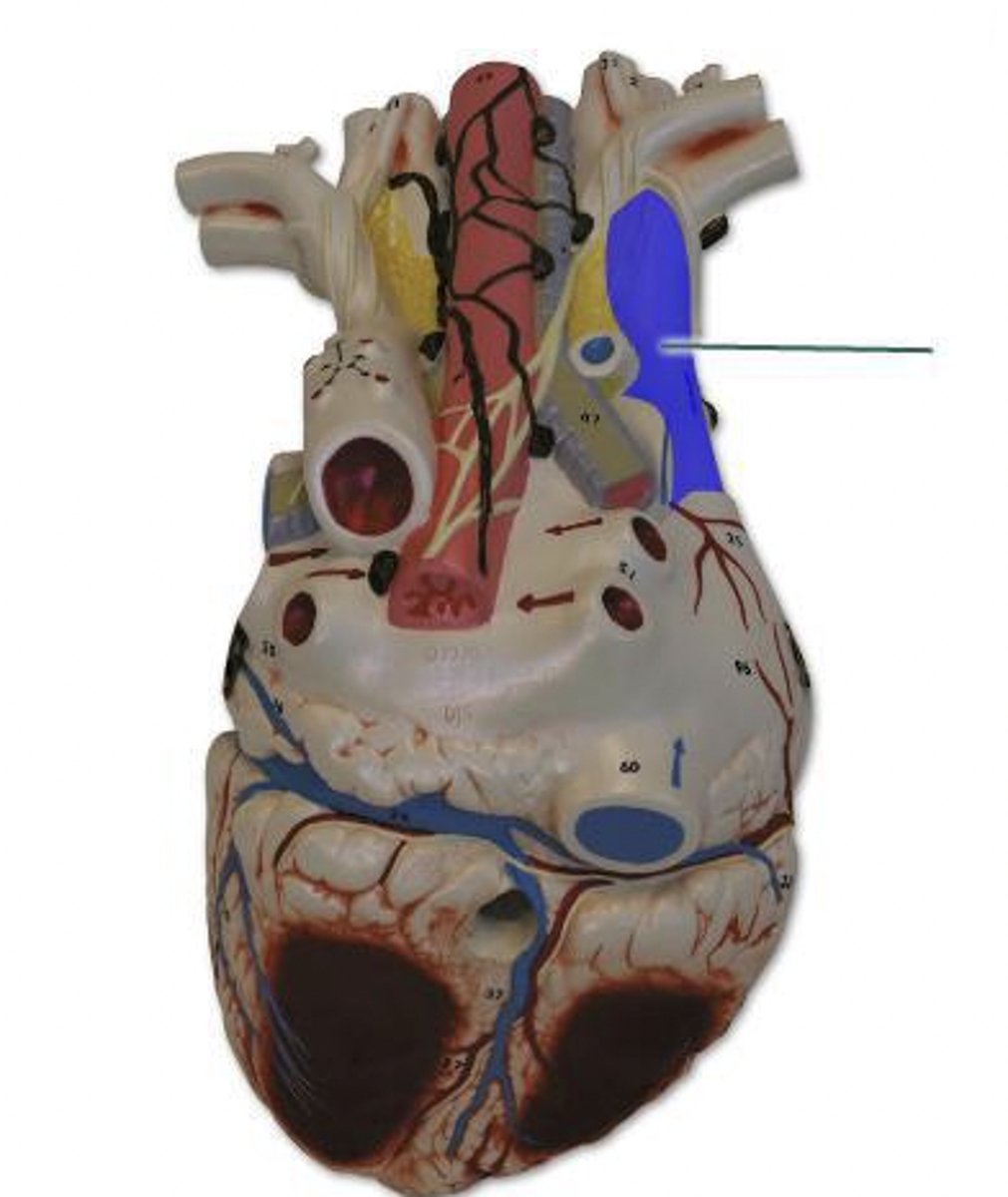
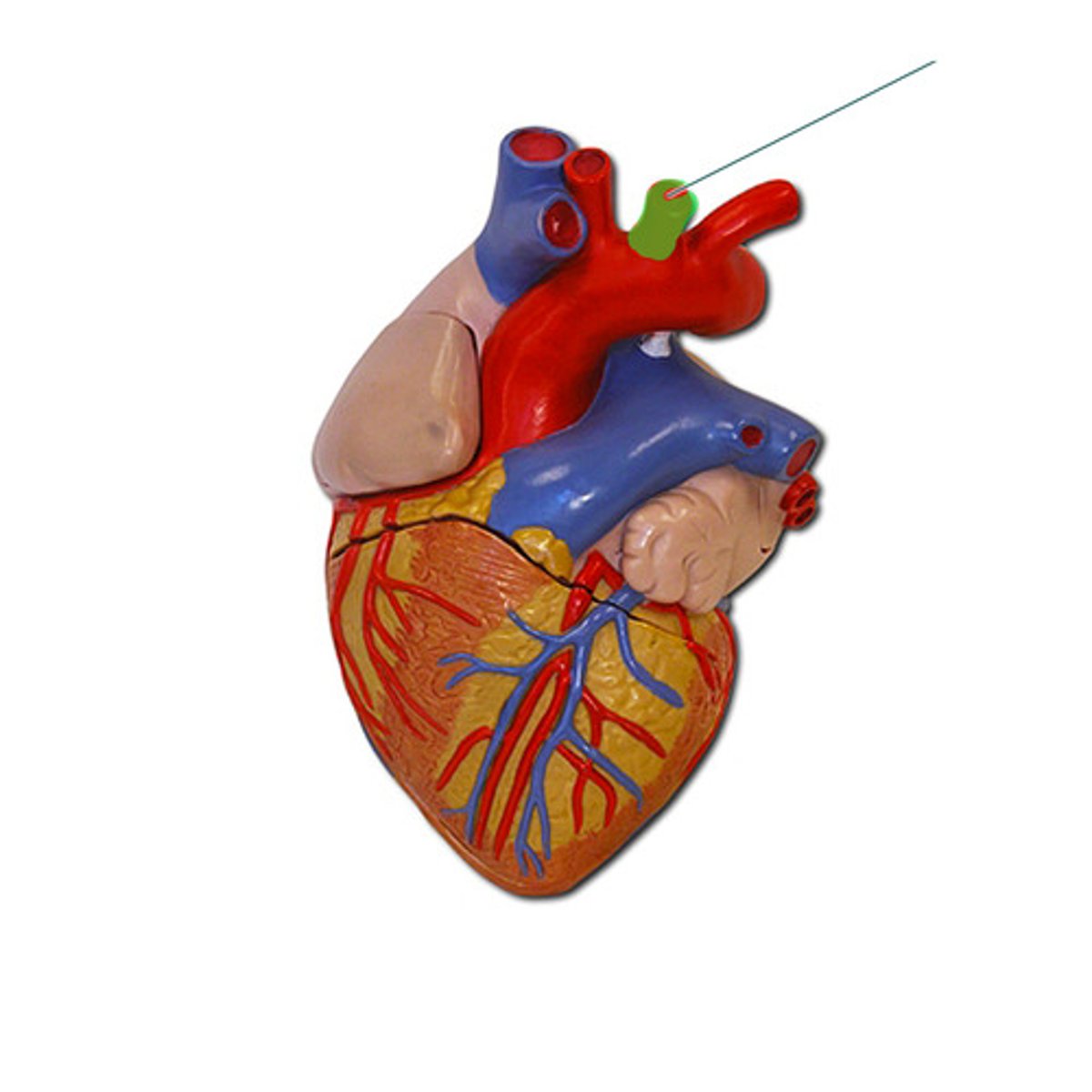
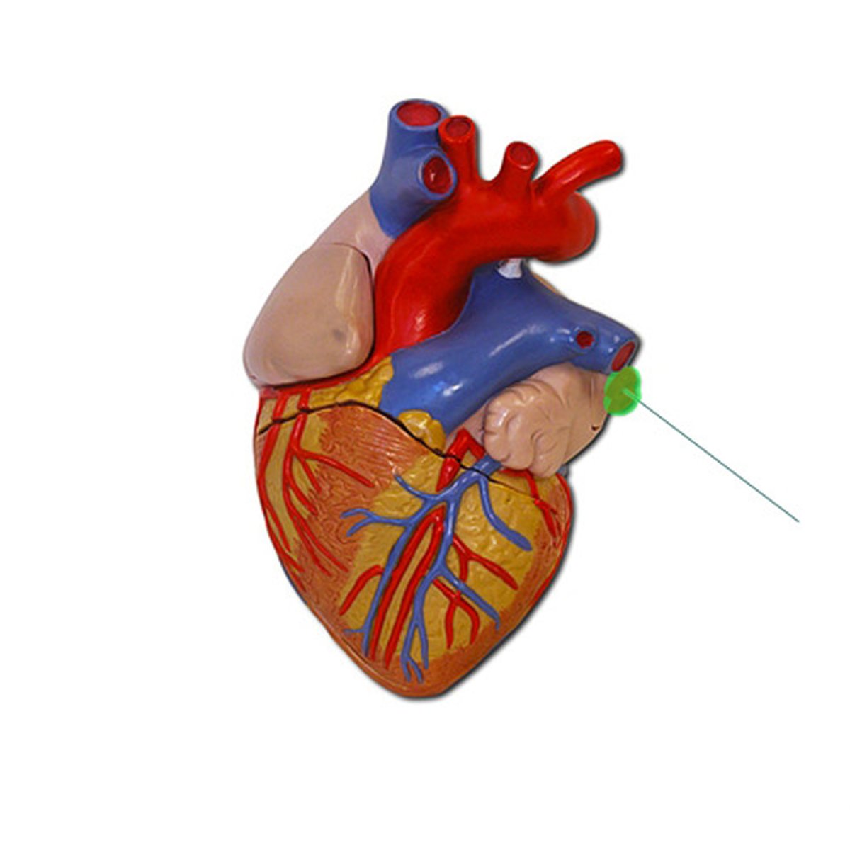
left pulmonary artery
Identify the highlighted structure.
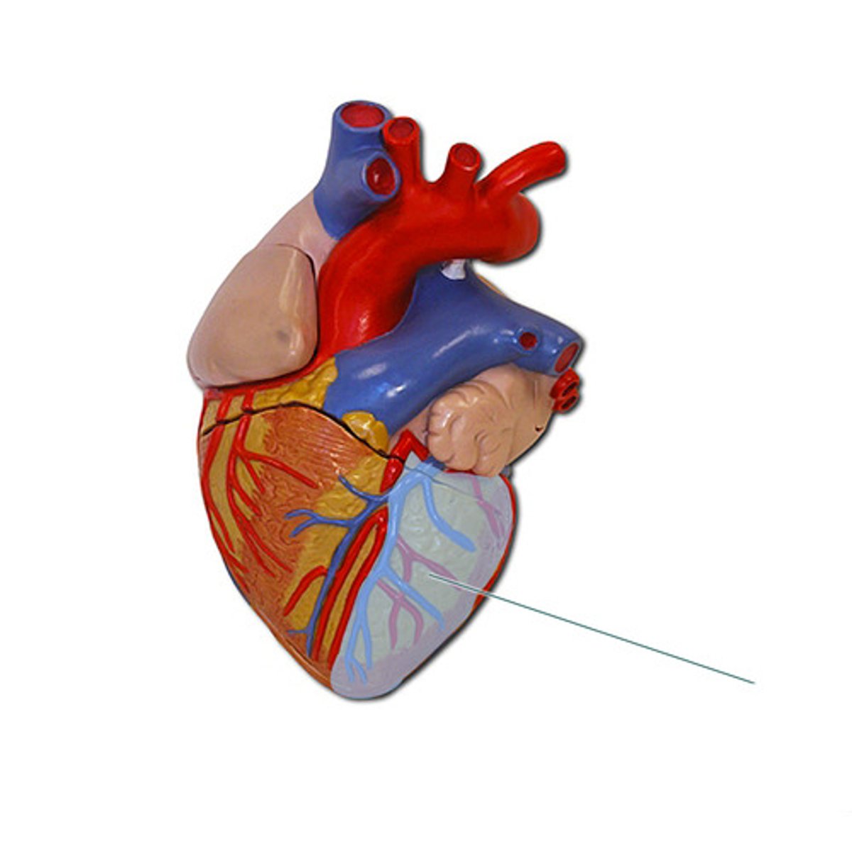
left ventricle
Identify the highlighted structure.
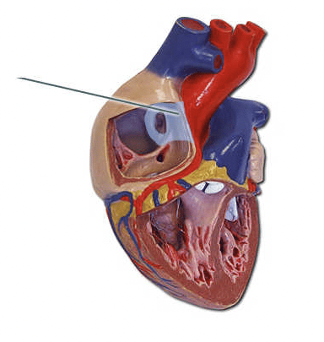
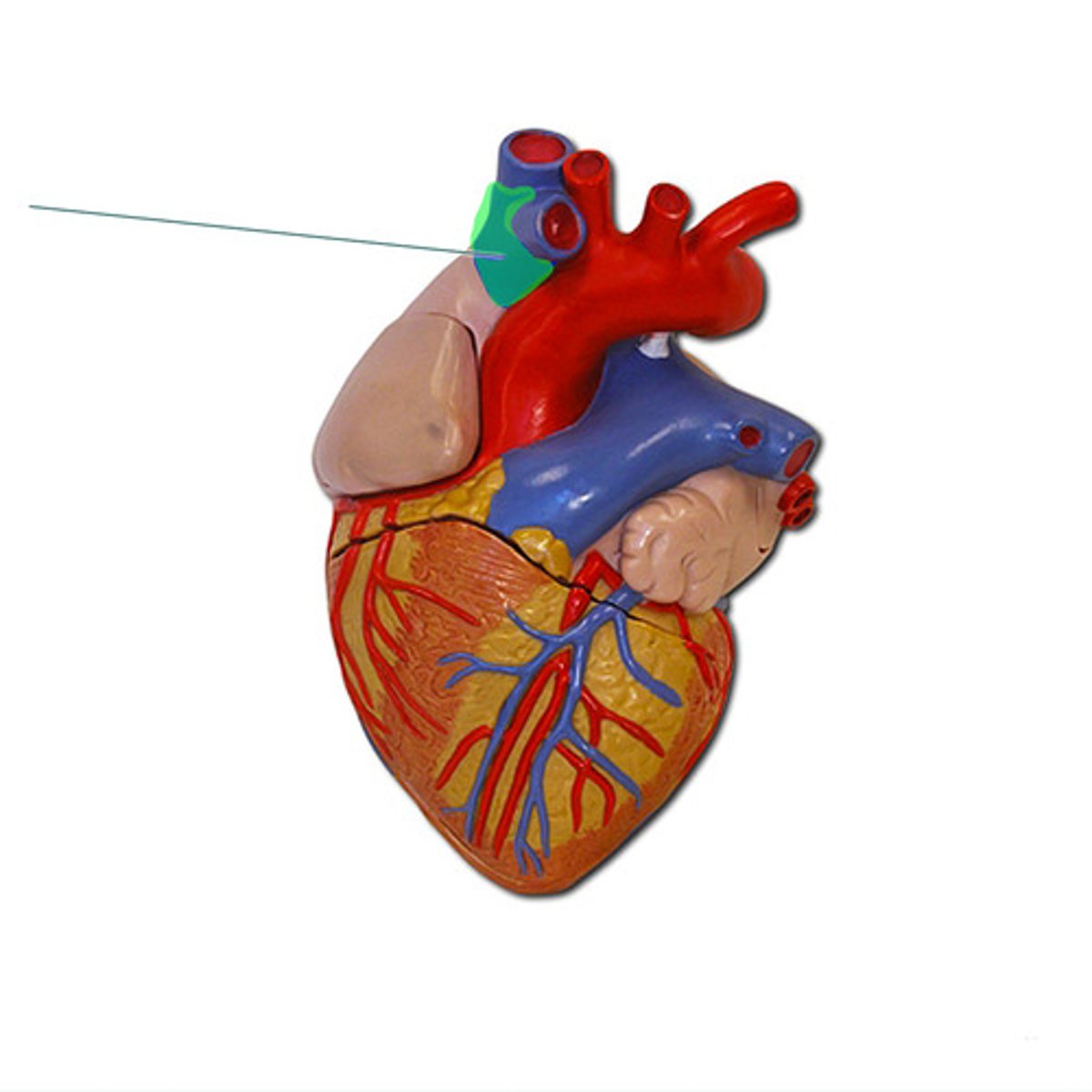
fetal ductus arteriosum
The highlighted structure is a remnant of which structure in the fetus?
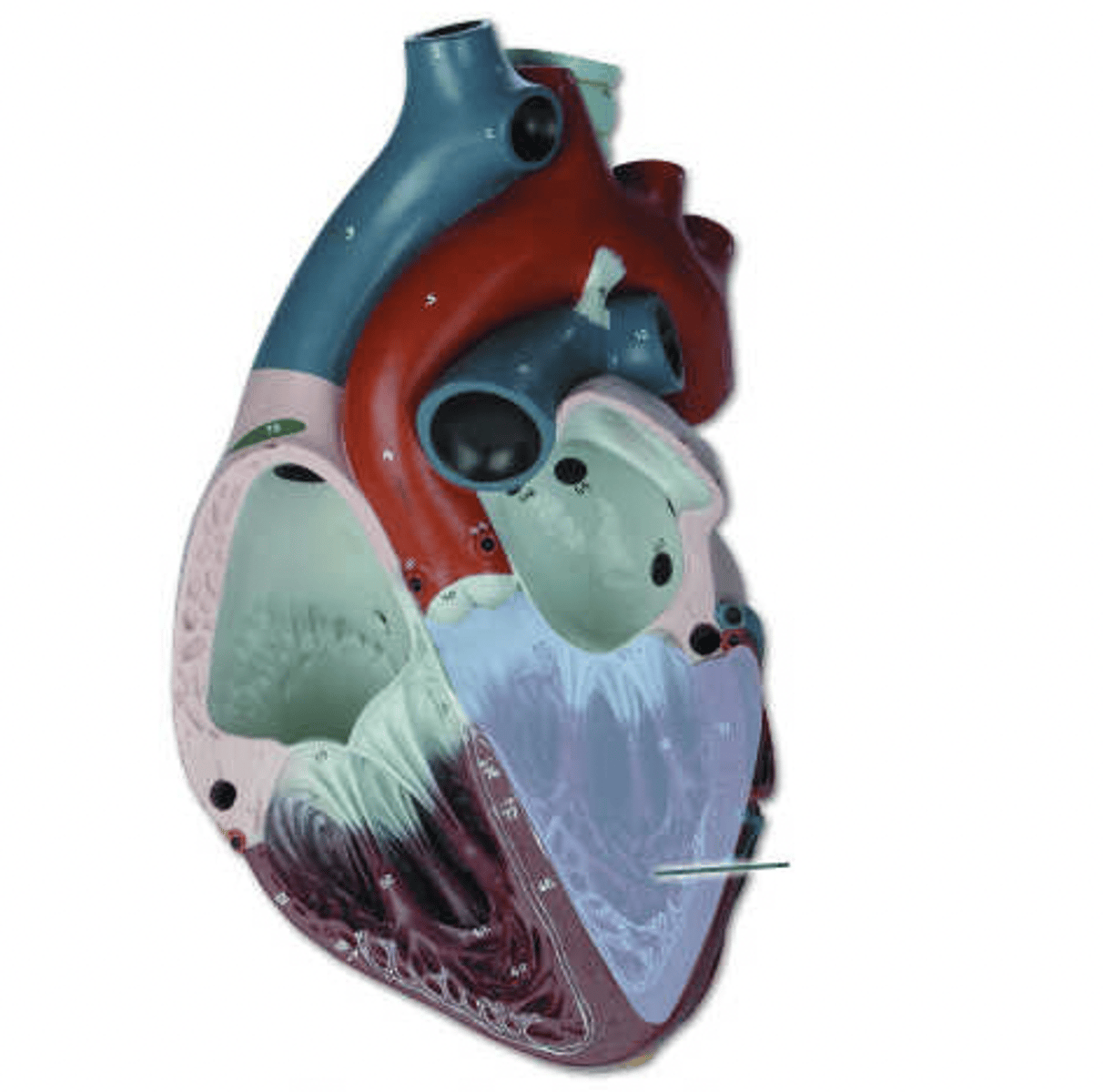
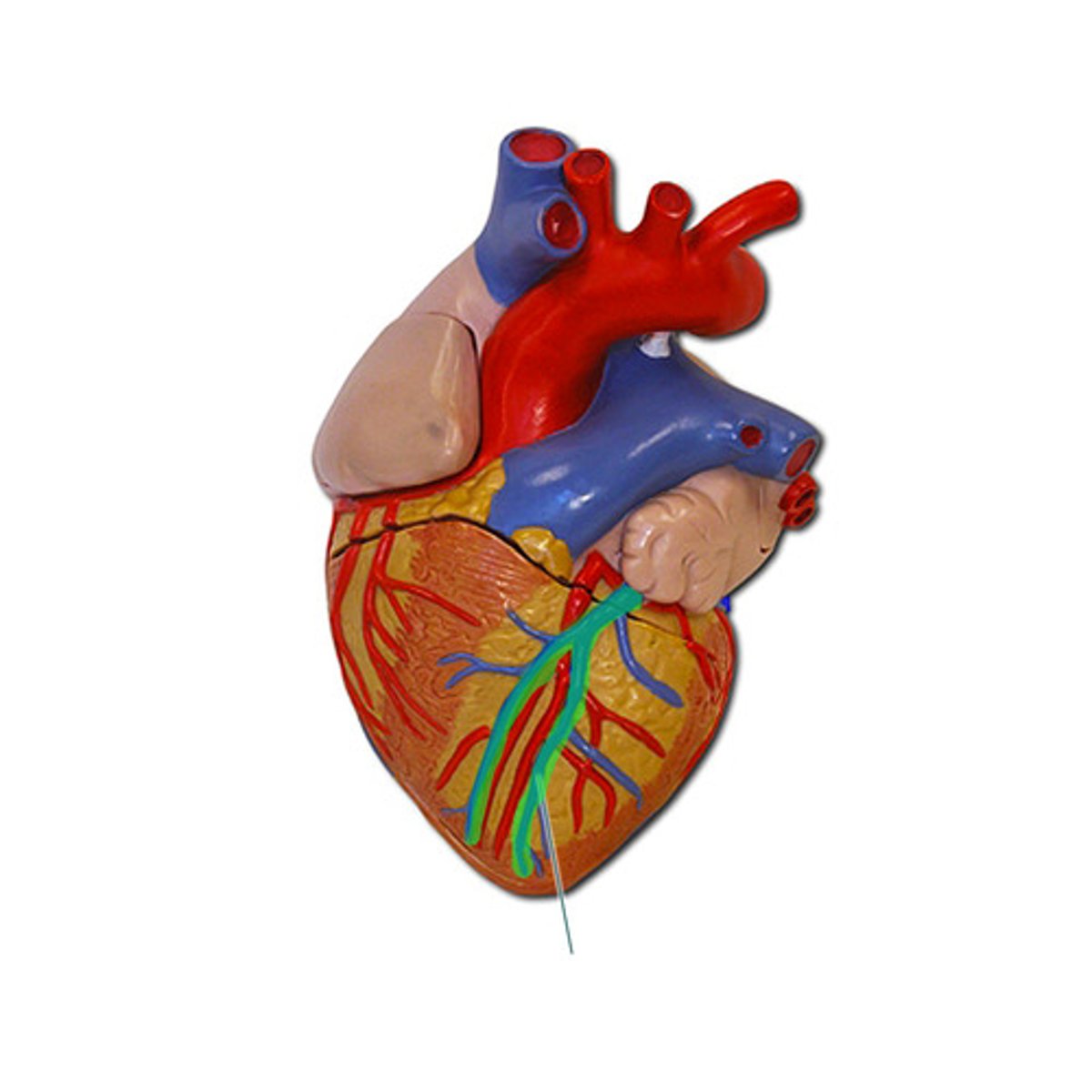
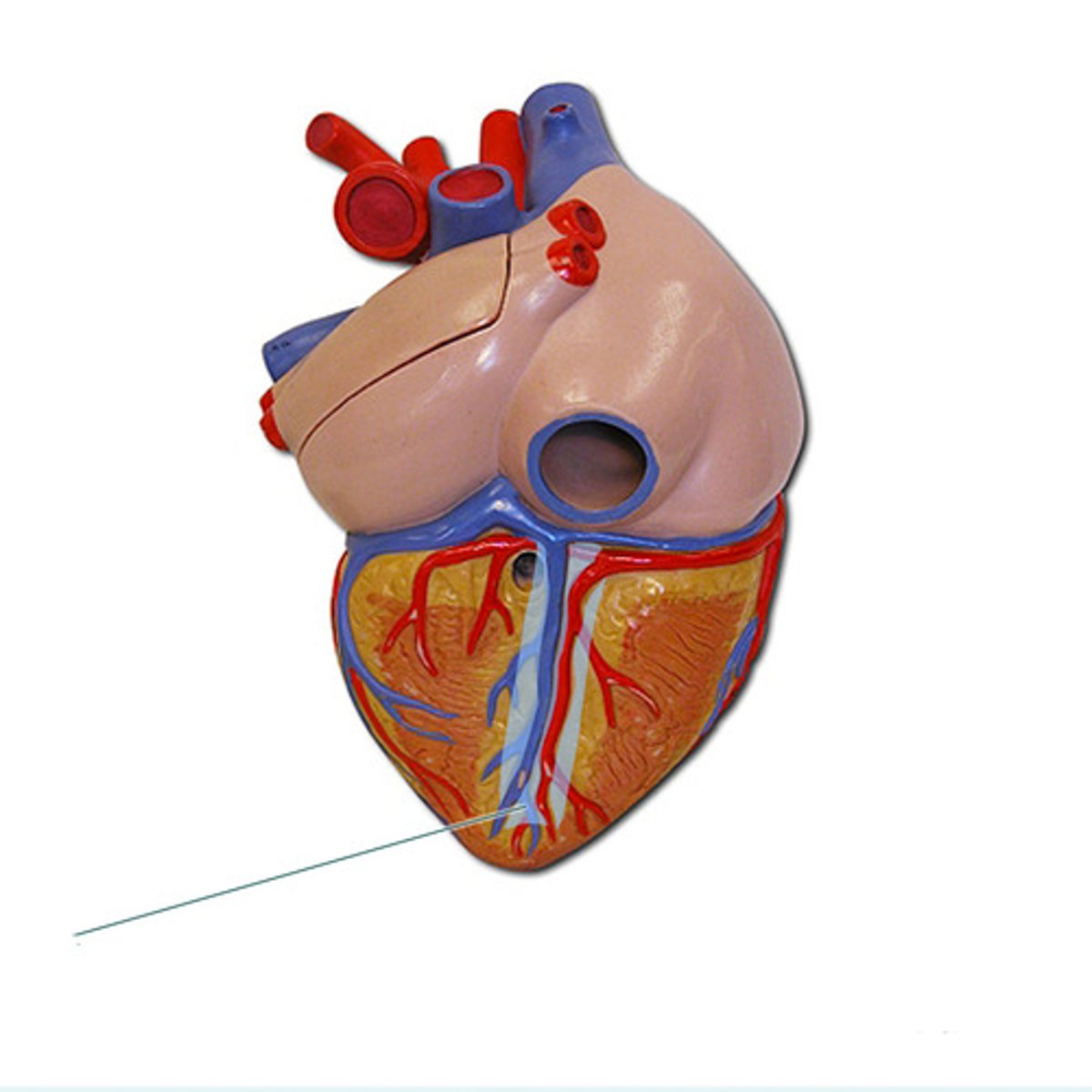
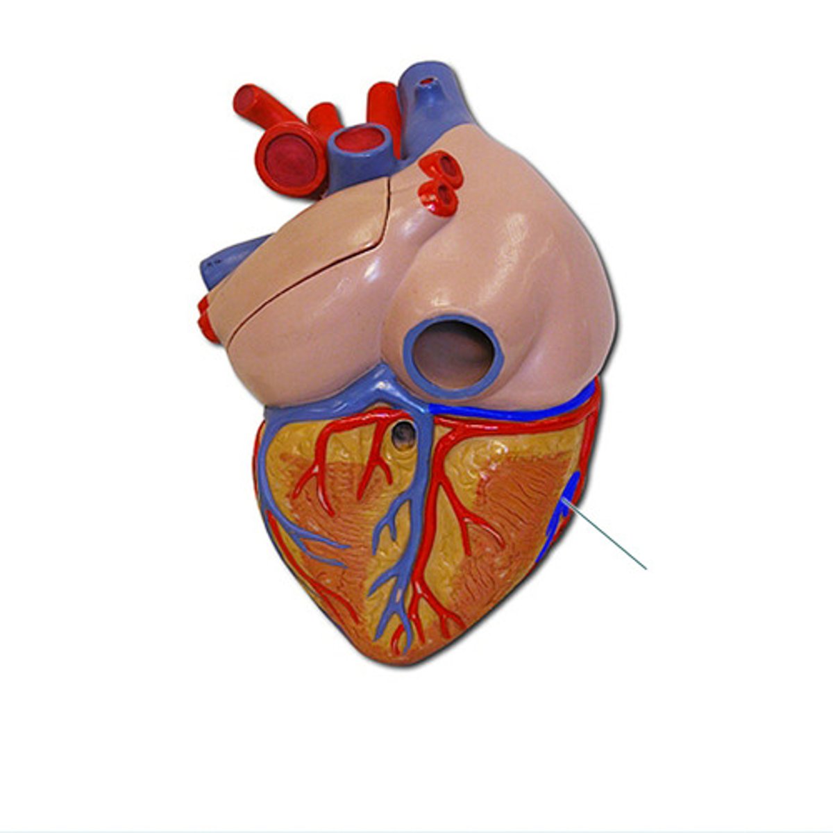
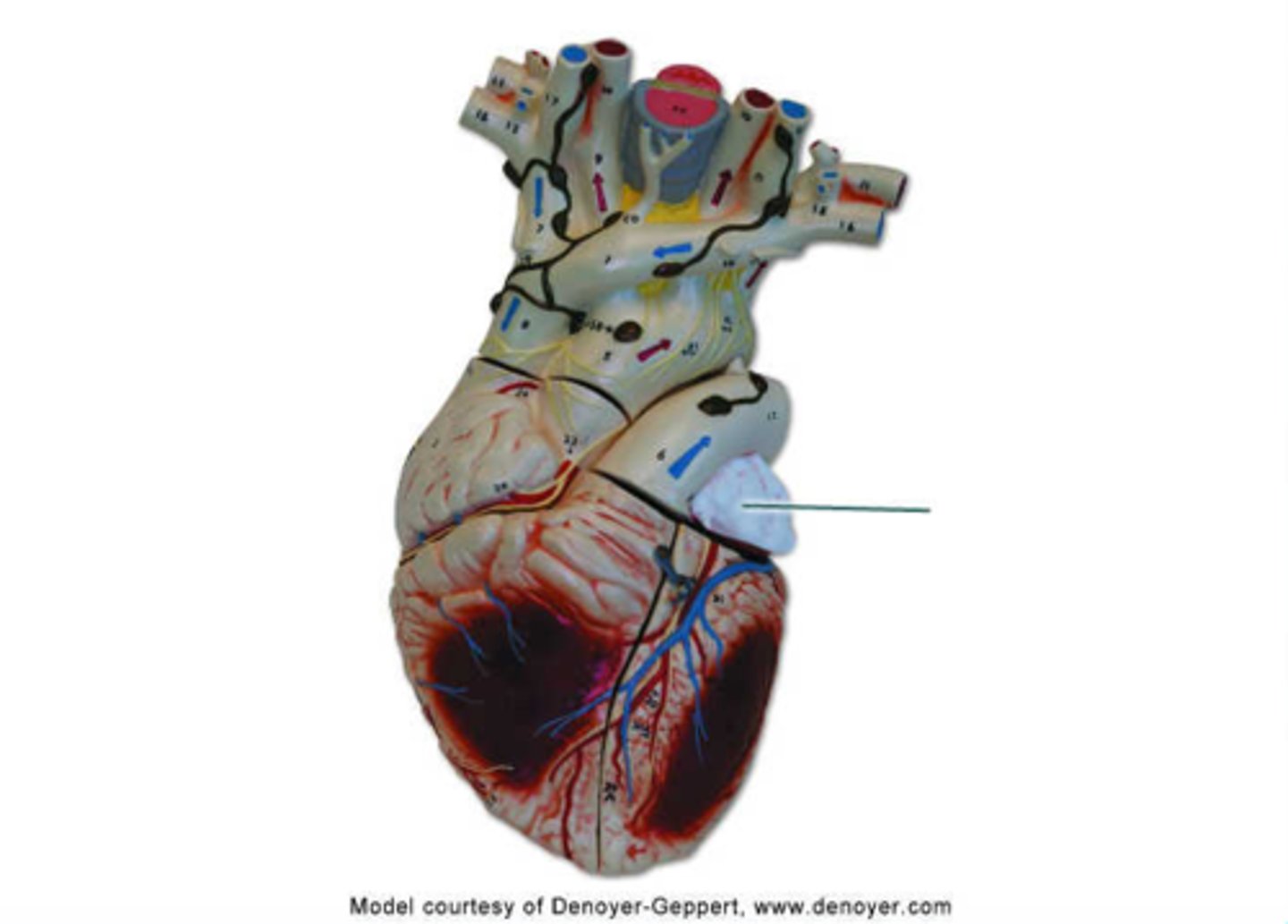
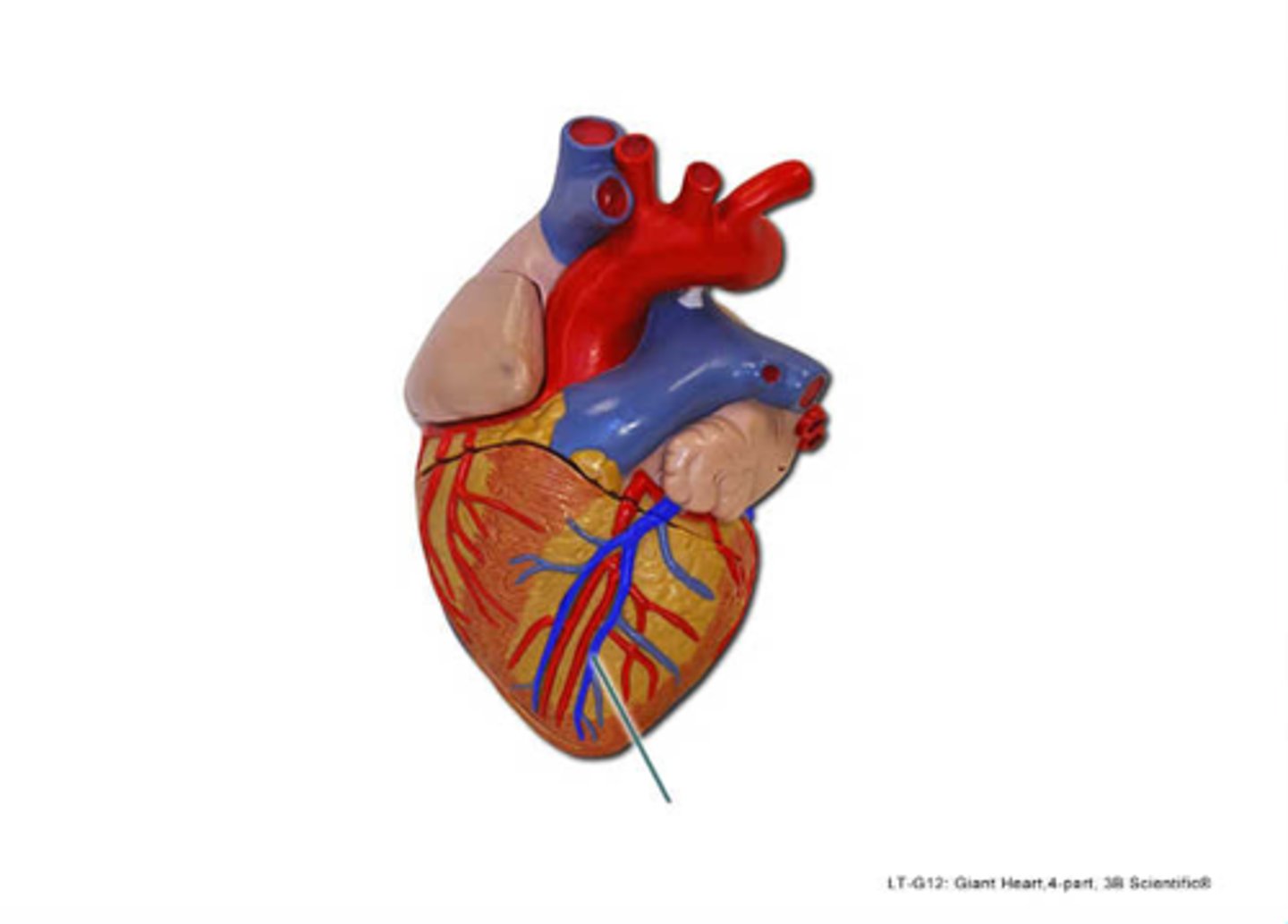
middle cardiac vein
Identify the highlighted vessel.
ventricular systole
Is the highlighted structure closed during atrial systole, or during ventricular systole?
deoxygenated blood (pulmonary arteries)
Do these vessels carry oxygenated blood or deoxygenated blood?
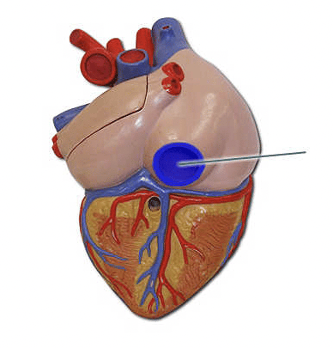
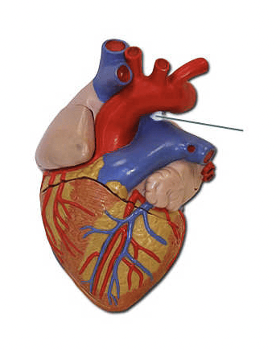
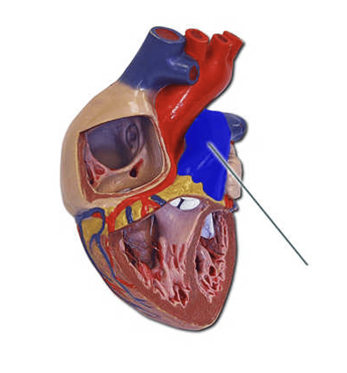
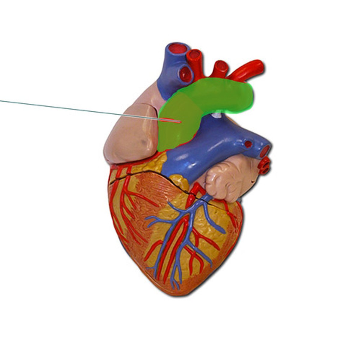
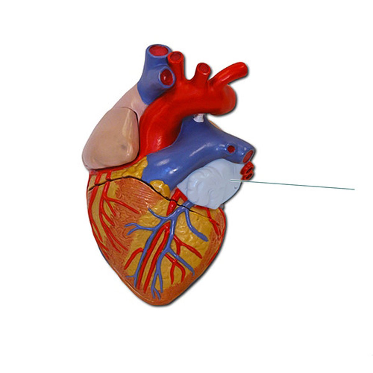
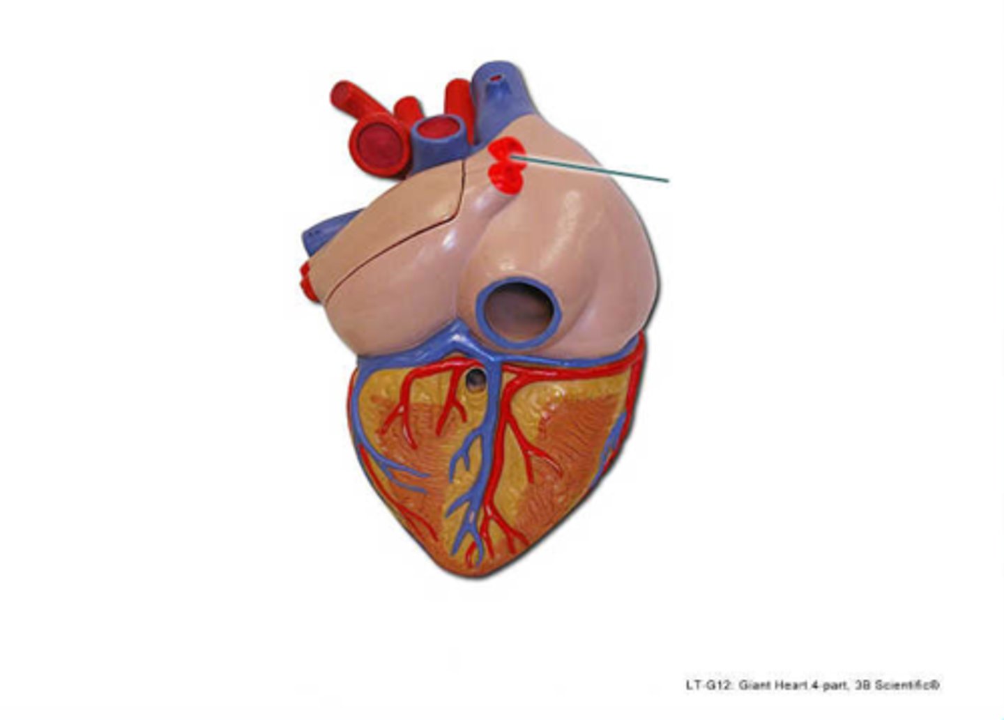
pulmonary trunk
Identify the highlighted structure.
right atrium
Identify the highlighted structure.
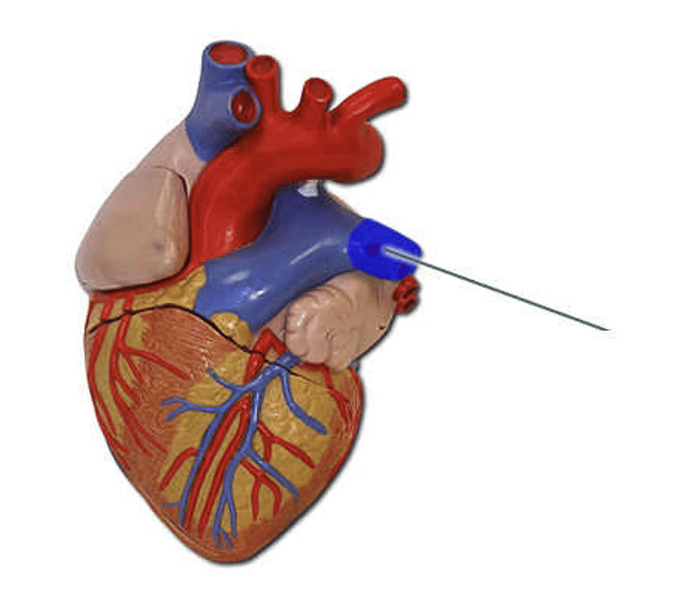
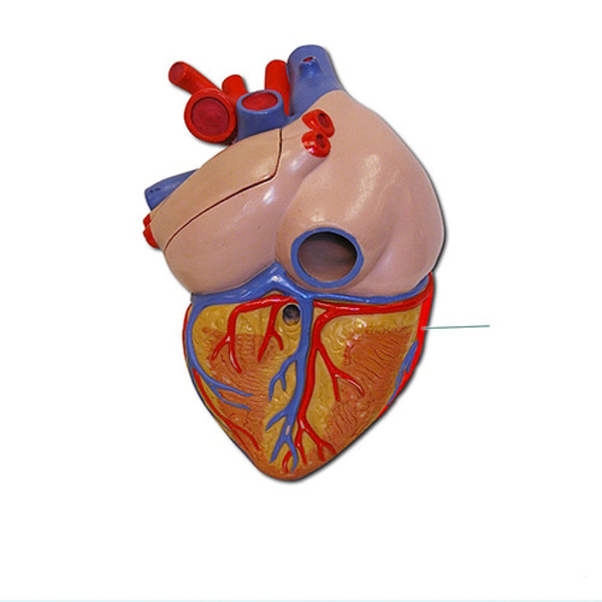
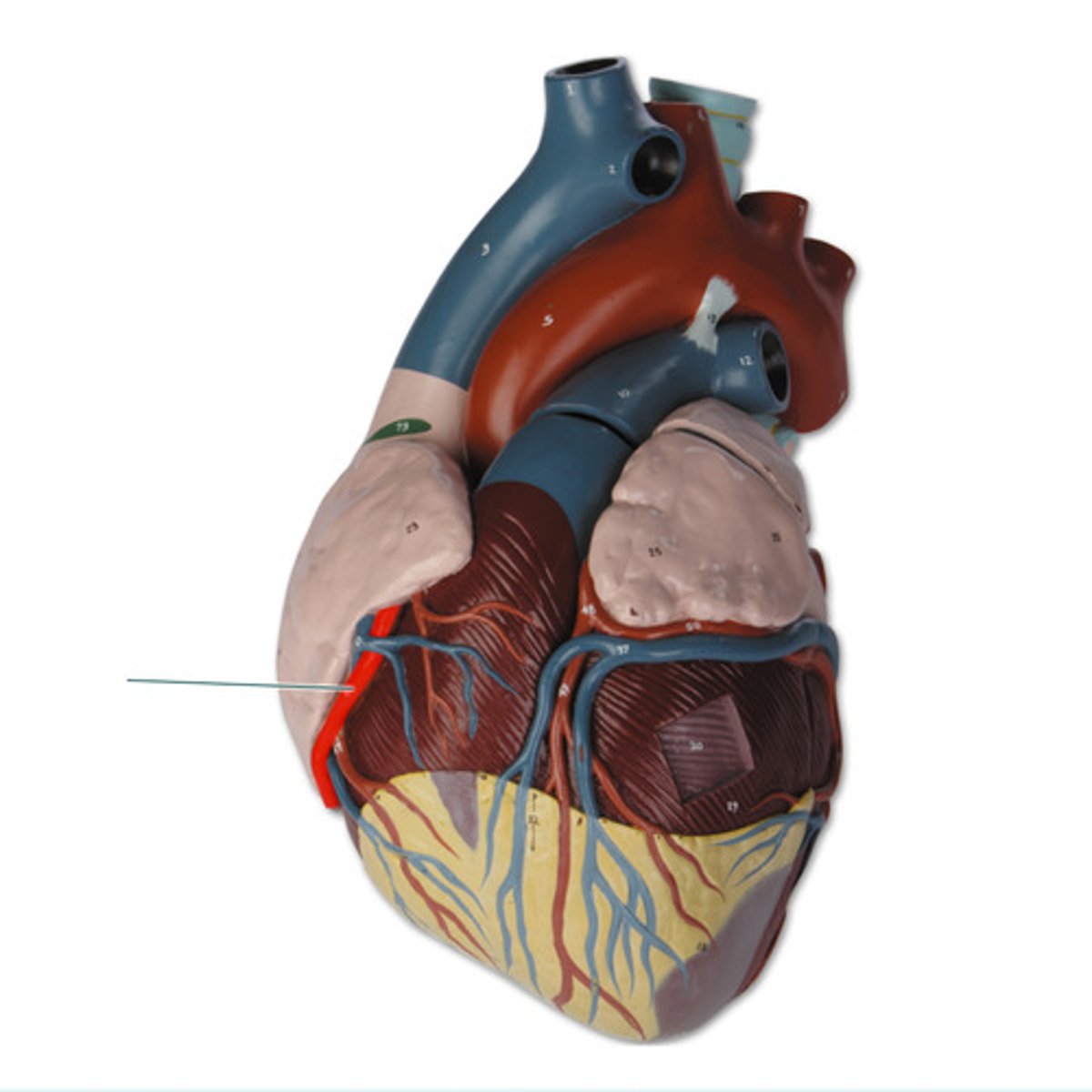
right coronary artery
identify the highlighted vessel.
right ventricle
identify the highlighted structure.
small cardiac vein
identify the highlighted vessel.
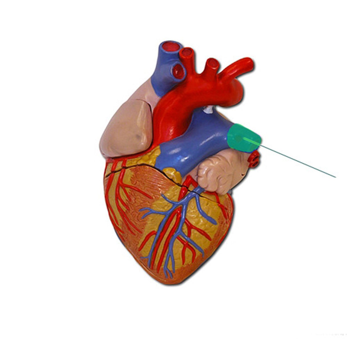
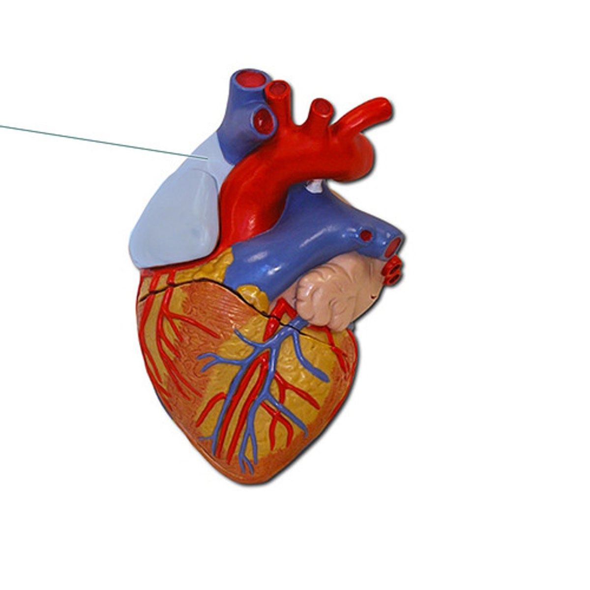
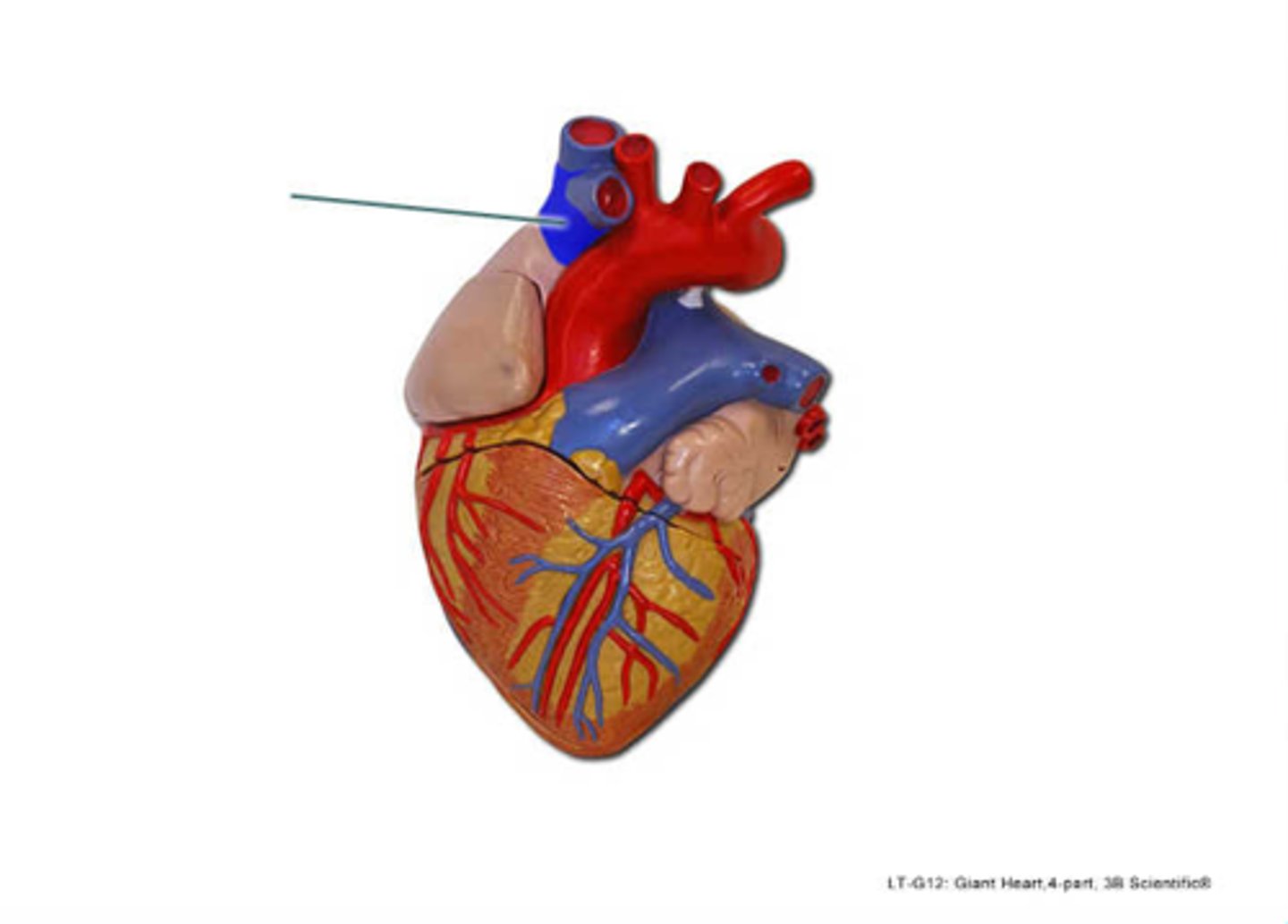
superior vena cava
identify the highlighted vessel.
Anterior Interventricular Artery
Heart

Anterior Interventricular Sulcus
Heart

Aorta
Heart
Aortic Semilunar Valve
Heart
Apex
Heart

Chordae Tendineae
Heart

Circumflex Artery
Heart
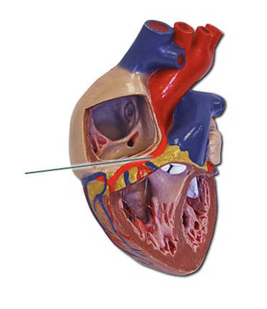
Coronary Sinus
Heart
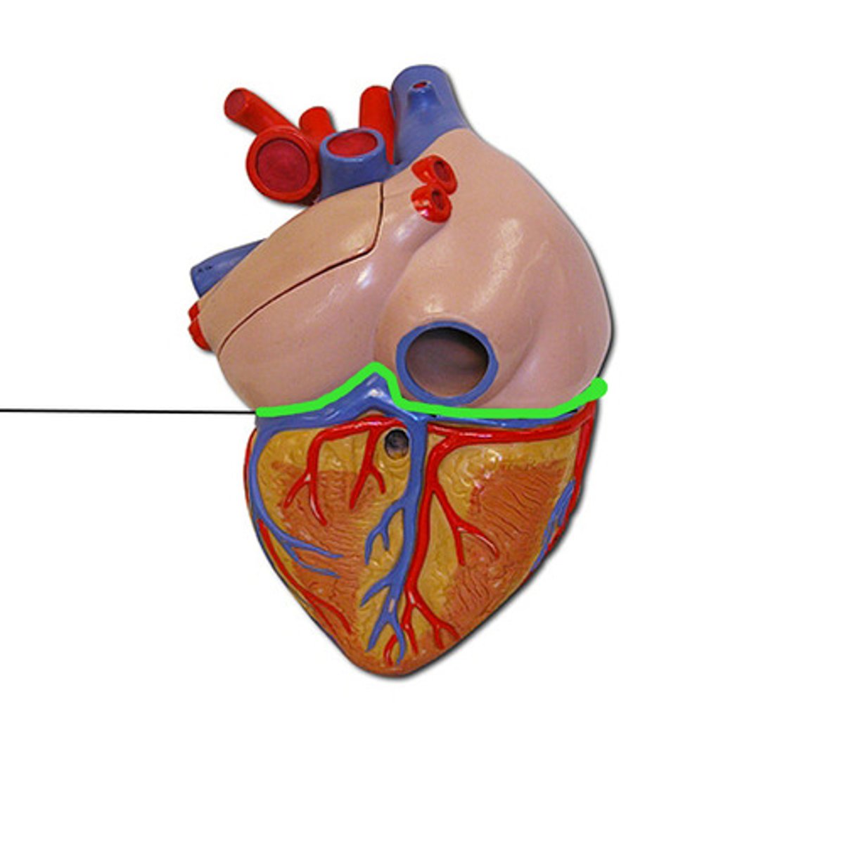
Coronary Sulcus
Heart
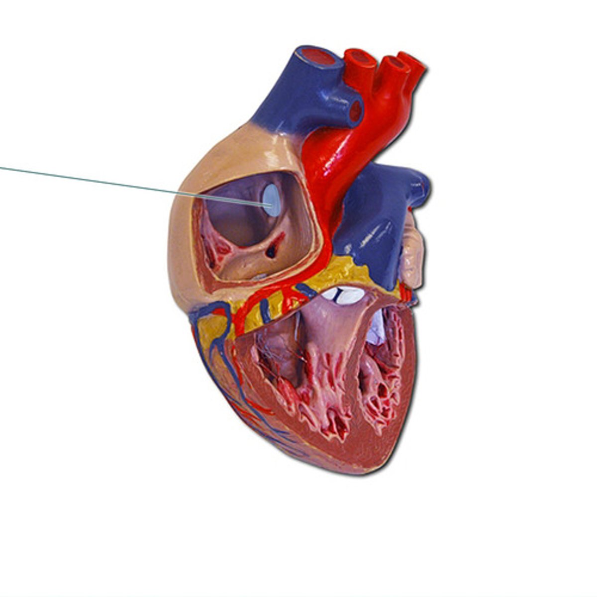
Fossa Ovalis
Heart
Great Cardiac Vein
Heart

Great Cardiac Vein
Heart
Inferior Vena Cava
Heart
Interventricular Septum
Heart
Left Atrium
Heart
Left Auricle
Heart

Left Common Carotid Artery
Heart
Left Coronary Artery
Heart
Left Pulmonary Vein
Heart
Left Ventricle
Heart
Marginal Artery
Heart
Middle Cardiac Vein
Heart

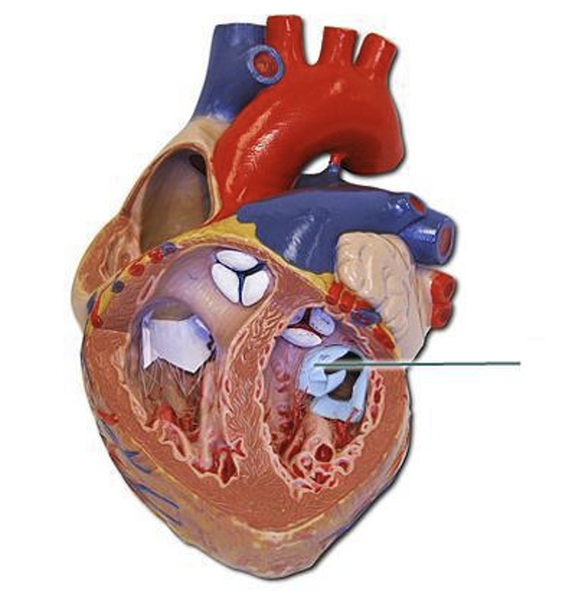
Mitral Valve
Heart
Papillary Muscles
Heart
Posterior Interventricular Artery
Heart

Posterior Interventricular Sulcus
Heart
Pulmonary Arteries
Heart
Pulmonary Semilunar Valve
Heart

Pulmonary Trunk
Heart
Right Atrium
Heart
Right Auricle
Heart

Right Coronary Artery
Heart
Right Coronary Artery
Heart
Right Ventricle
Heart
Small Cardiac Vein
Heart
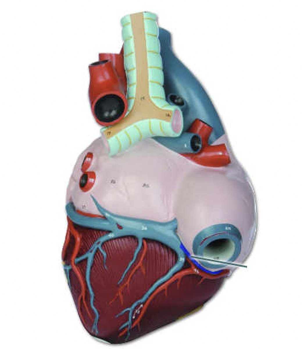
Superior Vena Cava
Heart
Tricuspid Valve
Heart

apex
ascending aorta
left auricle
right pulmonary veins
superior vena cava
trabeculae carneae
tricuspid valve
Name the ridged bundles of muscle found projecting inside the right atrium.
Pectinate muscles
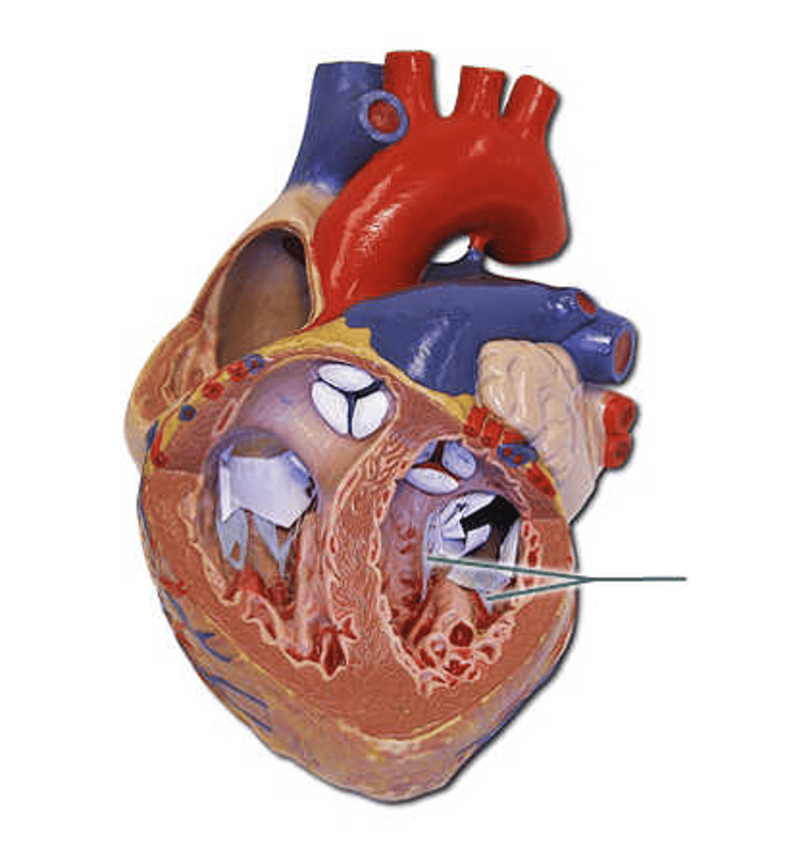
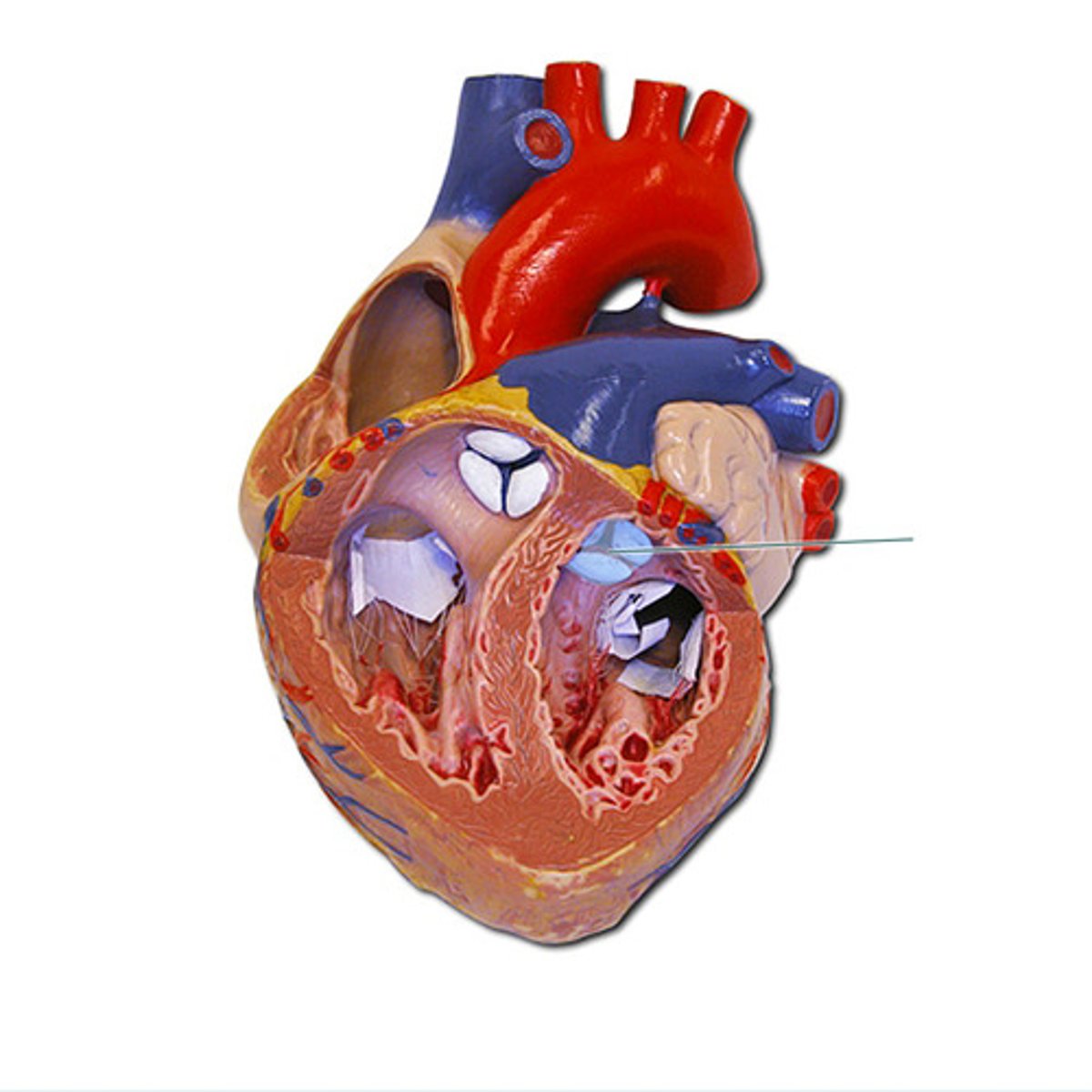
Identify the right atrioventricular valve.
Tricuspid valve
Identfiy the valve located at the exit of the right ventricle.
Pulmonary semilunar valve
The moderator band is found on both the right and left side of the heart.
False
Oxygenated blood flows through the right side of the heart.
False
great cardiac vein
papillary muscles
right auricle
What is the name of the "strings" that are attached to the two cuspid valves?
Chordae tendinae
Which side of the heart is larger and has thicker walls?
Left
Why do you think this side (your answer to Part B) is larger? What purpose does this serve?
The left ventricle of your heart is larger and thicker than the right ventricle. This is because it has to pump the blood further around the body, and against higher pressure, compared with the right ventricle.
Systolic pressure is:
Blood pressure inside large arteries when the ventricles contract
You should record the systolic number of a blood pressure reading:
When you first hear the heart beat
Which of the following is considered a "normal" blood pressure reading?
Systolic: 120
Diastolic: 80
Blood pressure is most often measured in the:
Brachial artery
Because of its accessibility, heart rate is most frequently measured at which pulse point?
Radial artery
A "normal" reading for a resting heart rate would be:
75
Specifically, what part of the intrinsic conduction system stimulates the atrioventricular (AV) node to conduct impulses to the atrioventricular bundle?
sinoatrial (SA) node
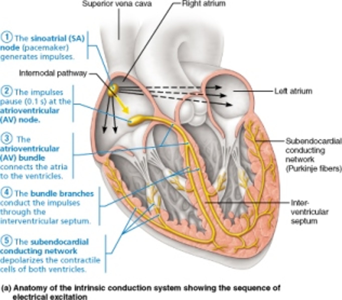
In sequential order, the components of the intrinsic conduction system, beginning at the SA node, are ________.
AV node, AV bundle, bundle branches, subendocardial conducting network (Purkinje fibers)
In the normal heart, the ________ has the highest rate of discharge and sets the rate of depolarization for the heart as a whole.
SA node
In a typical ECG, the __________ wave signals the depolarization of the atria immediately before they contract.
P
The QRS complex is associated with ___
depolarization of the ventricles
The T wave of the ECG represents ________.
repolarization of the ventricles
What is the outermost layer of the pericardium called?
fibrous pericardium
Another name for the epicardium is the ________.
parietal pericardium
Which letter points to the right coronary artery?
B
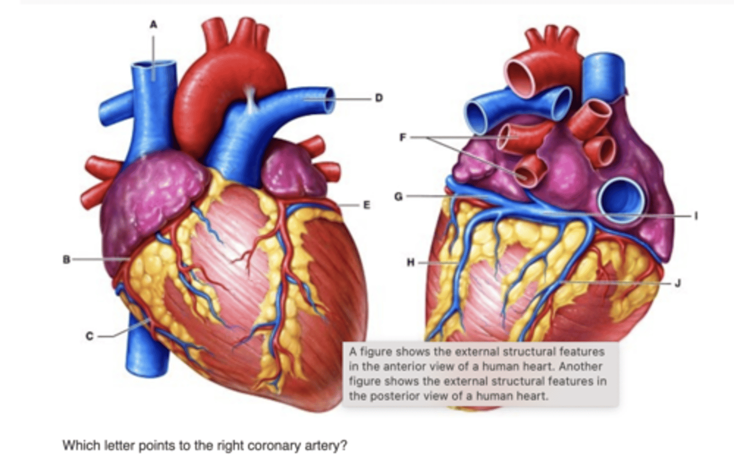
Which letter points to the great cardiac vein?
G

Which letter points to the left pulmonary artery?
D
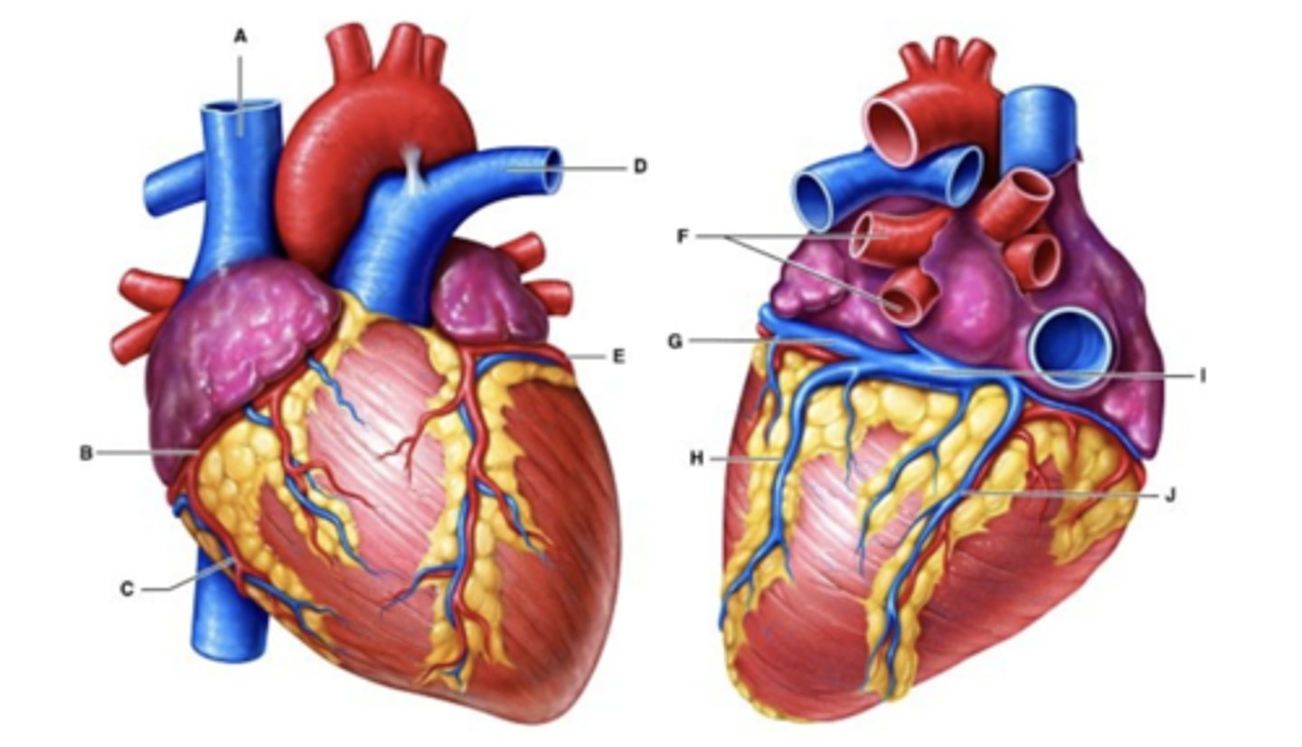
Which letter points to the right marginal artery?
C

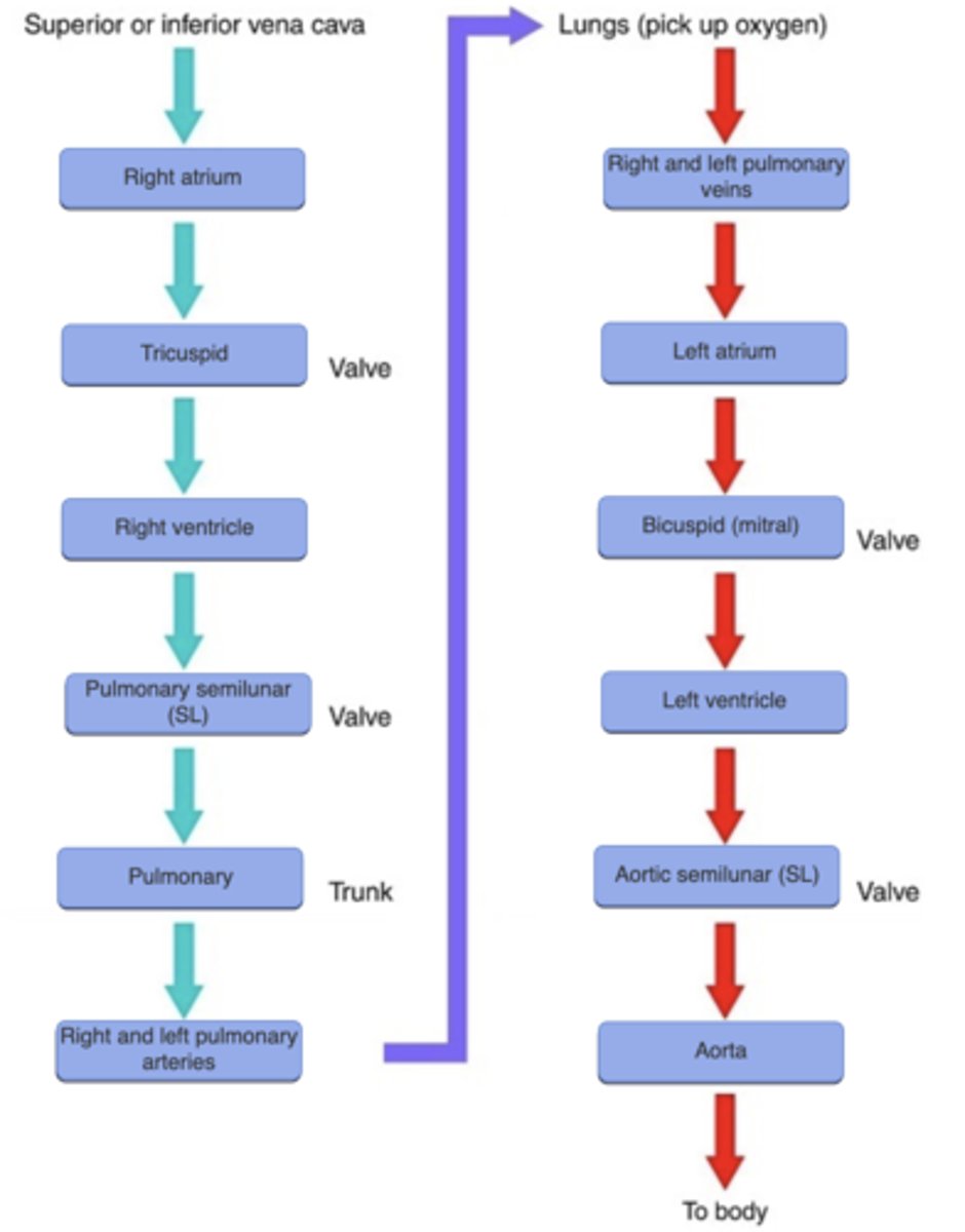
When blood travels from the left atrium to the left ventricle, it must travel through the ________.
bicuspid (mitral) valve
3 multiple choice options
What is the first blood vessel to carry deoxygenated blood out of the heart?
pulmonary trunk
The coronary sinus empties into the ________ of the heart.
right atrium