BIO 330 - Neuroanatomy EMU Winterhalter Summer 25 Lab 11
1/35
There's no tags or description
Looks like no tags are added yet.
Name | Mastery | Learn | Test | Matching | Spaced | Call with Kai |
|---|
No analytics yet
Send a link to your students to track their progress
36 Terms
What are the two paired arteries that supply blood to the brain?
internal carotid (anterior / front circulation)
vertebral arteries (posterior / back circulation)
If one of the arteries in the brain is damaged, why might there still be enough blood supply / flow in the brain?
the brain has an anterior circulation (internal carotid) and a posterior circulation (vertebral artery) allowing compensation of one is damaged
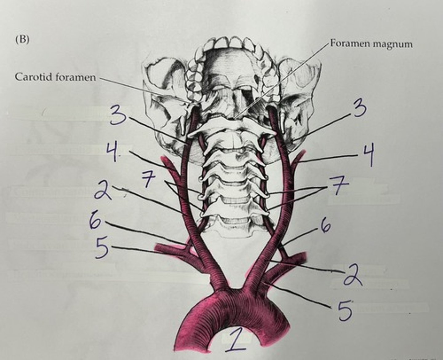
In this image of the arteries on the anterior neck, label the numbered arteries
1 = aorta (branches off heart)
2 = common carotid arteries, paired & 2 segemnts
3 = internal carotid artery
4 = external carotid artery
5 = subclavian artery (branches off aorta)
6 = vertebral arteries, paired
7 = transverse foramina
What type of head / facial structures does the external carotid artery supply blood to?
superficial (outermost layers) of head, face, teeth, mouth, and eyes
What does the internal carotid artery supply blood to?
a great amount of the brain
The transverse foramina (holes of the transverse processes) of the cervical spinal cord have which artery through them?
vertebral artery
What artery connects the two vertebral arteries and is located on the anterior surface of the spinal cord (cervical)?
anterior spinal artery (single, branches off aorta)
What structures of the spinal cord receive blood from the anterior spinal artery?
ventral horns, ventral columns, and lateral columns
What structures of the spinal cord receive blood from the posterior spinal arteries? (remember these are paired)
dorsal horns and dorsal columns
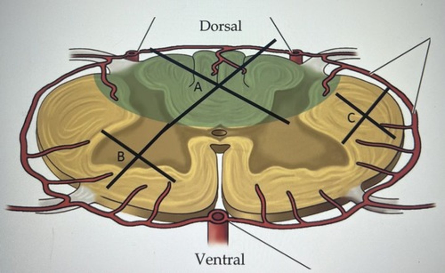
Describe the structures damaged and symptoms if there is a blockage of blood flow to letter A area. The black X shows what specific areas are included.
dorsal columns, some of right and left dorsal horns
bilateral fine touch & proprioception loss at the cord level and below
(tract and LMN damage)

Describe the structures damaged and symptoms if there is a blockage of blood flow to letter B area. The black X shows what specific areas are included.
right ventral horn and some of anterolateral system motor tracts
no motor control of right limbs and probably trunk muscles at only that cord level
pain & temp loss on left side of the body

Describe the structures damaged and symptoms if there is a blockage of blood flow to letter C area. The black X shows what specific areas are included.
LCT & Rubrospinal tract in lateral column
no motor control of left limbs at cord level and below
(tract damage only)
What is an aneurysm?
ballooning / filling of a weakened artery wall
What are 3 most common arteries to have aneurysms?
anterior communicating (AComm), posterior communicating (PComm), middle cerebral (MCA)
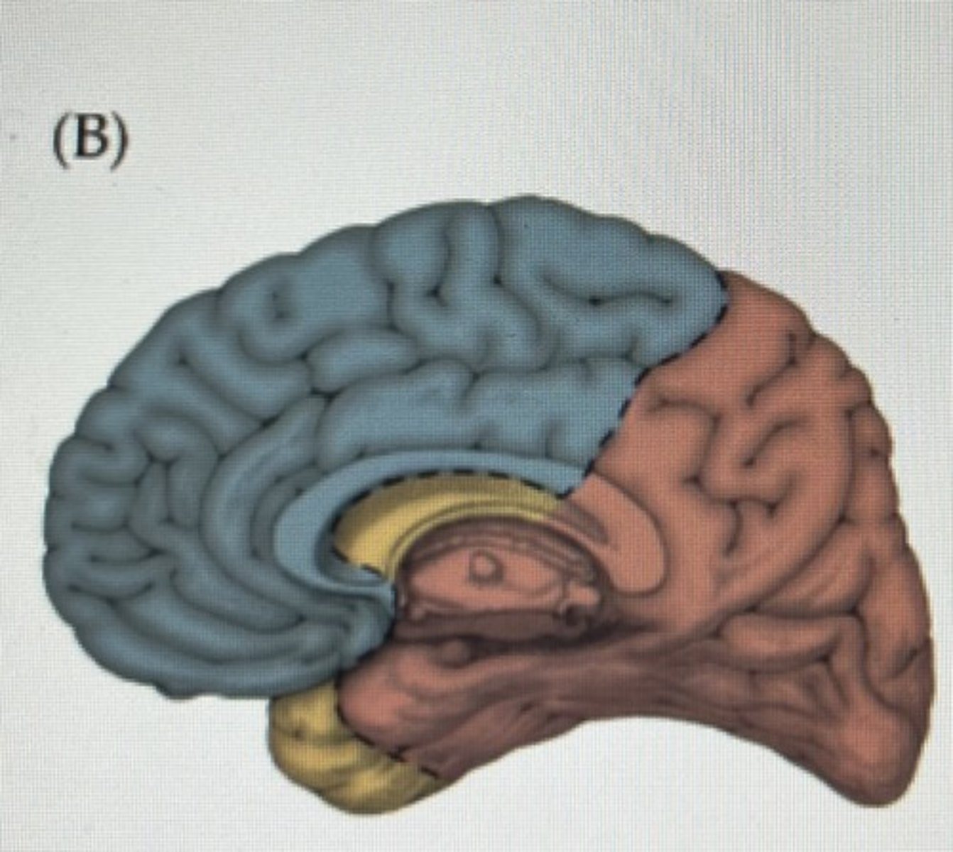
In this midsagittal cut of the brain name which artery supplies each color and what side of the brain is this?
blue = anterior cerebral artery
yellow = middle cerebral artery
redish = posterior cerebral artery
right side
Ozzy Osbourne had a stroke. His left foot is spastic and has lost all sensation. Name the likely artery and side.
right anterior cerebral artery
Name two structures supplied by the lenticulostriate arteries
internal capsule and caudate nucleus
Rihanna had a stroke in the thalamic artery to her left VPM. What type of sensation(s) is lost and on what side of the body?
right side of face ; loss of fine touch, pain, and temp
The _____________ arteries connect the internal carotid to the posterior cerebral arteries
PComm (posterior communication)
What artery and side supplies the right auditory cortex?
right middle cerebral artery
John Cena has "locked in syndrome". Name the likely artery where the stroke occurred
basilar artery
What artery and side supplies blood to Broca's area?
left middle cerebral artery
If the posterior cerebral artery is damaged, will a patient have problems with pupil constriction?
No, pupil constriction involves the EW nucleus, CN III, and ciliary ganglion. If midbrain structures specifically, these are damaged then yes, otherwise pupil constriction is good
If the right posterior cerebral artery is damaged, will a patient lose their Right visual fields from both eyes?
No, right visual cortex processes left visual fields
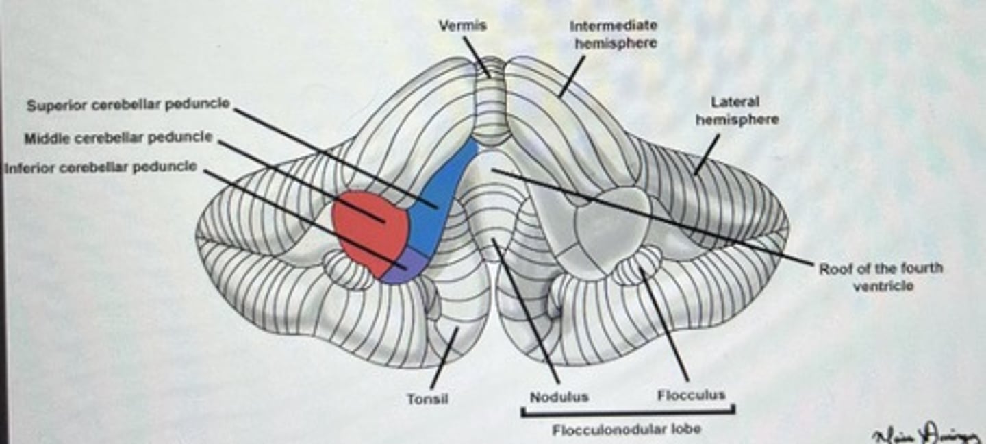
Be able to ID ALL structures of the cerebellum (reference lab 10 quizlet)
ignore roof of the fourth ventricle
also know arborvitae, folia, and the two lobes
Following a stroke, your patient has normal eye movements, speech, hearing, and vision. Their legs have flaccid paralysis and cannot sense pinpricks, but they can sense light touch with a cotton swab. What artery is damaged, and at which cord level?
anterior spinal artery ; lumbar cord
Your patient complains of a headache and double vision. Upon examination the left eye has ptosis and a blown pupil. Where could an aneurysm be? What cranial nerve(s) are likely damaged?
internal carotid, PComm, PCA, or SCA
CN III Oculomotor
Your patient can ONLY move their eyes up and down and blink. They cannot open or close their eyes. Limbs are rigid but still has some spinal reflexes. Breathing is labored. They can communicate by using a computer with eye blinks. Normal sensory capabilities and cognition.
What artery has failed and its location?
What is this phenomenon called?
Explain why they have the symptoms they do based on the structures that have no blood supply?
basilar artery, pons & medulla
locked in syndrome
LCT & ACT tracts on ventral side of pons and medulla are not receiving blood supply
Your patient complains of a headache and difficulty reading / looking at close objects. Upon examination, you notice the Right pupil points up & out. The Right eye also cannot look down well.
What cranial nerve(s) are affected and how does it relate to the symptoms?
What arteries could be damaged based on the symptoms?
CN IV Trochlear ; eyes cannot accommodate
PCA, SCA, PComm could be damaged
Your patient had a stroke. They are blind and cannot smell now. Blindness includes the loss of the pupil reflex. They also cannot feel or move the lower limbs.
What artery has failed?
What CNS structures/ areas are damaged?
Would these be some cognitive problems as well? Why or why not.
ACA
CNS structures damaged = CN I & II, corpus callosum
Yes. Frontal lobe damage results in loss of smell and vision
Your patient had a stroke and is blind. However, the pupil reflex is still intact.
What artery has failed?
What part of the brain does this artery supply?
Would there be problems in both arteries or only one?
PCA ; supplies visual cortex
both because they are fully blind
Your patient had a stroke in a small thalamic artery that supplies the Right VPL.
What are their symptoms?
loss of limb control on the left side of the body
sensory loss of fine touch, vibration, proprioception, pain, and temp
Your patient is paralyzed on their entire Right side (spastic paralysis). Although they have difficulty moving their mouth (since only left side muscles are functional), there is no speech or hearing problems.
What neural structure could be damaged due to the symptoms?
What artery and side could be damaged?
What is the term for paralysis on one side of the body?
internal capsule
left lenticulostriate arteries
hemiplegia
Your patient cannot feel or move their right arm, right hand, or right side of face. They are having difficulty hearing as well as difficulty understanding and generating speech.
What artery and side is damaged?
What neural structures/areas have been damaged to cause these symptoms?
What is the location(s) of the structures listed above?
left MCA
neural structures = left precentral gyrus, left postcentral gyrus, left Wernickes area, left Brocas area
frontal & parietal lobes
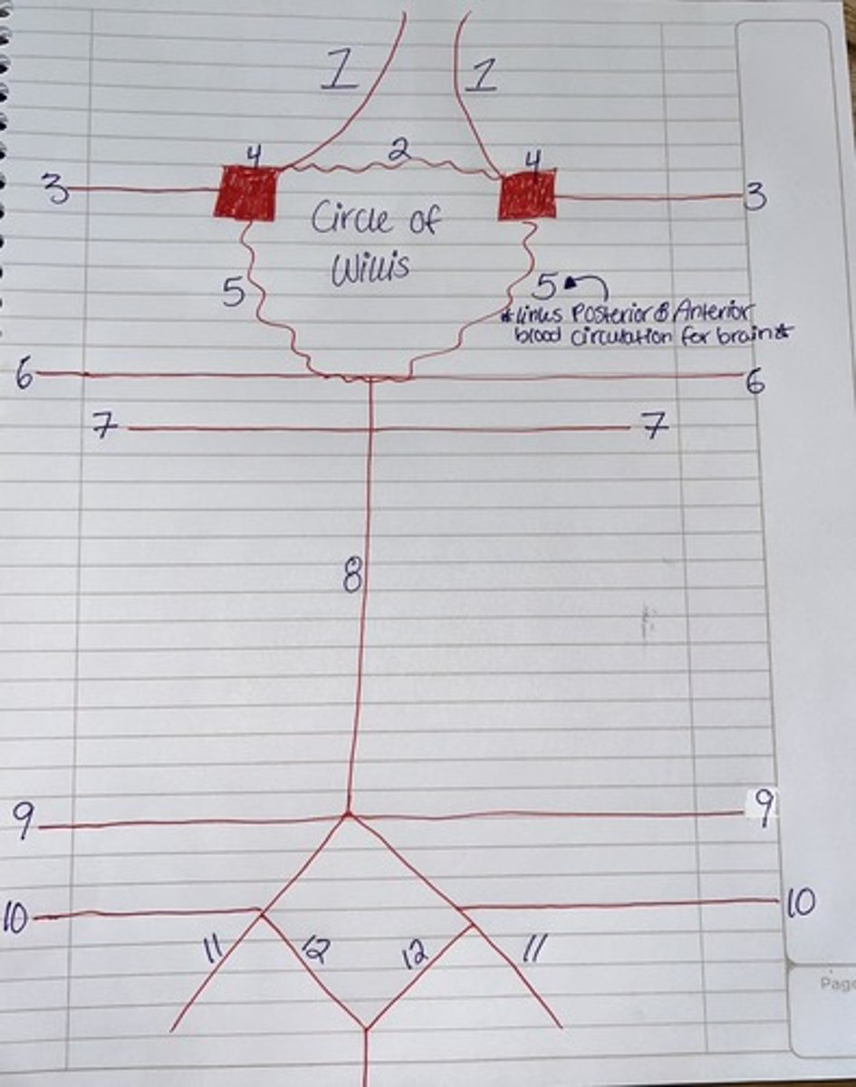
Label this schematic drawing of the cerebral arteries
1 = ACA (anterior cerebral artery)
2 = AComm (anterior communicating)
3 = MCA (middle cerebral artery)
4 = Internal carotid
5 = PComm (posterior communicating)
6 = PCA (posterior cerebral artery)
7 = SCA (superior cerebellar artery)
8 = basilar
9 = AICA (anterior inferior cerebellar artery
10 = PICA (posterior inferior cerebellar artery)
11 = vertebral artery
12 = anterior spinal artery
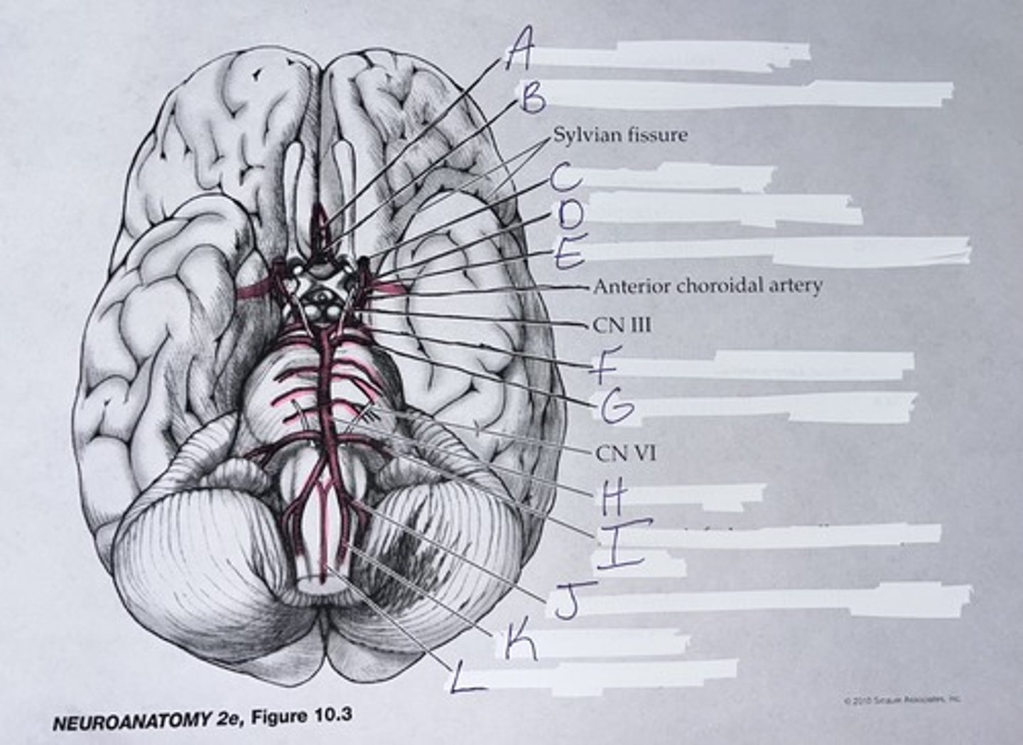
Label the cerebral arteries. I do not think we will have to ID them but definitely know what structures they are near and how to draw the schematic
A = ACA (anterior cerebral artery)
B = AComm (anterior communicating)
C = internal carotid
D = MCA (middle cerebral artery)
E = PComm (posterior communicating)
F = PCA (posterior cerebral artery)
G = SCA (superior cerebellar artery)
H = basilar artery
I = AICA (anterior inferior cerebellar artery)
J = PICA (posterior inferior cerebellar artery)
K = vertebral artery
L = anterior spinal artery