Primary Open-Angle Glaucoma and Clinical Assessment of the Optic Nerve Head - Management of Glaucoma Summer 2026
1/125
There's no tags or description
Looks like no tags are added yet.
Name | Mastery | Learn | Test | Matching | Spaced | Call with Kai |
|---|
No analytics yet
Send a link to your students to track their progress
126 Terms
What is the definition of POAG?
chronic, progressive optic neuropathy in ADULTS, in which there is a characteristic acquired atrophy of the optic nerve and loss of RGCs and their axons
POAG is associated with what? How to diagnose POAG?
Associated with an open anterior chamber angle by gonioscopy
What is NOT mentioned in the definition of POAG?
high IOP
True or False:
Treatment of glaucoma depends on the type that is present
true
What is the most prevalent form of glaucoma?
POAG -- 70% of glaucoma is of this type
What is the worldwide prevalence of POAG?
2.4%
What is the prevalence of POAG for adults aged 40+ in the US?
2.1%
Is the vision impairment from POAG permanent?
Yes -- similar to other forms of glaucoma
POAG is the leading cause of what?
irreversible blindness worldwide
True or False:
Most cases of glaucoma are diagnosed
false -- most cases go undiagnosed
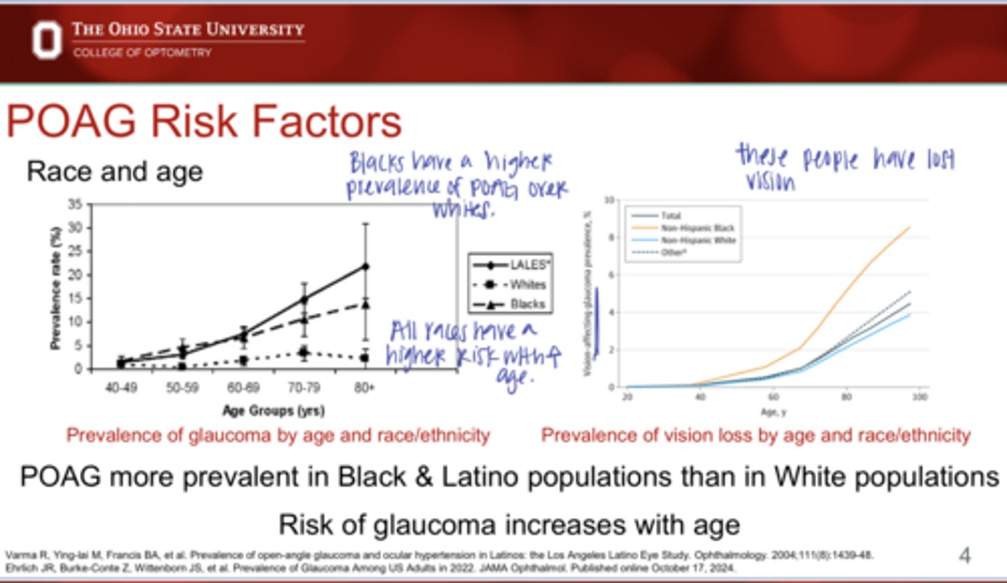
What are the risk factors of POAG?
More prevalent in the Black and Latino populations than in White populations
Risk increases with age
Family hx

Patients with a confirmed family hx of POAG have a (higher/lower) chance of having POAG themselves
Higher

What family members having POAG puts an individual at risk for POAG as found in the Baltimore Eye Study?
Parents
Siblings
1st degree relatives

(Siblings/parents) having POAG leads to a higher risk of your patient developing POAG
Siblings

_____ is the most significant risk factor for POAG
IOP

Is there individual variation in the susceptibility to the optic nerve to IOP-related damage? What is the result of this?
Yes -- using an IOP cutoff of >21mmHg has poor predictive value for glaucoma
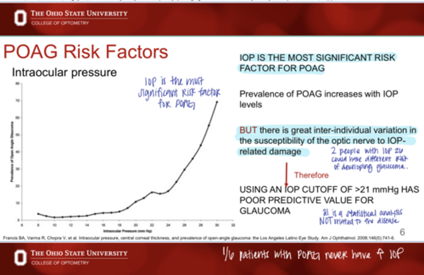
True or False:
2 people with an IOP of 26 could have different risk of developing glaucoma
true

______ of patients with POAG NEVER have an increased IOP measurement
1/6

What does central corneal thickness (CCT) affect?
IOP measurements with Goldmann
May be an independent risk factor for the conversion of ocular hypertension to POAG as seen in the OHTS study

(thicker/thinner) central corneal thickness leads to an increased risk of developing POAG
thinner
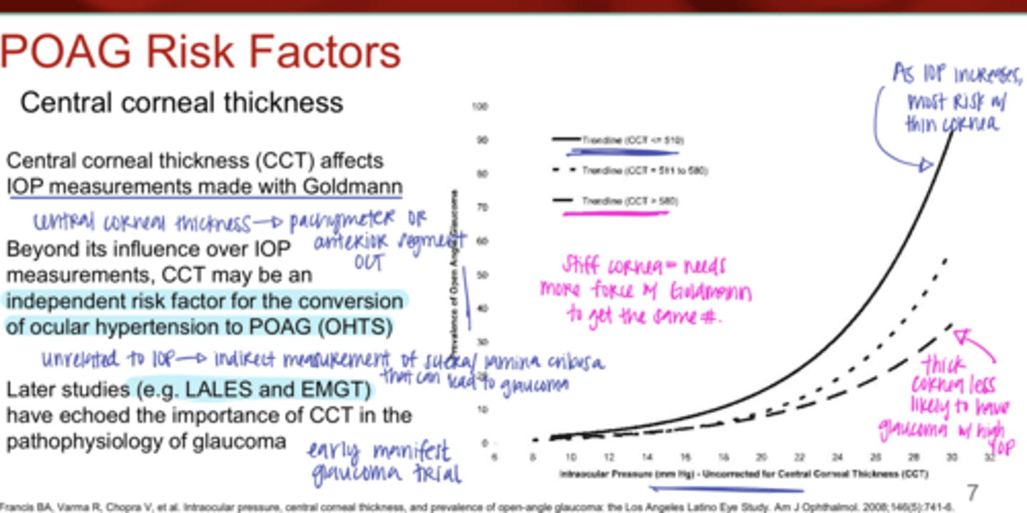
Why does a thick/stiff corneal lead to a DECREASED risk of developing POAG?
Will need more force (higher IOP reading) to get the same effect on the cornea

What studies have echoed the importance of CCT int he pathophysiology of glaucoma?
-LALES
-EMGT

Ocular Perfusion Pressure Equation
Ocular Perfusion Pressure (OPP) = Diastolic Presssure - IOP

What are some risk factors associated with POAG besides high IOP?
Low ocular perfusion pressure
Myopia
Low CSF pressure
Lack of exercise

What is characteristic of low ocular perfusion pressure?
-diastolic pressure <50mmHg
-systolic pressure <125mmHg

Can systemic hypertension and/or diabetes mellitus be linked to POAG?
Yes -- unsure of the correlation

Can migraines and peripheral vasospasm be linked to POAG?
Yes -- less O2 to the ONH

Why is low CSF pressure a risk factor for POAG?
CSF acts as a backstop for the lamina cribosa; force against IOP

____ is protective against POAG
Exercise
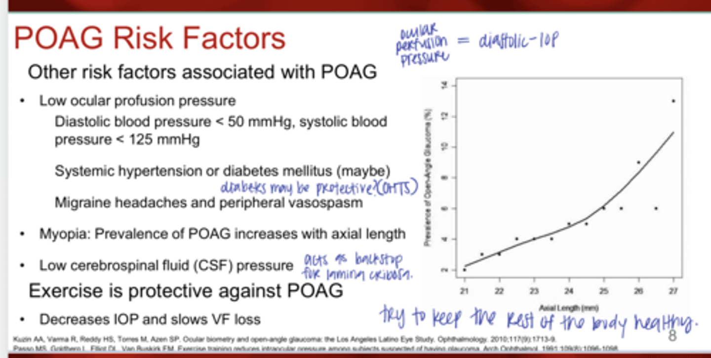
Why is exercise protective against POAG?
decreases IOP pressure and slows VF loss

What is the major contributing factor for the elevated IOP in primary open-angle glaucoma?
increased resistance to fluid flow through the TM

What is the Pathophysiology of POAG & Increased resistance to fluid flow through the TM?
1) Increased extracellular matrix beneath the inner wall of Schlemm's canal (JCT)
2) Loss of endothelial cells causing thickening of trabecular lamellae beams/sheets
3) Plaques form in the corneoscleral beams and JCT meshwork

Uveoscleral outflow may (increase/decrease) in POAG
increase

Why will Uveoscleral outflow be increased in POAG?
AH may be redirected from the TM, an area of abnormally high resistance, to the uvea, a region where flow is less dependent on IOP

What are the 1st line medications for POAG & what do they target? Why?
Prostaglandins -- target the uveoscleral outflow pathway and maximize outflow through a pathway that is NOT subject to increased resistance in glaucomatous eyes
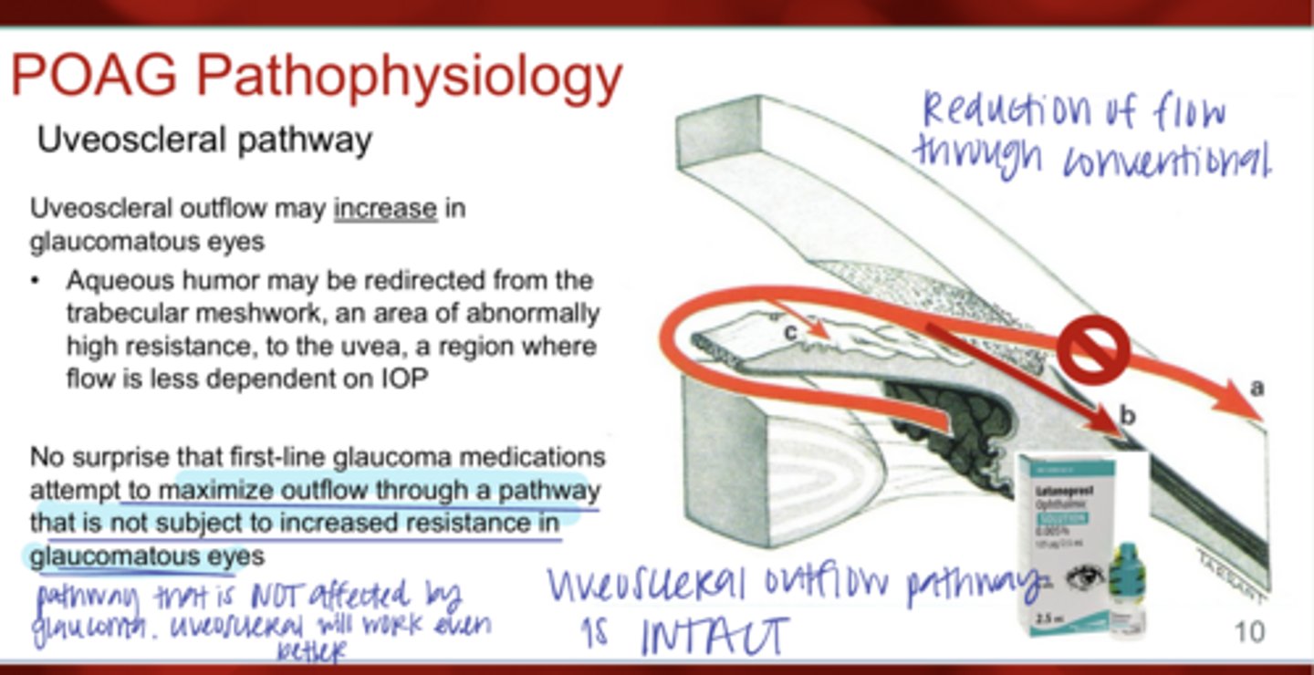
Is the uveoscleral pathway INTACT in POAG eyes?
Yes
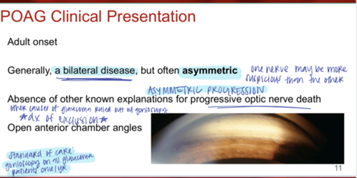
What is the clinical presentation of POAG?
-adult onset
-bilateral disease
-often asymmetric
-absence of other known explanations for progressive optic nerve death
-open anterior chamber angles

Since POAG is asymmetric, can one optic nerve be more suspicious than the other?
Yes

Since POAG is asymmetric, can one optic nerve atrophy progress faster than the other?
Yes

POAG is a diagnosis of _______
exclusion -- rule out other causes with gonioscopy

What is the standard of care on all glaucoma patients?
gonioscopy 1x/yr
What is the most important structure in POAG and all other types of glaucoma?
optic nerve head
With glaucoma, it's all about the _____, not the _____
donut, hole

We should be more concerned about the appearance of the ____ over the appearance of the ONH cup
ONH rim

_______ is the most important clinical tool that you have in assessing the ONH
Stereopsis

What does stereopsis help evaluate?
depth of the ONH
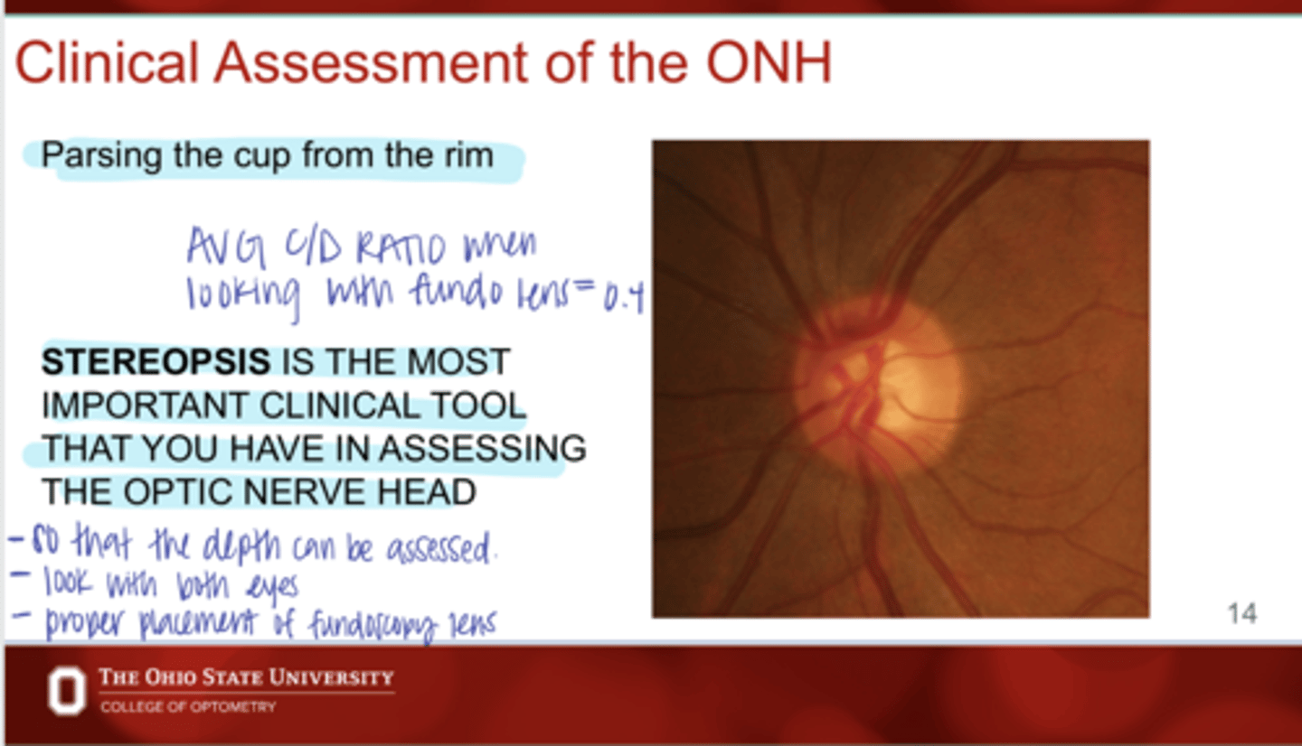
What is the avg C/D ratio when looking with stereoposis/fundo?
0.4R
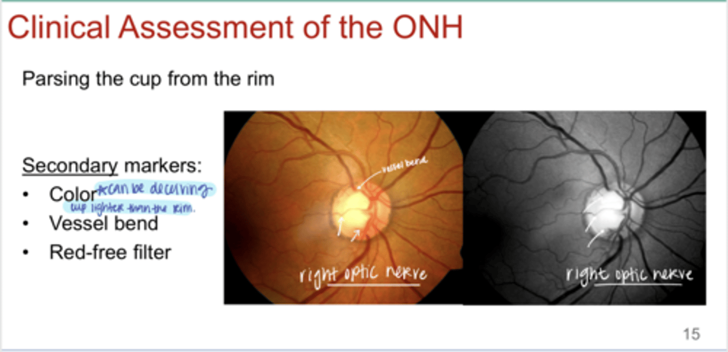
What are secondary markers that can help distinguish cup from rim?
-color
-vessel bend
-red-free filter
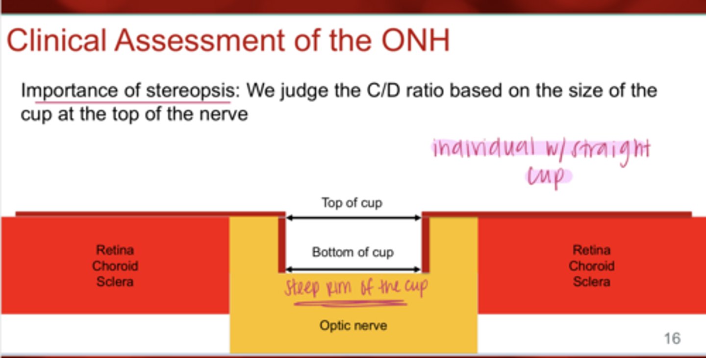
REVIEW: What is the importance of stereopsis in the clinical assessment of the ONH?
we judge the C/D ratio based on the size of the cup at the top of the nerve

Straight Cup w/ Steep Rim (Pic)
Straight Cup w/ Steep Rim (Pic)
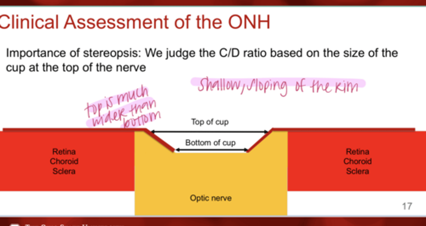
Shallow Sloping of the Cup (Pic)
Shallow Sloping of the Cup (Pic)
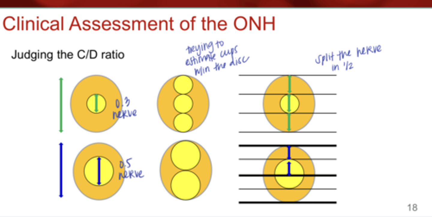
Judging the C/D Ratio (Pic)
Judging the C/D Ratio (Pic)

True or False:
Optic nerve size will affect cup size
true

The size of the optic nerve disc can influence what?
Can influence the appearance of the nerve & how the retinal ganglion cell axons are distributed.
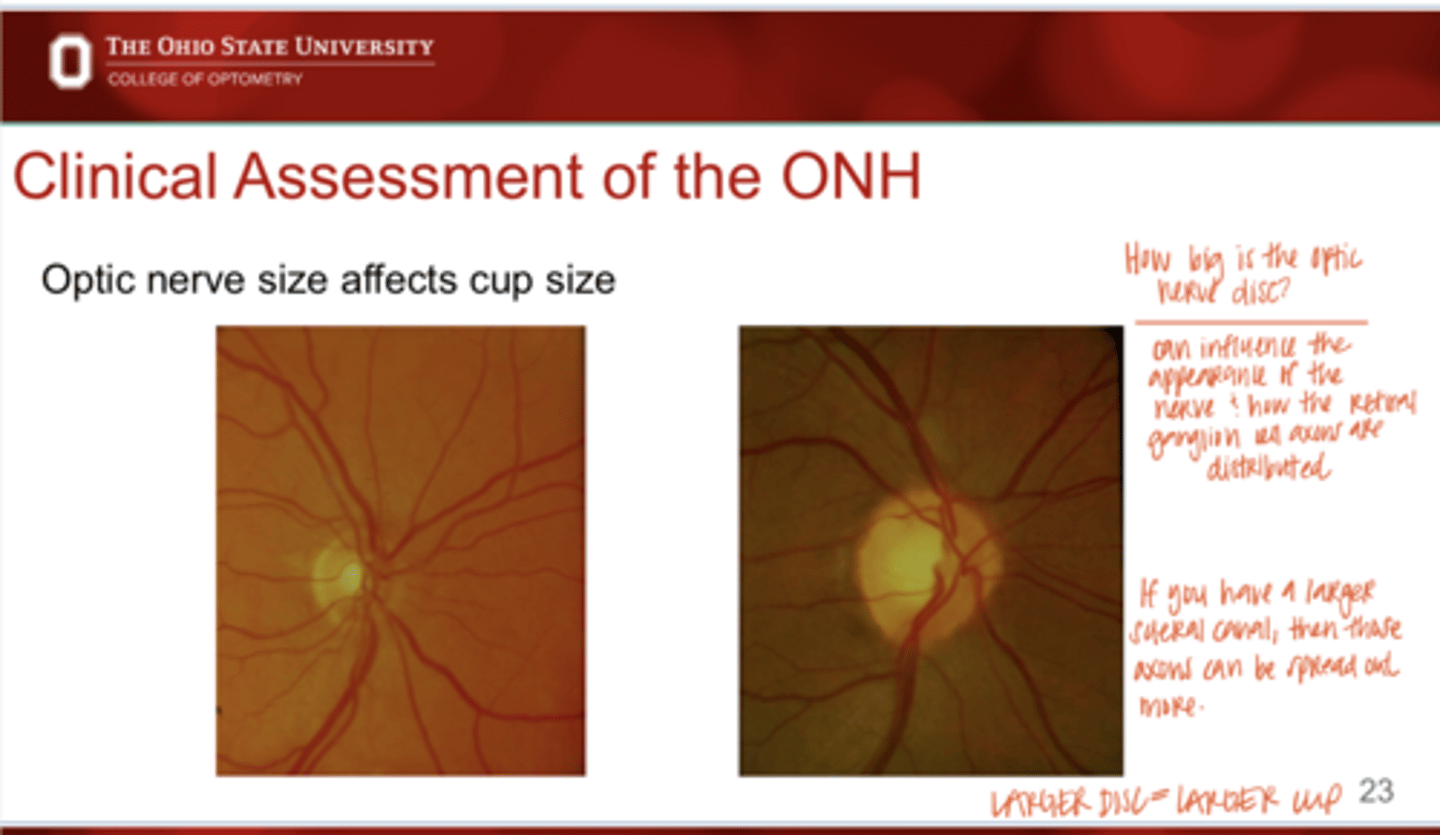
If you have a larger scleral canal at the optic nerve disc, then those axons will appear how?
Axons will be more spread out

What population is known to have large C/D ratios?
African Americans
What is the measurement for a SMALL OPTIC NERVE DISC AREA?
<1.6mm^2
What is the MEDIAN measurement for a OPTIC NERVE DISC AREA?
1.6-1.9 mm^2
What is the measurement for a LARGE OPTIC NERVE DISC AREA?
>1.9mm^2

A moderate-large C/D can have a _____ OCT
spurred
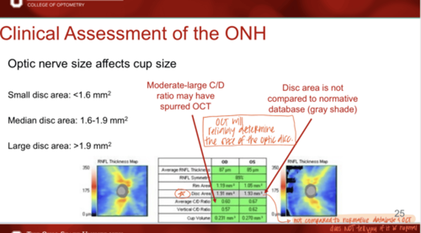
Is the disc area compared to normative data on an OCT?
No -- will show up as a grey shade in the print out
What is the measurement for a SMALL VERTICAL NERVE DISC DIAMETER?
<1.5mm
What is the measurement for an AVG VERTICAL NERVE DISC DIAMETER?
1.8mm
What is the measurement for a LARGE VERTICAL NERVE DISC DIAMETER?
>2.2mm
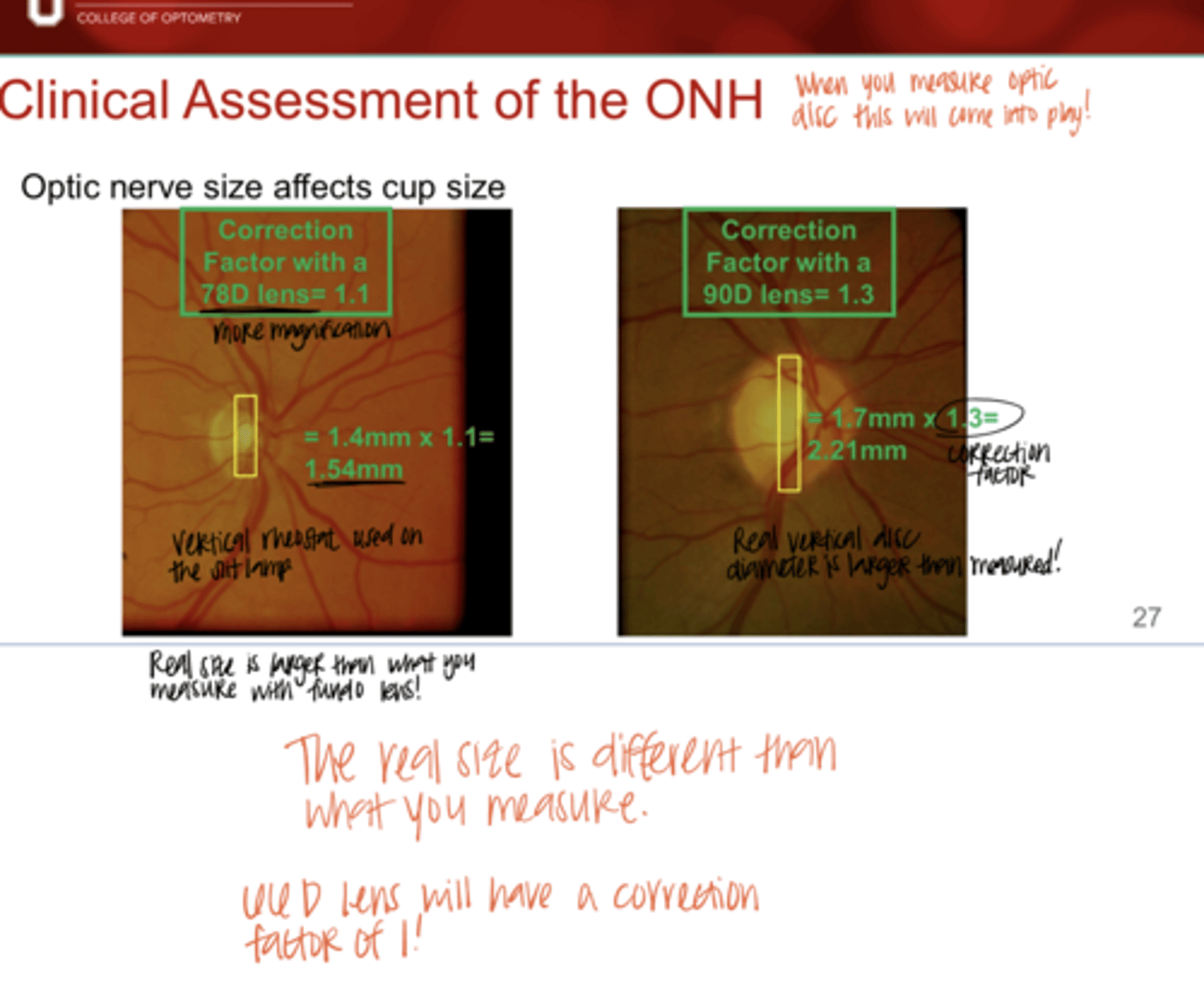
Correction factor on a 78D fundo lens?
1.1x

Correction factor on a 90D fundo lens?
1.3x

Is the real size of the nerve that you measure with a 78/90D fundo lens different than what is measured?
Yes

Once you identify the cup, what rule should be followed in order to assess the nerve?
ISNT rule
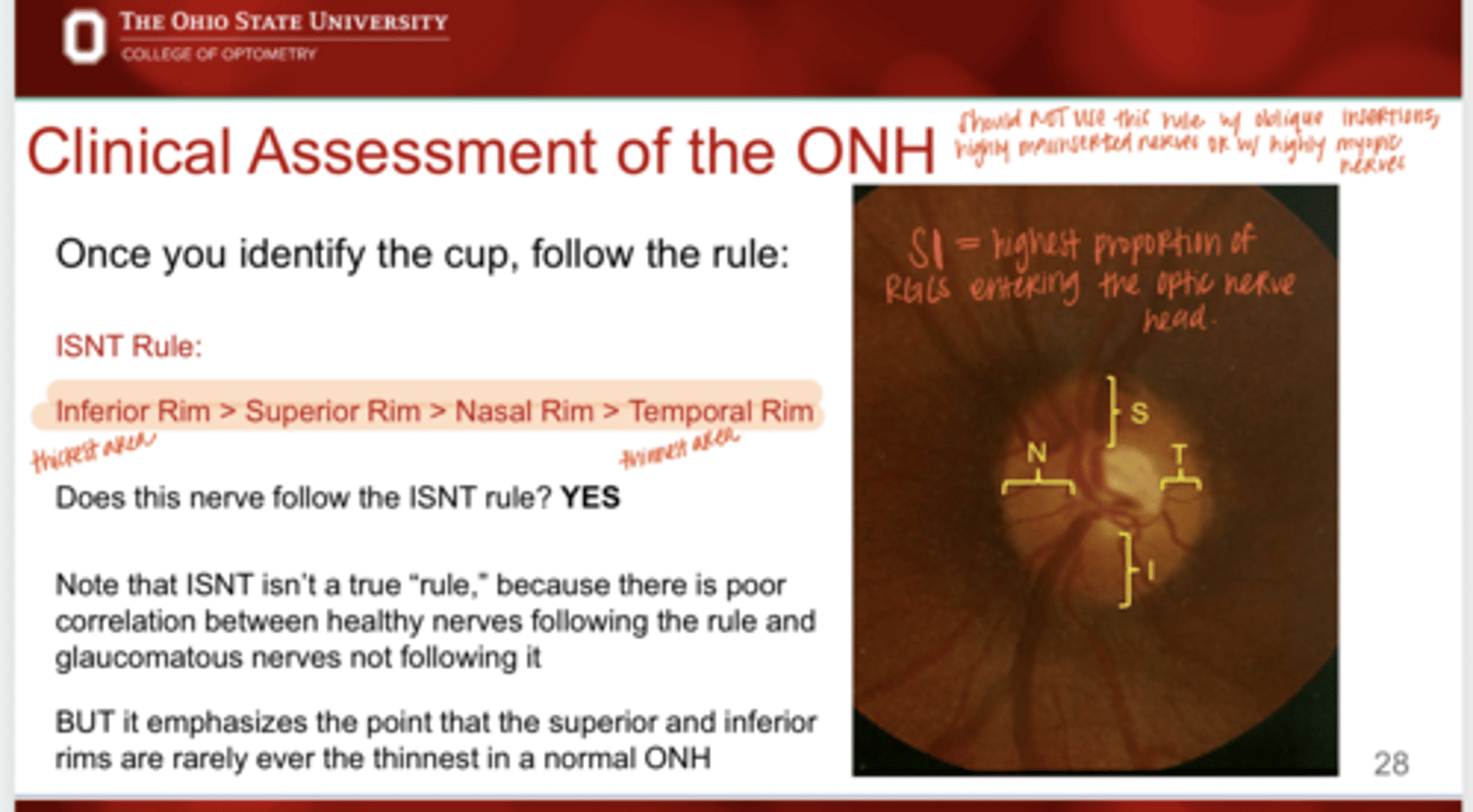
What does the ISNT rule suggest?
Inferior Rim (thickest) > Superior Rim > Nasal Rim > Temporal Rim (thinnest)
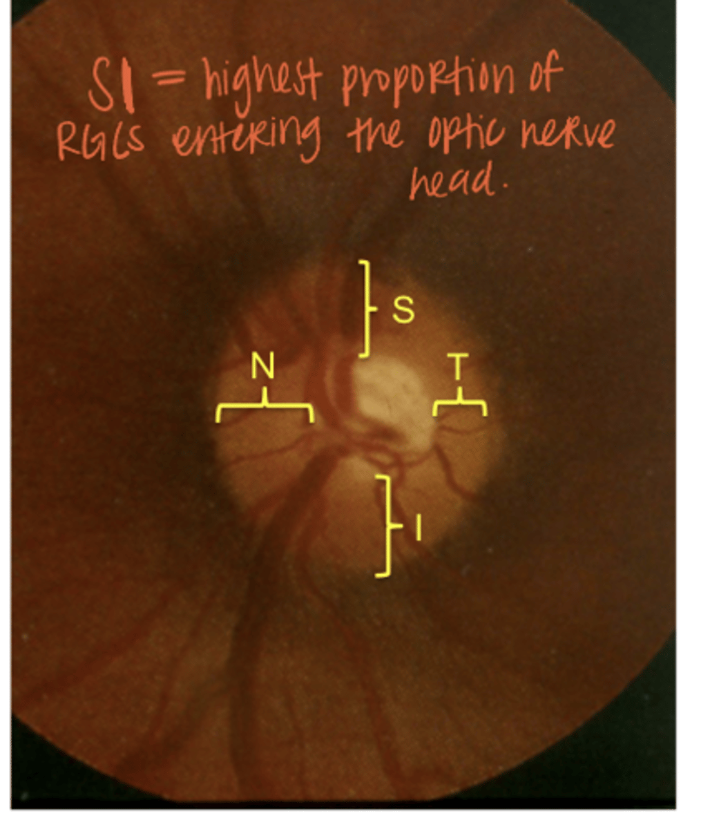
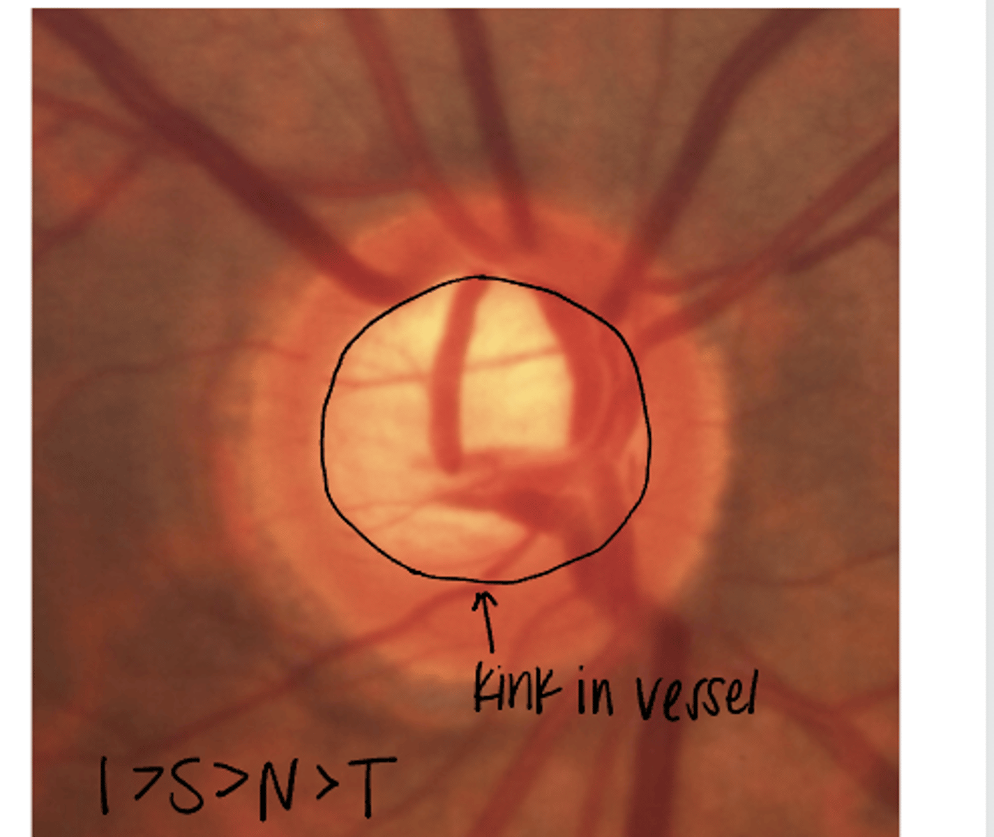
Does this nerve follow the ISNT rule?
YES

Is the ISNT rule a true rule? What is the main purpose of the rule?
No -- There is poor correlation between healthy nerves following the rule and glaucomatous nerves not following it. BUT it emphasizes the point that the superior and inferior rims are rarely ever the thinnest in a normal ONH

The ISNT rule should not be followed in what situations?
-obliquely inserted nerves
-highly malinserted nerves
-highly myopic nerves

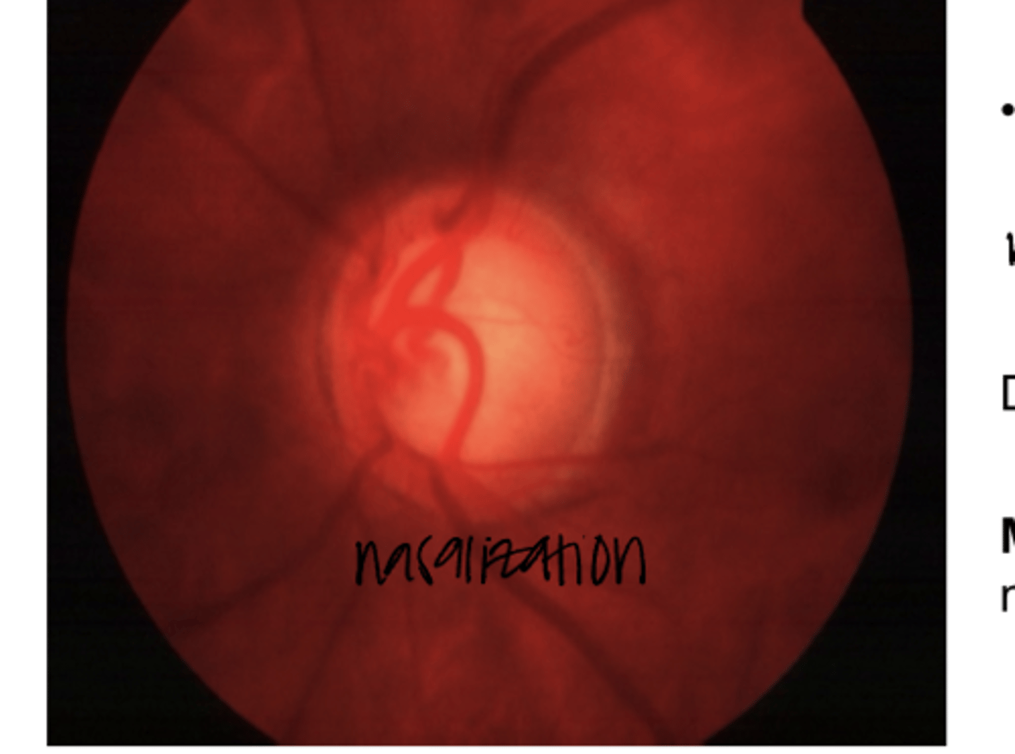
______ of the disc vessels makes it difficult to assess the nasal rim tissue
Nasalization

Is the nasal rim typically damaged in glaucoma?
No -- this is the last tissue to be damaged (late in the disease)
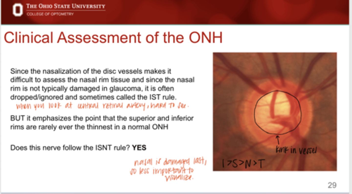
True or False:
N is sometimes dropped from the ISNT rule
true
Does this nerve follow the ISNT rule?
Yes
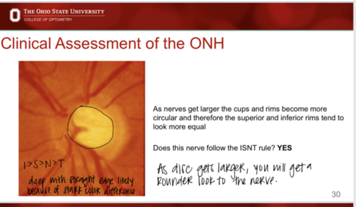
As nerves get larger, the cups/rims become more circular and therefore the superior and inferior rims tend to look more ______
equal
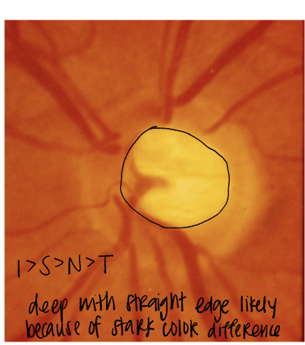
Does this nerve follow the ISNT rule?
Yes
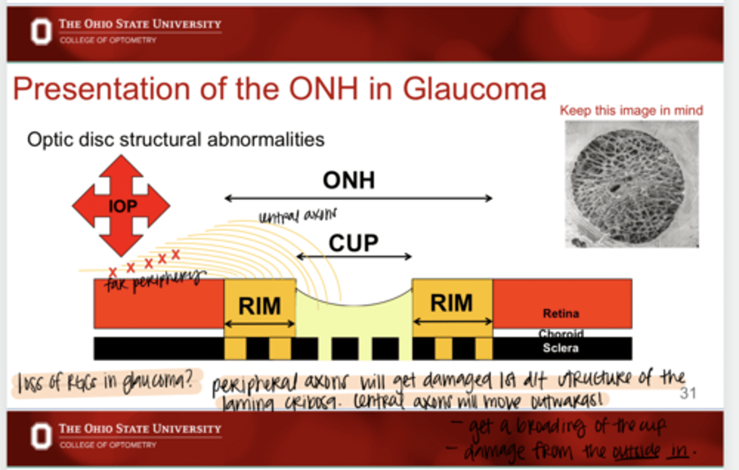
(Peripheral/Central) RGCs will get damaged first in glaucoma
Peripheral

Why do the peripheral RGCs get damaged 1st in glaucoma?
d/t the structure of the lamina cribosa; central axons will move outwards

Healthy Nerve Diagram (Pic)
Healthy Nerve Diagram (Pic)
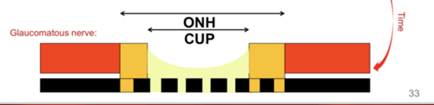
Glaucomatous Nerve Diagram (Pic)
Glaucomatous Nerve Diagram (Pic)
Does this nerve follow the ISNT rule?
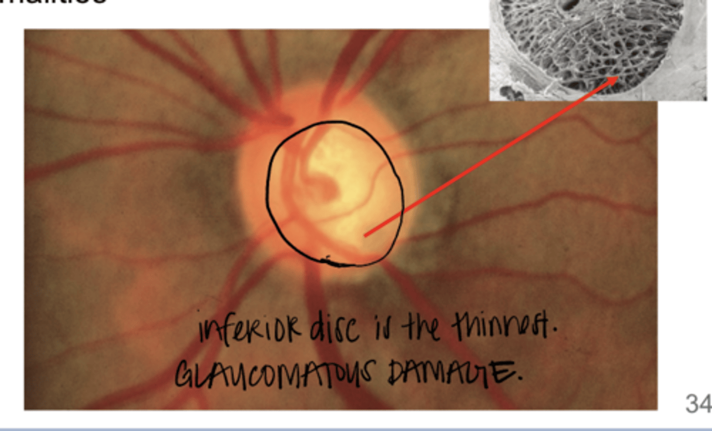
No -- the inferior neuroretinal rim is the thinnest causing vertical elongation of the cup
Does this nerve follow the ISNT rule?
No -- thin inferior neuroretinal rim & vertical elongation of the cup
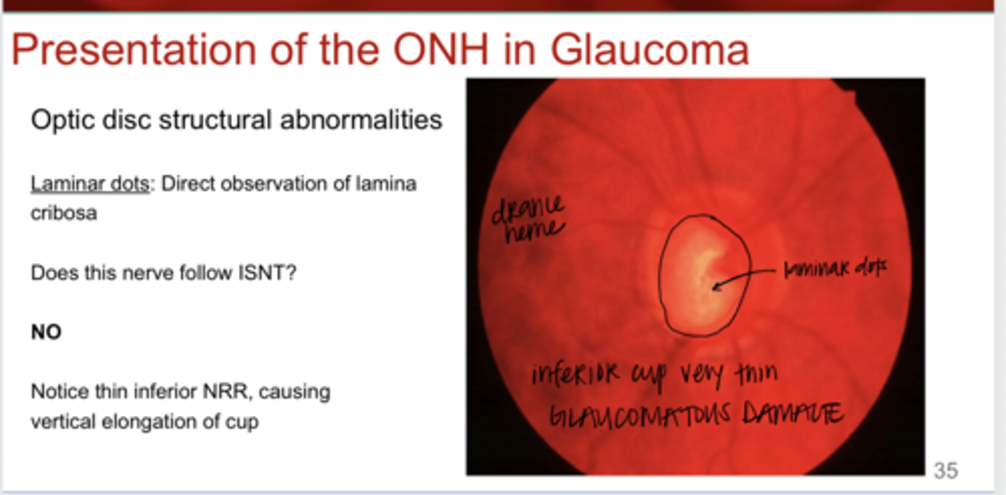
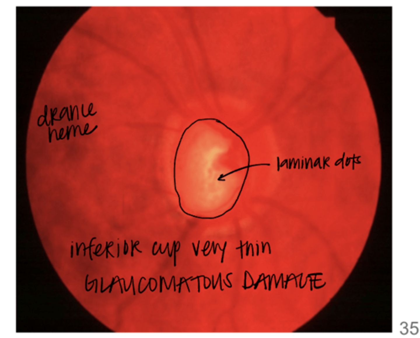
When are laminar dots observed?
direct observation of the laminate cribosa
Does this nerve follow the ISNT rule?
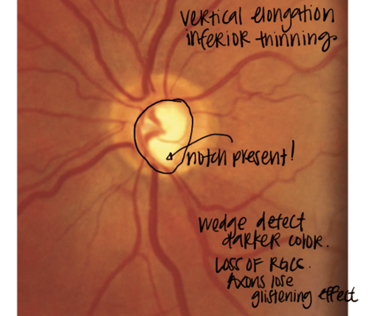
No -- thin inferior NRR causing vertical elongation of the cup. This dramatic, focal area is a notch.
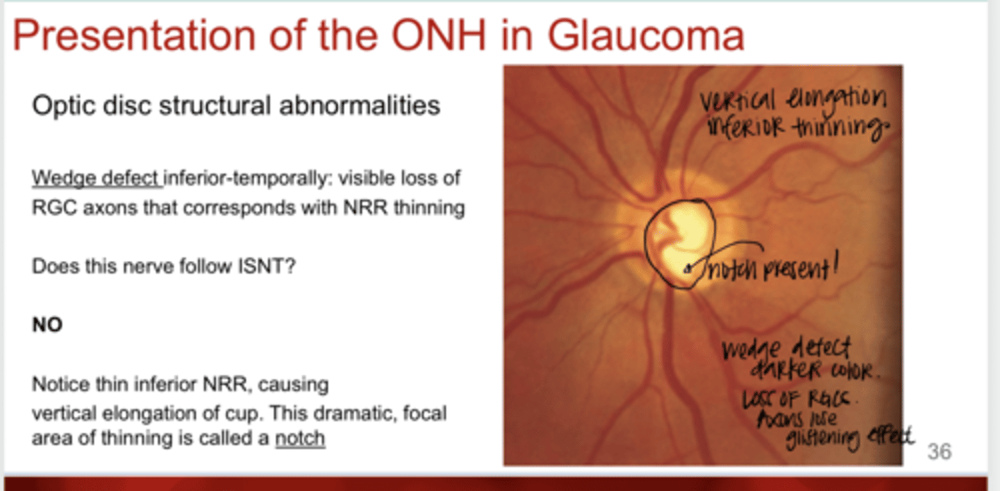
What is a wedge defect?
Visible loss of RGC axons that correspond to NRR thinning

Where is the wedge defect in this pic?
inferior-temporally
Does this nerve follow the ISNT rule?
Maybe? But entire NRR is thinned in advanced glaucomatous optic neuropathy
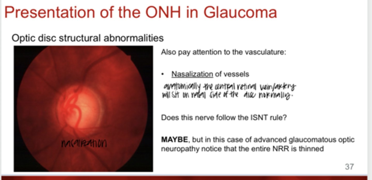
What is nasalization of vasculature in glaucoma?
Refers to the inward and nasal displacement of the central retinal vessel trunk on the optic nerve head. As the optic cup enlarges due to glaucoma, the retinal vessels, which normally enter the optic disc symmetrically, shift nasally as surrounding nerve tissue deteriorates
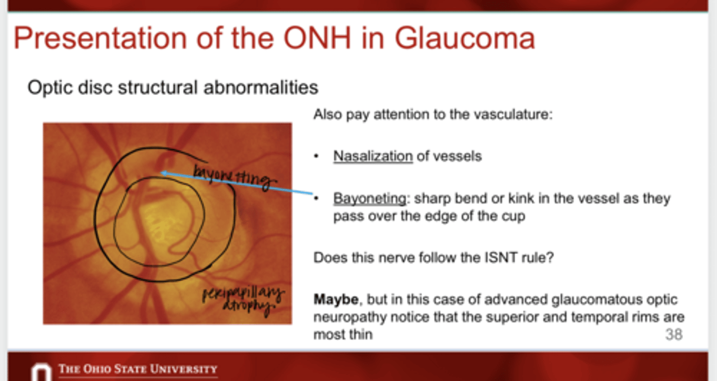
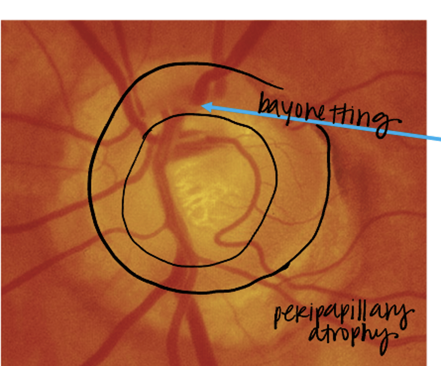
What is bayonetting of vessels in glaucoma?
sharp bend or kink in the vessel as they pass over the edge of the cup
Does this nerve follow the ISNT rule?
Maybe? But in this case of advanced glaucomatous optic neuropathy the superior and temporal rims are most thin
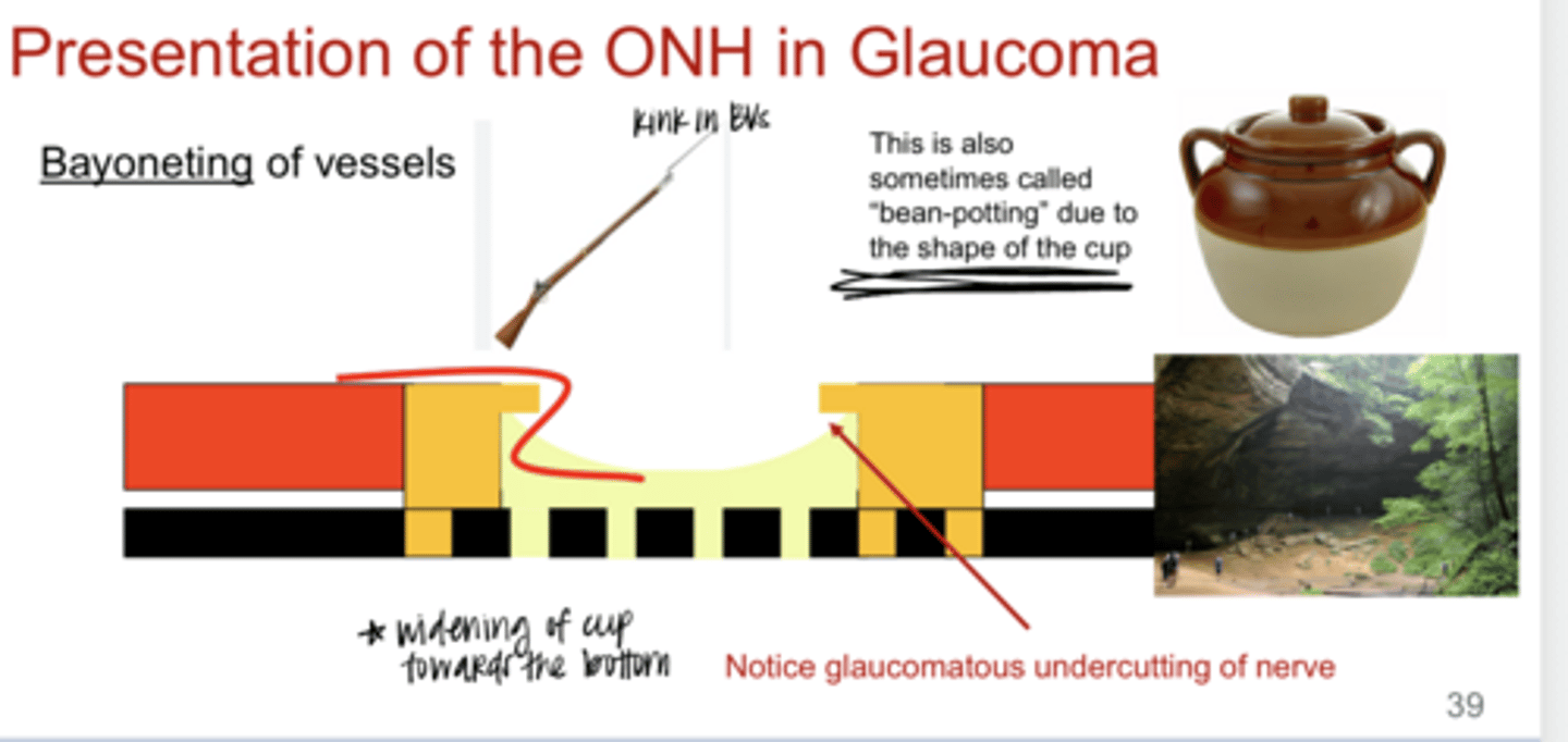
Bayoneting of Vessels Diagram (Pic)
**glaucomatous undercutting of the nerve
Bayoneting of Vessels Diagram (Pic)
**glaucomatous undercutting of the nerve

This shape of the cup is sometimes referred to as _______
bean potting

What is a circumlinear vessel of the ONH?
curves to follow the contour of the physiological cup
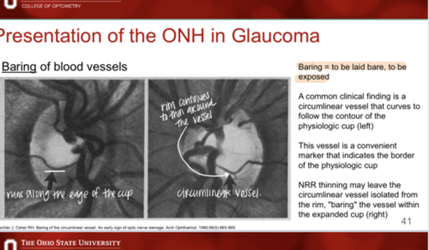
What are circumlinear vessels helpful for?
indicates the border of a physiological cup

NRR thinning can leave what when a circumlinear vessel is present?
Can leave the vessel isolated from the rim, "baring" the vessel within the expanded cup

What is a drance hemorrhage?
disc hemorrhage secondary to glaucoma

What does a drance hemorrhage indicate?
that damage will or has happened in that area
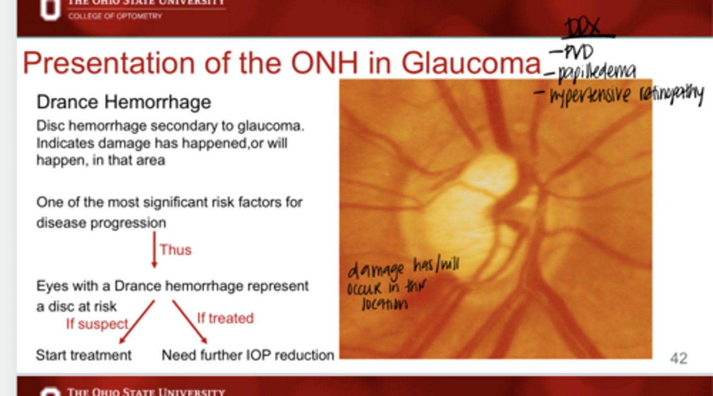
What is one of the most significant risk factors for disease progression?
drance hemorrhage