BS3054 L6 - Cyclic Nucleotides
1/27
There's no tags or description
Looks like no tags are added yet.
Name | Mastery | Learn | Test | Matching | Spaced | Call with Kai |
|---|
No analytics yet
Send a link to your students to track their progress
28 Terms
Secondary Messengers
Intermediate chemicals that help transduce a chemical signal
- e.g. cAMP, cGMP, inositol triphosphate, Ca2+, diacylglycerol
What is cAMP
- second messenger
- product of reaction catalysed by adenylyl cyclase
- ATP (+ adenylyl cylase) --> cAMP
Adenylyl cyclases
- 10 isoforms
- M1 and M2 membrane bound domains
- C1 and C2 domains in the cytosol
- ATP binding site sits in-between C1 and C2 domain
- Mg2+ binding site = Mg2+ is a cofactor
- activity dependent on the Galpha subunit it receives signals from
How do isoforms of adenylyl cyclase cause specifictiy
- different isoforms respond differently to signals from G-proteins
- allows tissue specificity - not all tissues will respond to a signal and some will respond stronger than others
effectors of cAMP mediated signalling
- Protein Kinase A
- cyclic nucleotide gated channels
- cyclic nucleotide regulated GEFs
What is PKA
- protein kinase A
- serine-threonine kinase
- phosphorylates proteins
- modulated by cAMP
Structure of PKA
- two regulatory subunits
- two catalytic subunits
- 4 isoforms of R subunit (RI-alpha, RI-beta, RII-alpha, RII-beta)
- 3 isoforms of C subunit (C-alpha,beta,gamma)
w
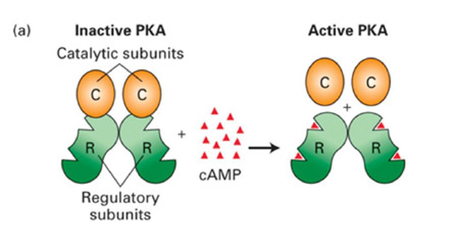
What happens when cAMP binds PKA
- cAMP binds to regulatory subunit dimer = releases the catalytic subunits from the complex
- the catalytic subunits are now active and can phosphorylate
What are the two classes of PKA
PKAI
PKAII
PKA I vs PKA II
PKA I - cytosolic
- cAMP binds to PKA and causes dissociation of the catalytic subunit = can phosphorylate substrate
PKA II - membrane bound
- docked to AKAP
- substrate binds to AKAP and then cAMP causes release of the R subunits = allows substrate to be phosphorylated
- AKAP localises PKA to specific cellular targets
What is AKAP
- A-kinase anchoring protein
Examples of PKA targets
- GPCRs, ion channels, cytoskeletal proteins, protein phosphatase and kinase inhibitors, transcription factors
Phosphodiesterase
enzyme that degrades cAMP, producing 5'AMP, to terminate signalling
- breaks phosphodiester bond
- regulated by kinases
Are there only one type of PDE (phosphodiesterase)
- 11 different families of PDEs in mammals
- some hydrolyse both cGMP and cAMP
- some preferentially hydrloyse cAMP or cGMP
guanylyl cyclases
- guanylyl cyclase catalyses GTP to cGMP
- PDE will convert cGMP to 5'GMP
What are the two types of guanylyl cyclase
1. particulate GCs = membrane bound
2. soluble GCs = in cytosol/ NO sensitive
particulate guanylyl cyclases
- transmembrane, ligand-activated homodimers
soluble guanylyl cyclases
- activated by NO and CO
- NO is an extremely potent activator
- nitric oxide binds haem group in guanylyl cyclase heterodimer
Nitric oxide
- vasodilator
- gaseous compound - only stable for seconds so made as and when needed
- detectable at very small amounts
NOS
- nitric oxide synthase
- synthesises NO
types of NOS
nNOS = neuronal + skeletal muscle = communication
iNOS = inducible = produces high NO concentrations that can exhibit direct toxic effects = immune defence
eNOS = endothelial = vasodilation
biosynthesis of Nitric oxide by NOS
- L-arginine is turned into L- citrulline and nitric oxide by NOS
Roles of NO
- activates soluble guanylyl cyclases
- nitrosylation of proteins
- direct toxicity (NO is a free radical)
major targets of cGMP
- cyclic nucleotide gated channels
- modulation of PDE activity
- activation of PKG
what is PKG
cGMP dependent protein kinase
PKG monomer
Regulatory domain
- Leucine zipper (pseudo-substrate)
- Nucleotide binding sites
Catalytic domain
- ATP site
- Kinase
= exist as a soluble homodimer
what is a pseudo-substrate
- substance that mimics the real substrate of an enzyme
- often part of enzyme own structure
- blocks enzyme active site and inhibits activity
How does NO cause vasodilation
- NO activates soluble guanylyl cyclase
- increases levels of cGMP within smooth muscle cells of blood vessel walls
- rise in cGMP leads to activation of PKG
- PKG activates SERCA pump to move calcium ions from cytoplasms to ER
- cGMP can activate potassium channels causing hyperpolarisation = closes VGCC = promoting relaxation