Test 3 - Appendicular Skeleton
1/16
There's no tags or description
Looks like no tags are added yet.
Name | Mastery | Learn | Test | Matching | Spaced | Call with Kai |
|---|
No analytics yet
Send a link to your students to track their progress
17 Terms
The 4 bones of the wrist in the distal row (lateral to medial) are
Trapezium, Trapezoid, Capitate, Hamate
What is identified in the photo (#2, #4, #5)
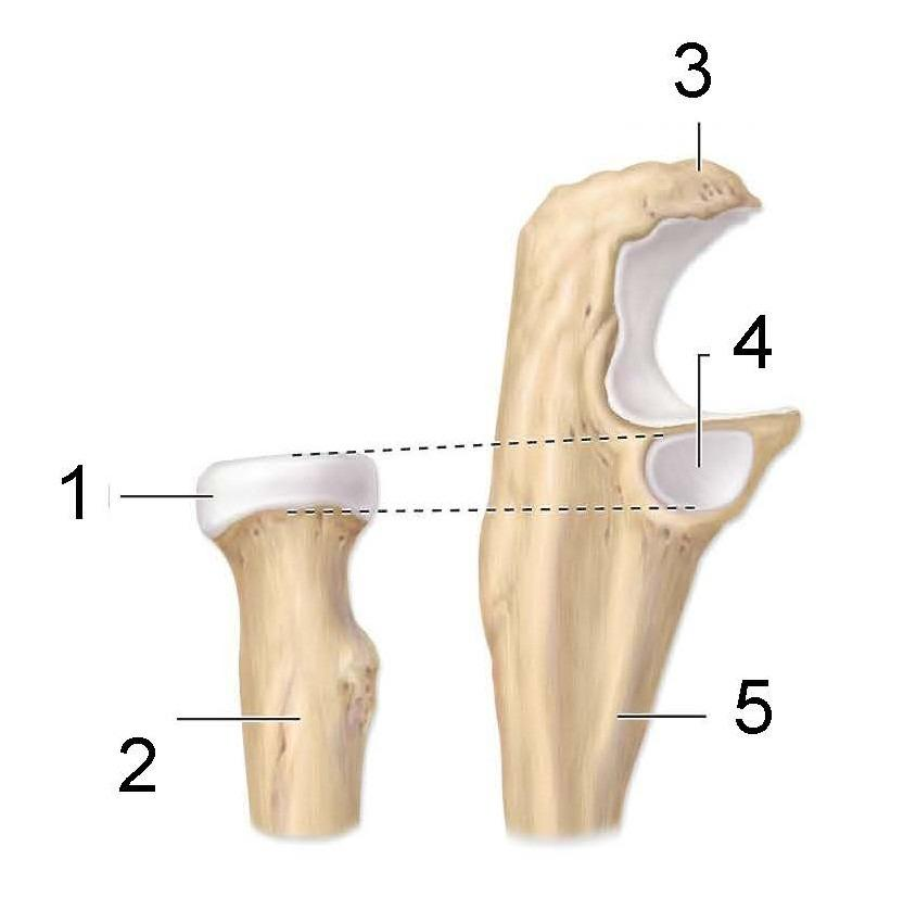
Proximal end of radius, radial notch, proximal end of ulna
What is identified in the photo (#1–#5)
Medial malleolus, fibular notch, Lateral malleolus, Interosseous borders, Shafts
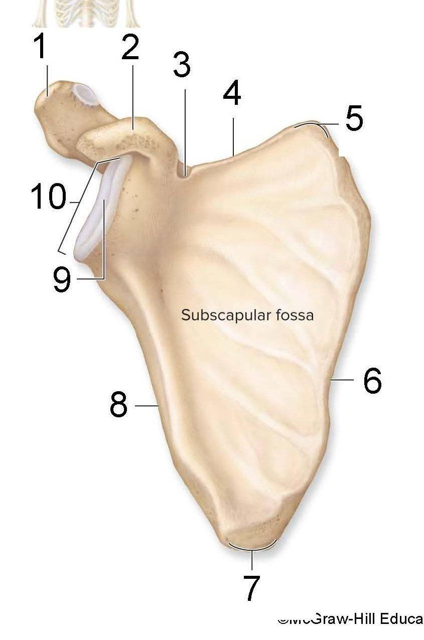
What is identified in the photo (#1, #2, #9) and is this a right or left scapula
Acromion process, Coracoid process, Glenoid cavity, Right
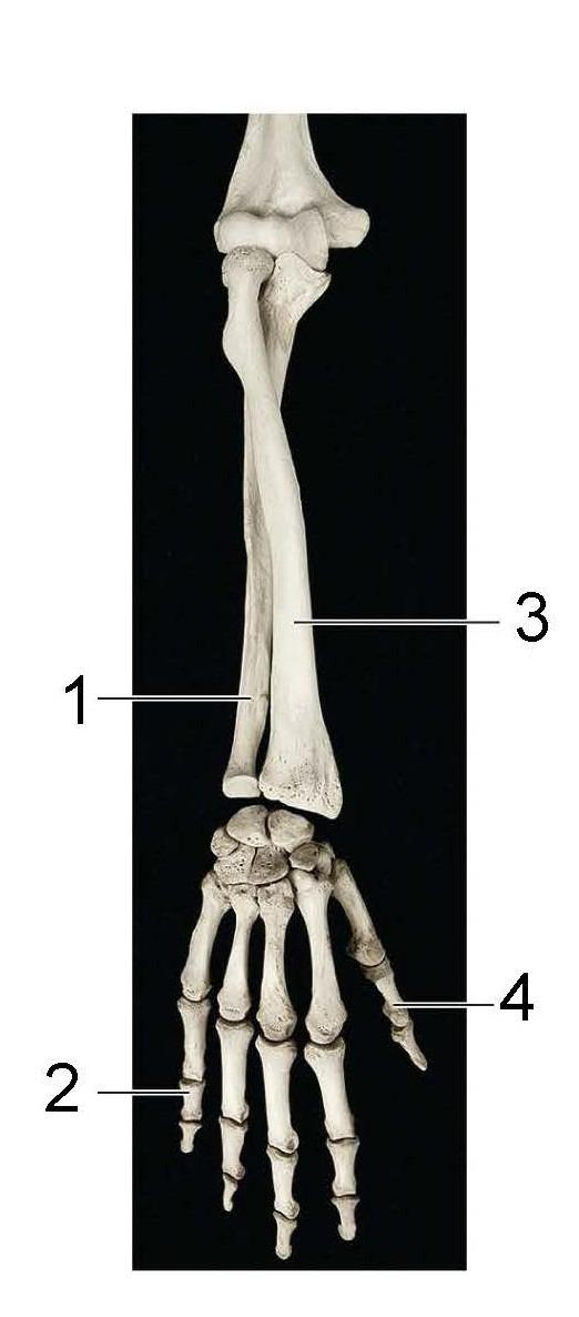
What is identified in the photo (#1, #3) and anatomical position
Ulna, Radius, Pronation
What is identified in the photo (#1–#6)
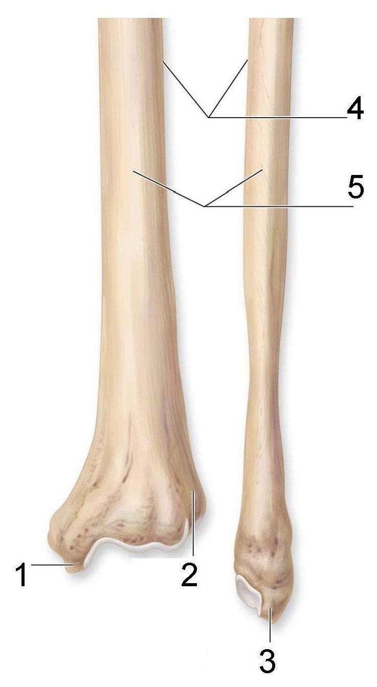
distal end of radius, Styloid process of radius, Ulnar notch, Head of ulna, Styloid process of ulna, distal end of ulna
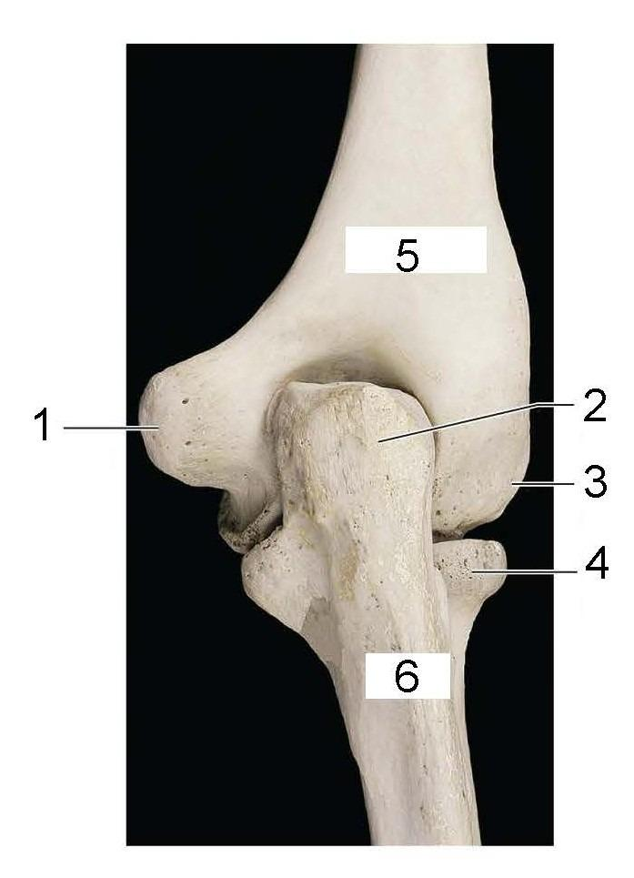
What is identified in the photo (#2) and what joint is this
Olecranon process, Elbow joint
The 3 arches of the foot are
Medial longitudinal arch, Lateral longitudinal arch, Transverse arch
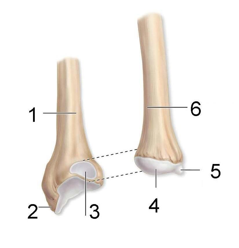
What is identified in the photo (#1–#6)
Lateral epicondyle, Capitulum, Head of radius, Medial epicondyle, Trochlea, Humerus
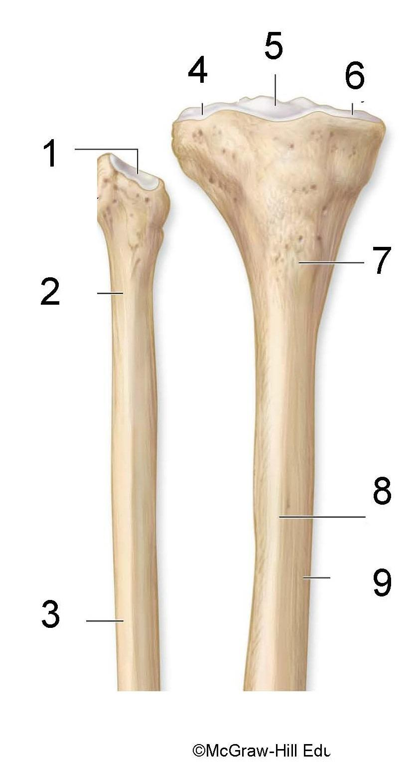
What is identified in the photo (#1–#9)
Articular facet, Neck of fibula, fibula, Lateral condyle, Intercondylar eminence, Medial condyle, Tibial tuberosity, Anterior border, tibia
Select ALL the bones of the ankle
Intermediate cuneiform, Navicular, Medial cuneiform, Calcaneus, Talus, Cuboid, Lateral cuneiform
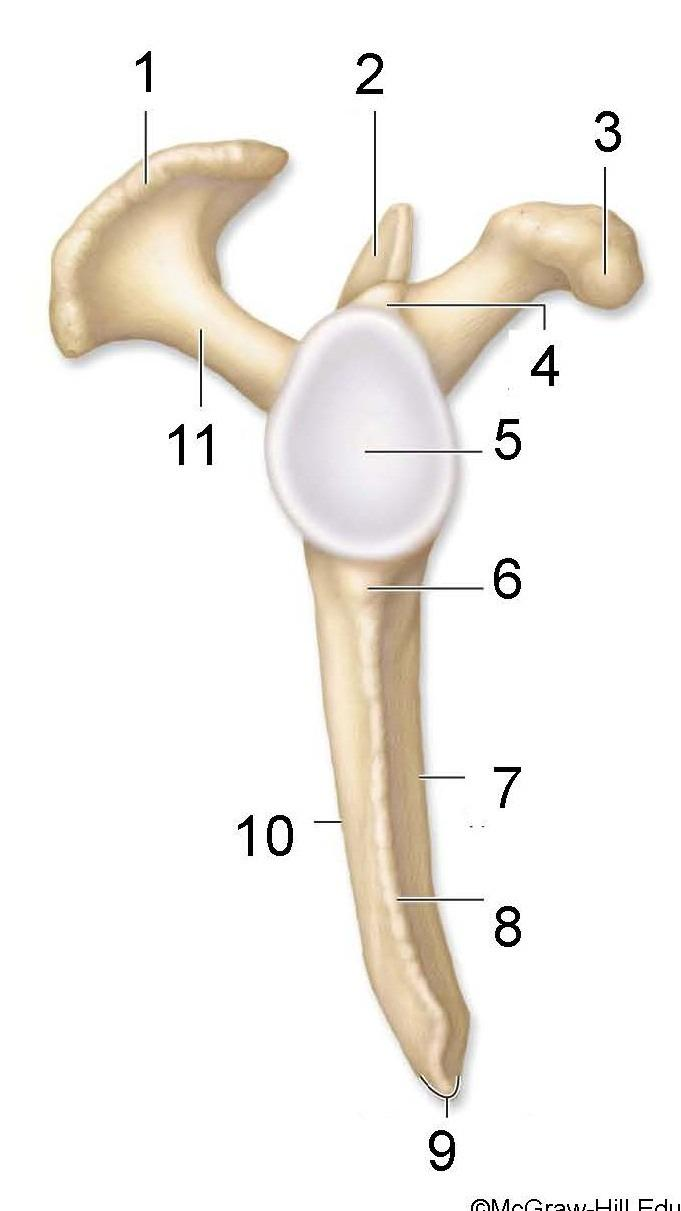
What is identified in the photo (#1, #3, #11)
Acromion process, Coracoid process, Spine of scapula
The 4 bones of the wrist in the proximal row (lateral to medial) are
Scaphoid, Lunate, Triquetrum, Pisiform
What is the name of the membrane that connects the radius and ulna
Interosseous membrane
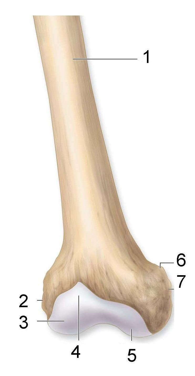
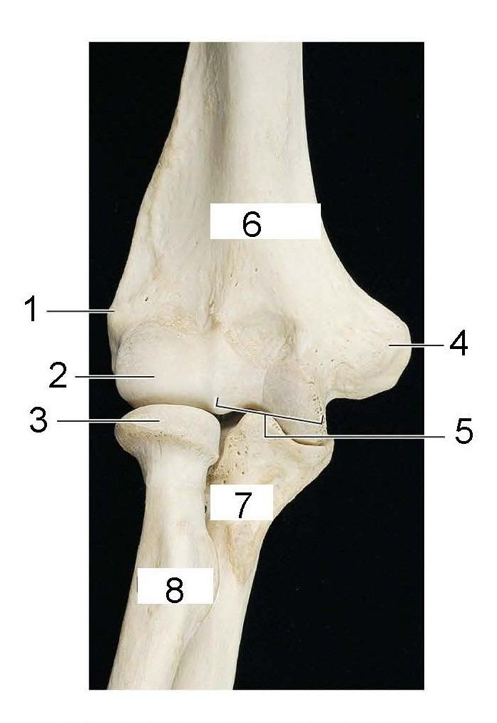
What is identified in the photo (#1–#7)
Shaft of femur, Lateral epicondyle, Lateral condyle, Patellar Surface, Medial condyle, Adductor tubercle, Medial epicondyle
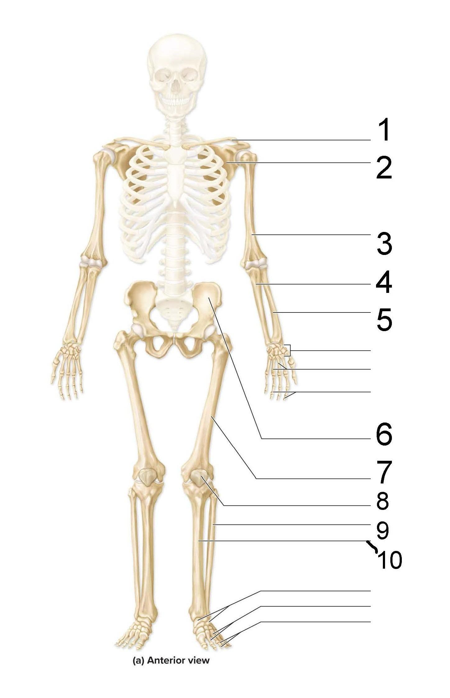
What is identified in the picture (#1–#10)
Clavicle, Scapula, Humerus, Ulna, Radius, Hip bone, Femur, Patella, Fibula, Tibia
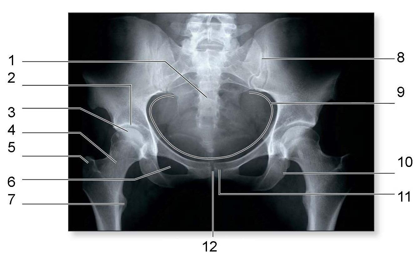
What is identified in the photo 2,3,4,5,6,7,10
Acetabulum, Head, Neck, Greater trochanter, Obturator foramen, Lesser trochanter, Ischial tuberosity