digestive system practical
1/29
There's no tags or description
Looks like no tags are added yet.
Name | Mastery | Learn | Test | Matching | Spaced | Call with Kai |
|---|
No analytics yet
Send a link to your students to track their progress
30 Terms
alimentary canal/gi tract - 6
mouth
pharynx
esophagus
stomach
small intestines
large intestines
accessory structures - 1 def, 5. structures
def. secrete their products into the canal
teeth
salivary glands
gallbladder
liver
pancreas
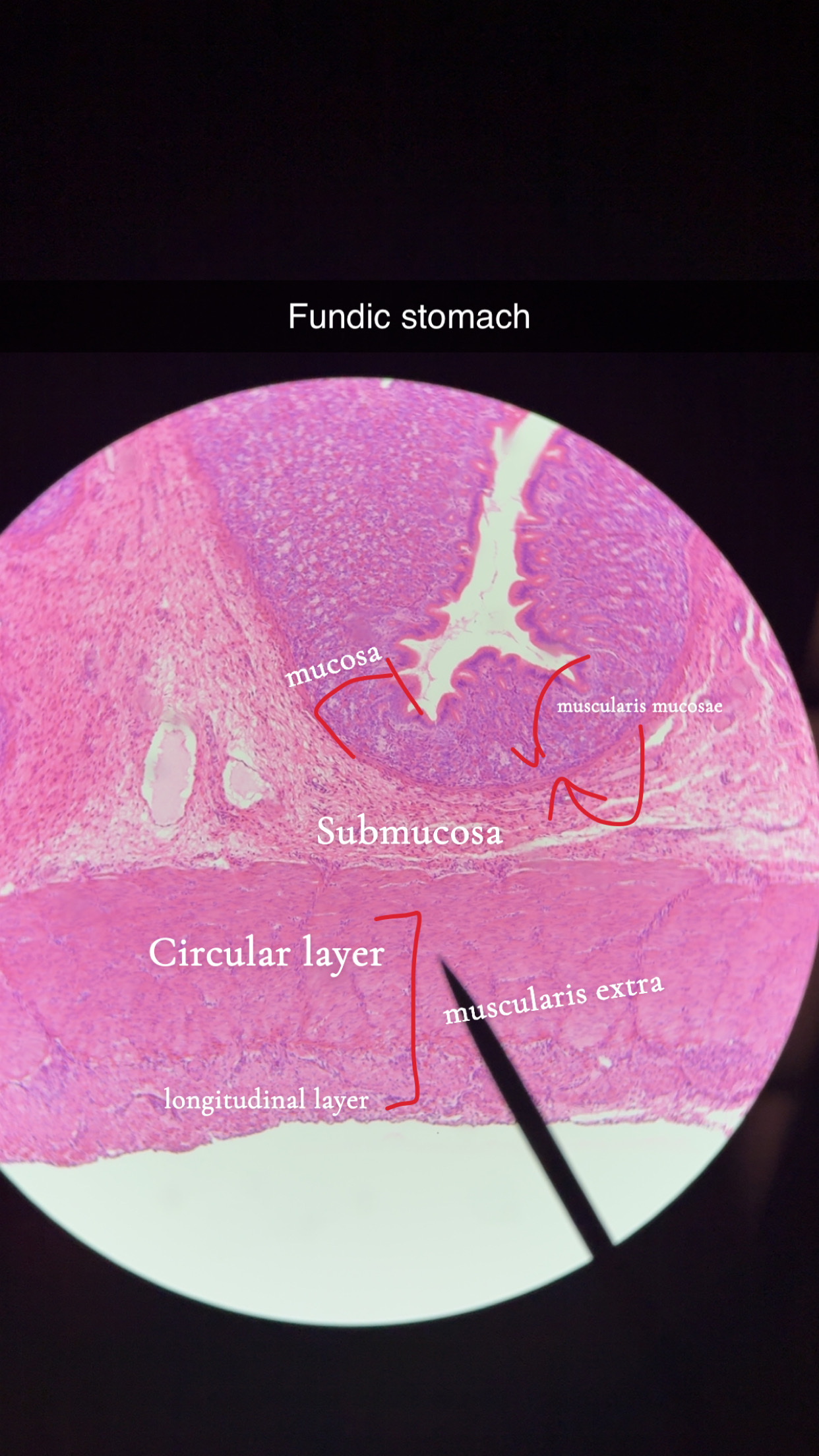
mucosa - 3 subdivisions
Epithelium [innermost] -> strat. squamous in mucosas/anus/mouth epithelium, simple columnar in the rest of the canal
Lamina Propria -> areolar CT w/ blood vessels, lymphoid follicles, MALT
Muscularis Mucosae [outermost] -> smooth muscle
mucosa function [sap]
secrete mucus, digestive enzymes, hormones
absorb end products
protect against disease
submucosa tissue/function - 3
tissue: CT, blood/lymph vessels, and nerve fibers
function:
blood vessels absorb and transport nutrients,
elastic fibers maintain the shape of each organ
muscularis externa sub/tissue - 2
circular -> inner layer of smooth muscle
longitudinal -> outer layer of smooth muscle
muscularis function
peristalsis of digested food
serosa 2 layers
CT -> areolar CT
epithelium -> simple squamous epithelium
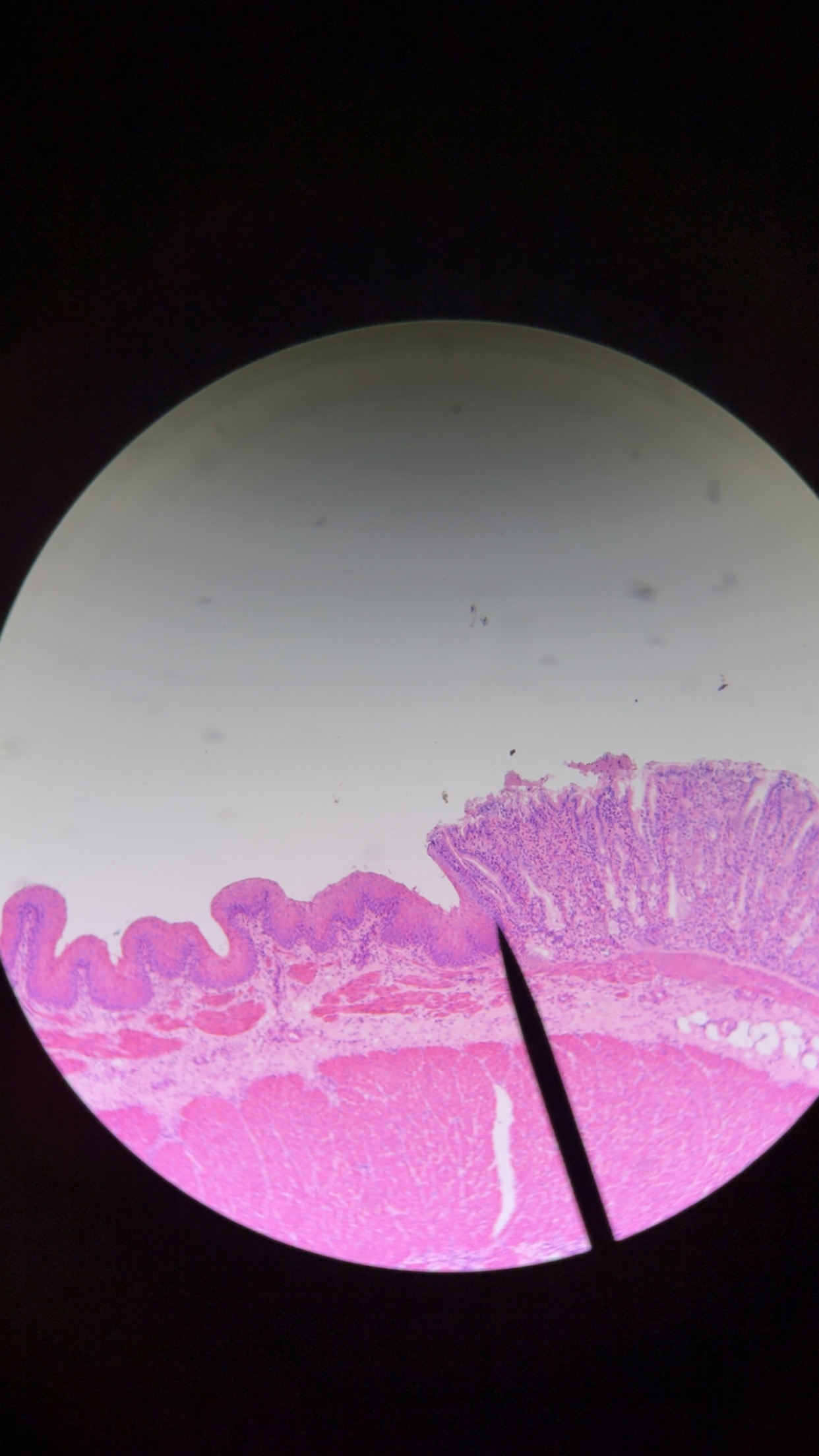
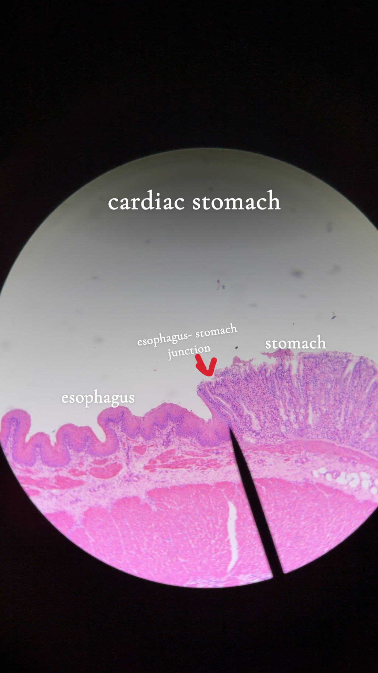
gastroesophageal junction - 3
controls food passage into the stomach
esophagus: stratified squamous (to withstand abrasion)
stomach: simple columnar (for secretional products)
stomach - 2
located in upper left quadrant
has a 3rd muscle layer that allows it to churn adn pummel food
mesentery - 3
double layer of peritoneum (a sheet of 2 serous membranes fused together) that extend from the organs to the body wall
2 types:
greater omentum
lesser omentum
greater omentum
extends from the greater curvature of the stomach adn covers most of the abdominal organs
lesser omentum
extends from the liver to the lesser curvature of the stomach
cardiac stomach - 3
surrounds cardiac orifice where food enters the stomach
stratified squamous: esophagus
simple columnar: stomach
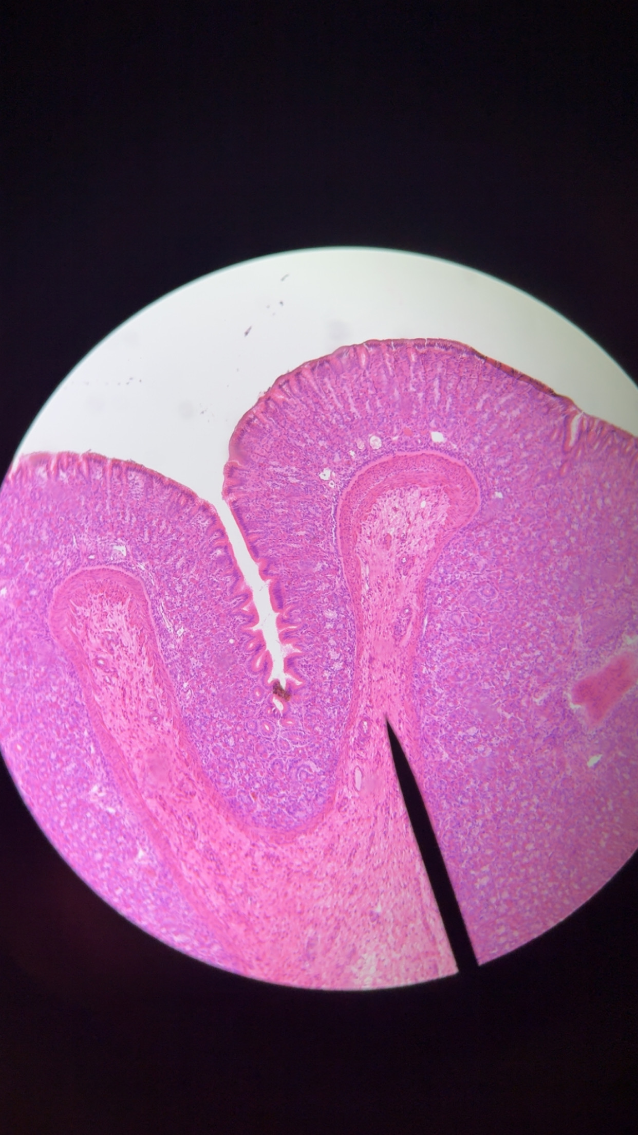
fundic stomach- anatomy & 2 cells
dome shaped area
parietal cells- HCL, intrinsic factors, red stained
chief cells- pepsinogen, blue stained
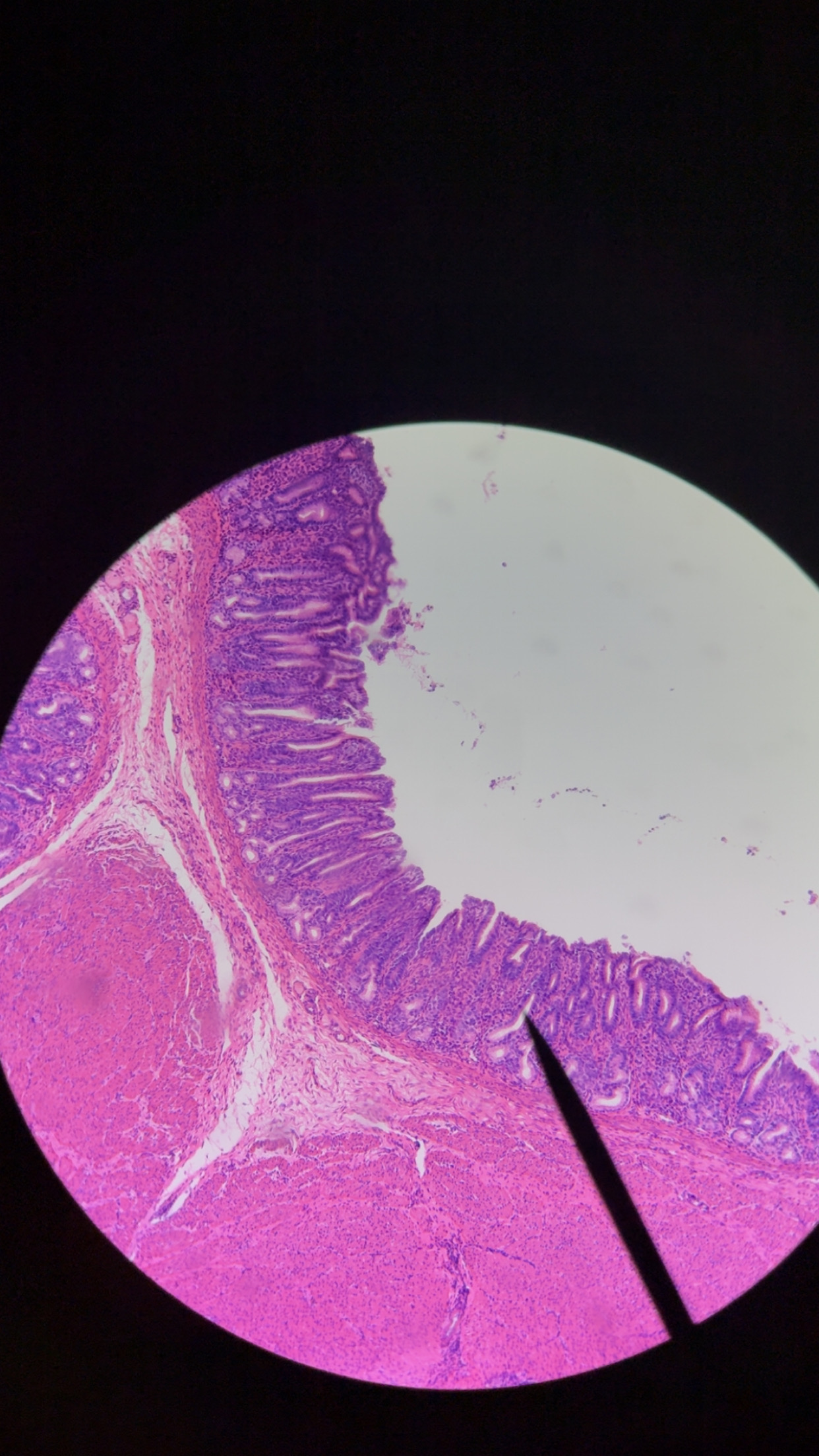
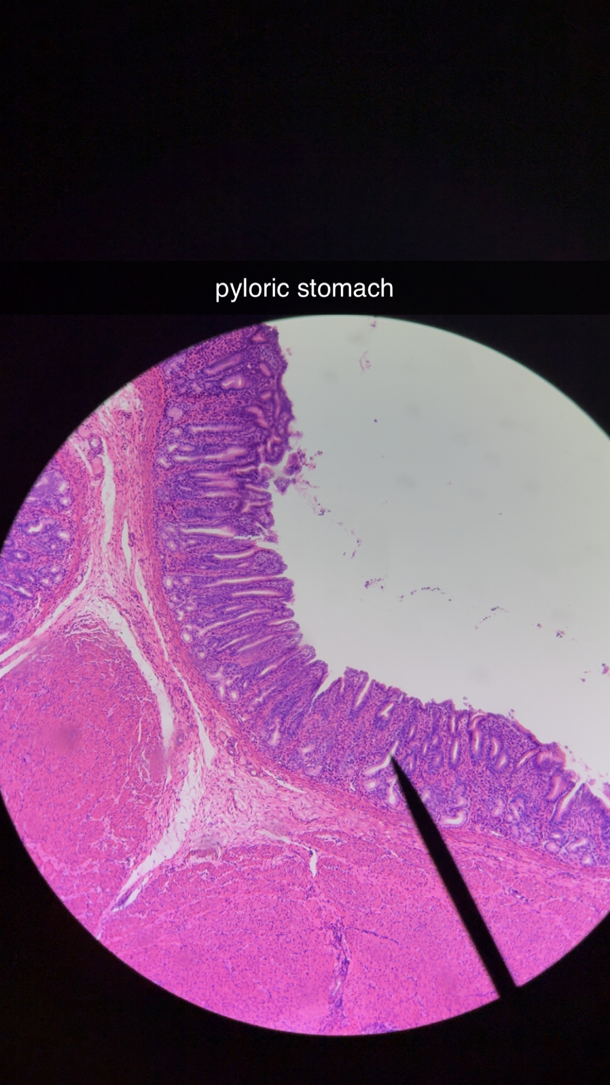
pyloric stomach - 3
stomach exits into duodenum
most digestive activity occurs here
has deep gastric pits and glands
mucosal glands
secrete a viscous mucus that prevents the stomach itself from being digested
chief cells - 2
produce pepsinogen
blue
parietal cells - 2
secrete HCL and intrinsic factors
red
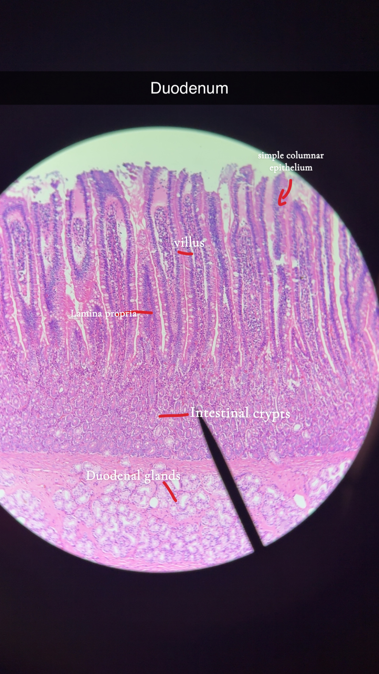
3 Structures that inc Absorptive Surface of Small Intestine Mucosa:
microvilli
villi
circular folds
circular folds
force chyme to spiral through intestine, slowing its progress
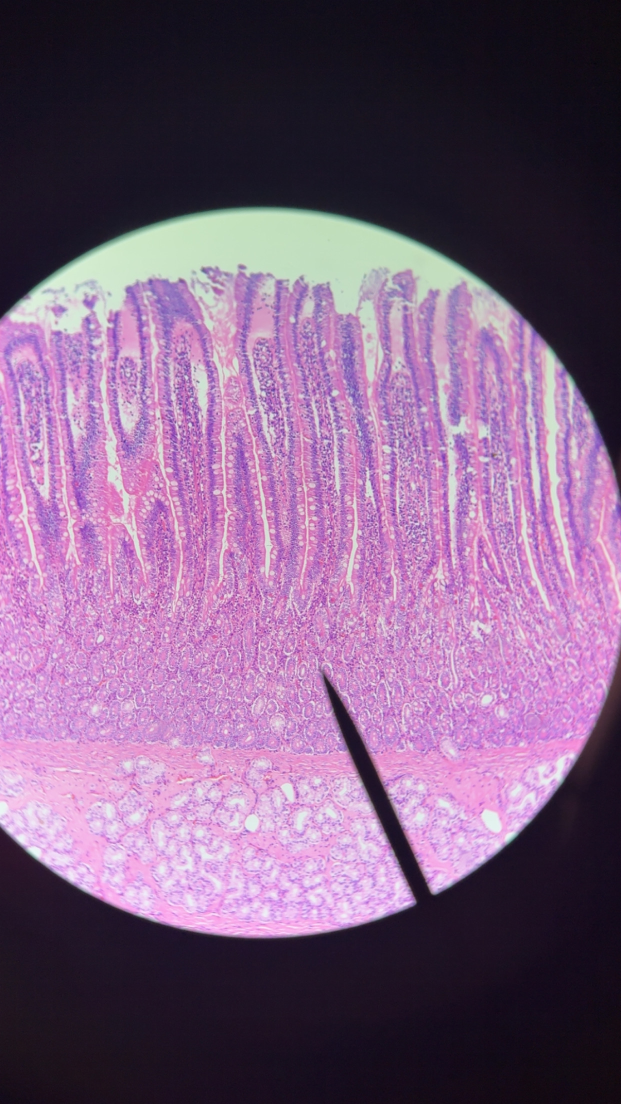
duodenum features - 4
long villi
brunner’s gland
duodenal glands : produe mucus
intestinal crypts : produce intestinal juice
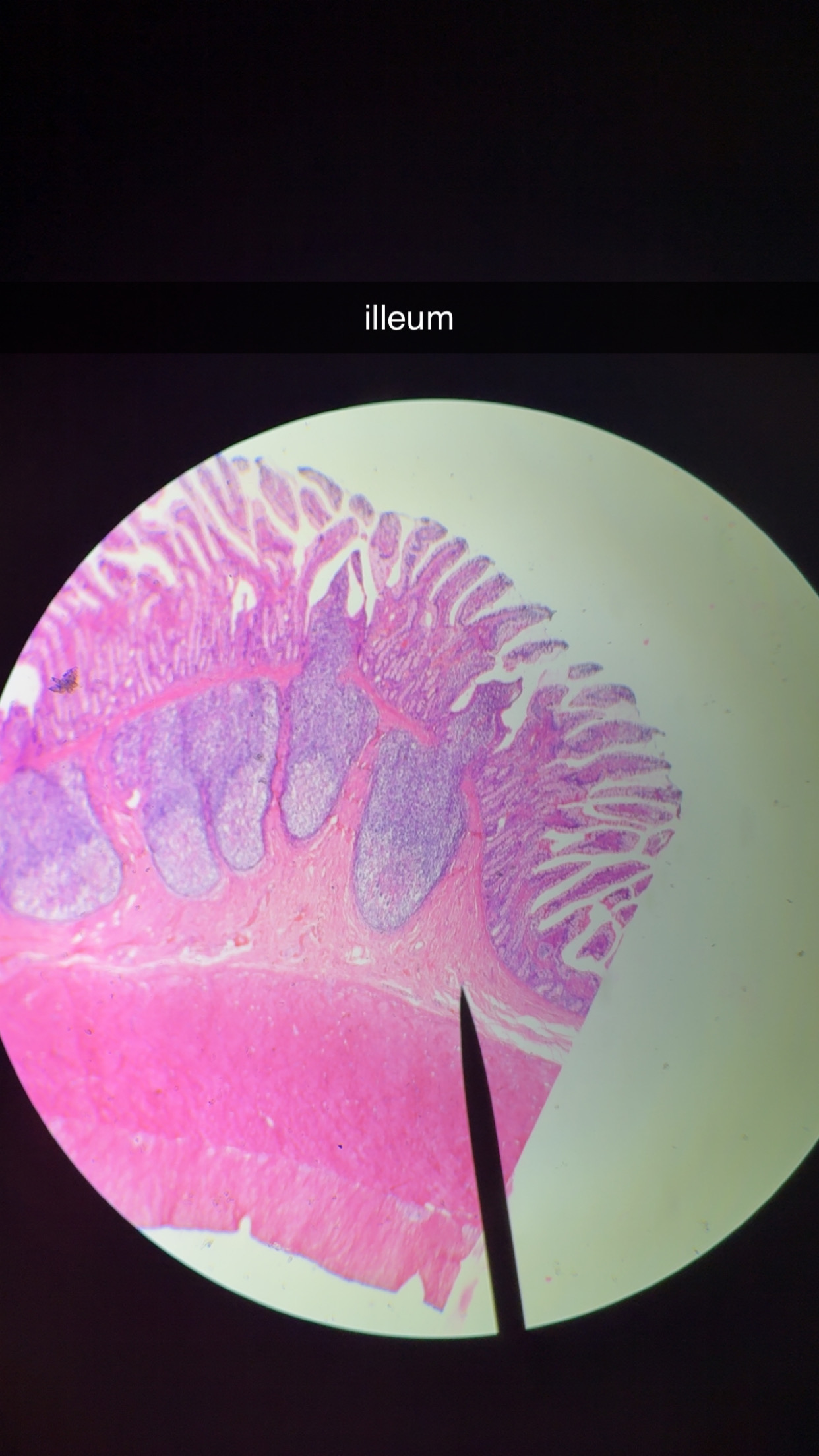
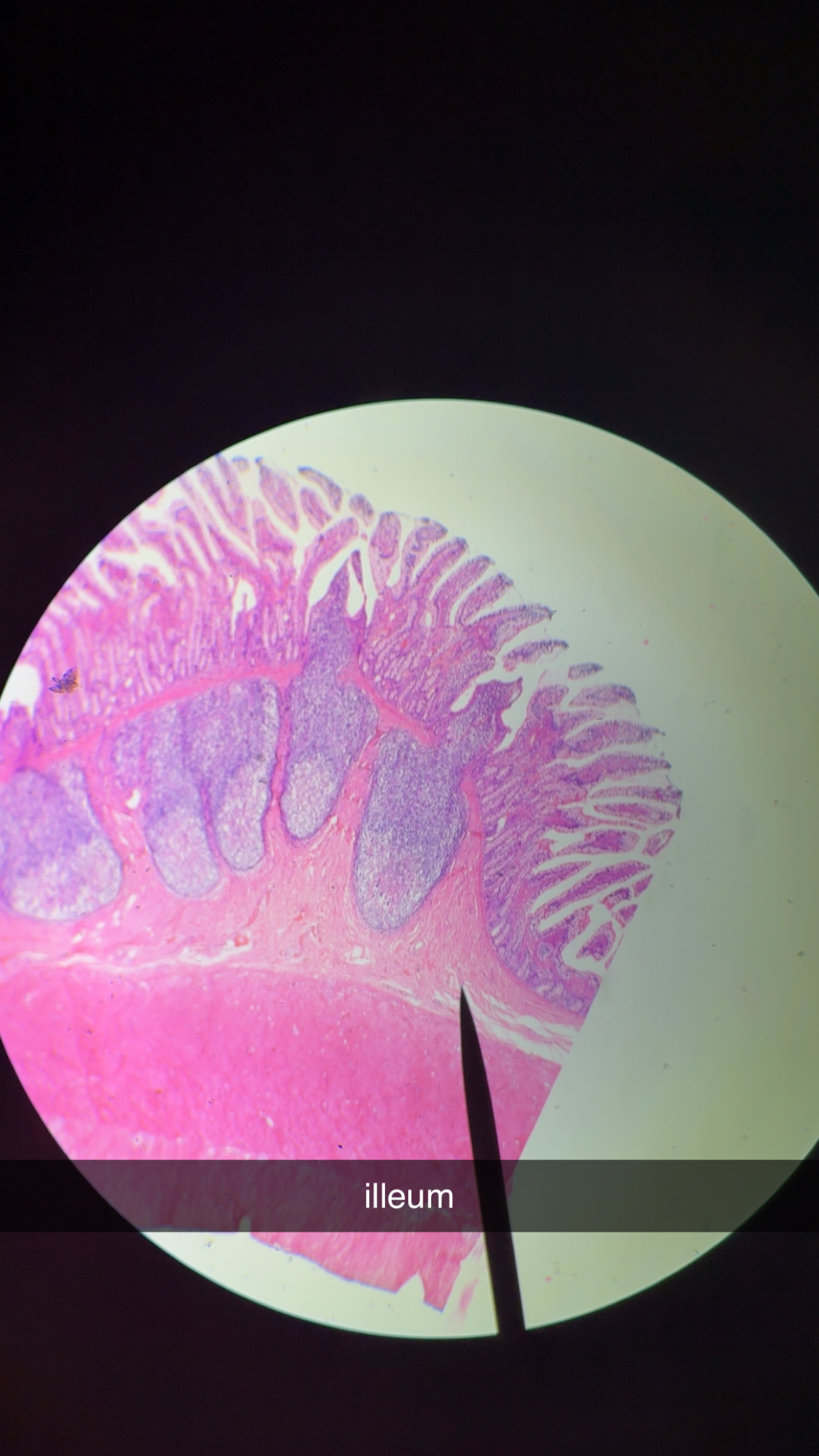
ileum cells - 4
Peters Patches- lymphoid follicles that inc along length of canal
Mucosa-Associated Lymphoid Tissues (MALT)
villi are shorter
large intestine 5 subdivisions: (cacra)
cecum, appendix, colon, rectum, and anal canal.
anal canal 2 sphincters - 2
- a voluntary external anal sphincter composed of skeletal muscle
- an involuntary internal anal sphincter composed of smooth muscle
only opened during defecation
large intestine 3 functions
1. Consolidate and propel unusable fecal mater towards anus.
2. Absorb water
3. Absorption of vitamins such as B and K which are produced by gut bacteria.
bile - 3
emulsifies fats, breaking up gat globules into small droplets
without it, very little fat digestion or absorption
Bile flows in the opposite direction toward the bile ducts.
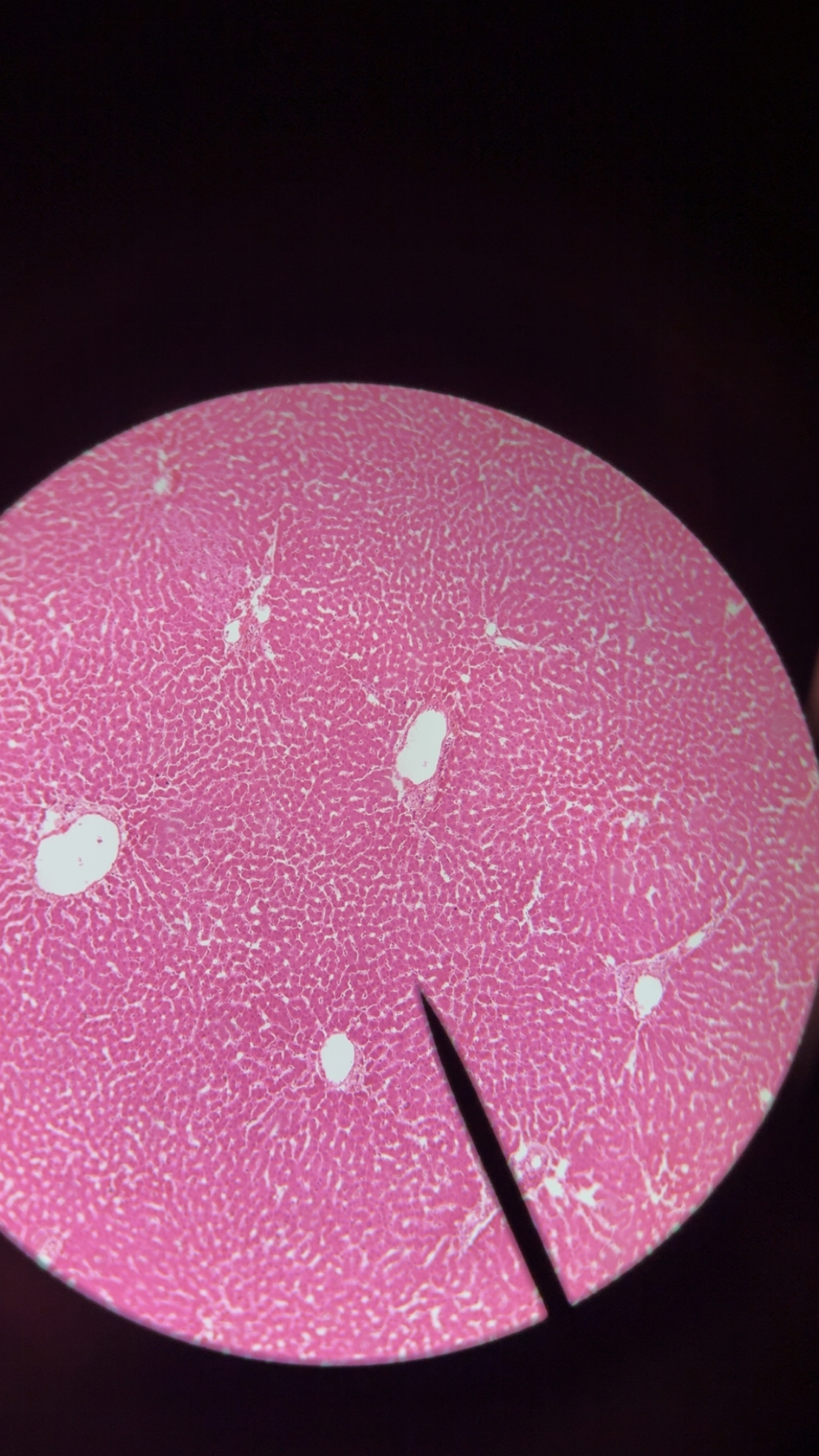
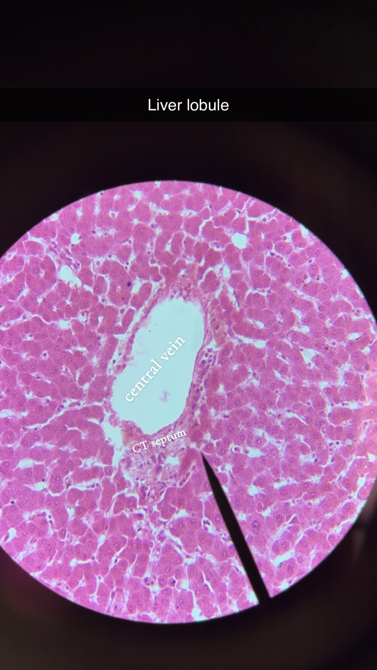
liver lobule 3 components
Hepatic artery branch (oxygenated blood)
Hepatic portal vein branch (nutrient-rich blood)
Bile duct
digestion
food being broken into smaller diffusible molecules
absorption
digested products passing through epithelial cells into blood for distribution