Week 4 Principles of Radiation Detection and Image Formation
1/33
Earn XP
Description and Tags
Explain the fundamental principles of radiation detection and image formation, including the roles of noise, resolution, contrast, and imaging geometry. ● Compare major radiation detector types (film, screen–film, CR, DR, gas-filled detectors) and evaluate their strengths, limitations, and clinical applications ● Interpret key detector performance measures especially DQE, efficiency factors, and temporal response and discuss how they influence image quality and patient dose
Name | Mastery | Learn | Test | Matching | Spaced | Call with Kai |
|---|
No analytics yet
Send a link to your students to track their progress
34 Terms
LO
Explain the fundamental principles of radiation detection and image formation, including the roles of noise, resolution, contrast, and imaging geometry.
● Compare major radiation detector types (film, screen–film, CR, DR, gas-filled detectors) and evaluate their strengths, limitations, and clinical applications
● Interpret key detector performance measures especially DQE, efficiency factors, and temporal response and discuss how they influence image quality and patient dose
Principles of radiation detection and image formation
Desirable characteristics of radiation detectors
Detective Quantum Efficiency (DQE)
Principles of image formation
Noise, Resolution, Contrast, Magnification, Speed (sensitivity)
Scatter
Radiographic detection techniques
Film
Screen-film radiography
Computed Radiography (CR)
Digital Radiography (DR)
Aim of diagnostic imaging
Produce images of optimum quality for diagnosis and management / treatment of disease
Examination is justified
Expected to impact clinical management of patient
Intervention
Do nothing
Factors influencing the image quality and patient dose
X-ray beam characteristics (CS, Exposure factors, beam filtration)
The patient (stillness, thickness)
The detector and imaging (CR or DR, Quantum Detection Efficiency (QDE), THe display system)
Practitioner skill and perception
Image noise
Images may contain useful information known as the signal
Background noise, which conceives useful information
Signal-to-noise ratio (SNR)
Image noise
is random variation in the recorded signal from pixel to pixel.
Image noise is proportional to the no. of quanta involved in forming the recorded signal
Ability to detect an object depends on the contrast of the object and the noise in the image
Imaging system geometry
Focus Receptor Distance (FRD, AKA SID)
Focus Object Distance (FOD, SOD)
Object Receptor Distance (ORD, OID)
Magnification
Minimal magnification and unsharpness is best
Unsharpness is magnified by increasing OID
Magnification reduced by keeping receptor close to patient
Minimise patient receptor (OID) distance
Typical FRD (SID) is 100cm for tabletop, 180 for standing chest
Magnification factor SID/SOD
Unsharpness
Penumbra - All images has some blurring approx. 0.3mm
Influenced by factors such as:
Movement of patient
OID
Brightness and contrast of display monitor
Background lighting
Unshapness = Focus x OID / SID - OID
Geometric unsharpness (penumbra)
Minimising geometric unsharpness
Fine focus should be used
small OID
SID large as possible
Magnification and sharpness
Large magnification as a result of large OID means reduced sharpness
Small magnification due to small OID increases sharpness
Resolution / definition
The ability of a system to distinguish two close objects or a specific part of anatomy
Measured objectively using a test object/phantom
Normally expressed in terms of line pairs / mm
Resolution depends on
all the elements in the imaging chain
Focus size
Source, object, detector geometry
Monitor display
Spatial frequency
The ability to see features in the image that are small or close together, upper limit is usually 3.5lp/mm
Image contrast
Low contrast vs high contrast (more blackening)
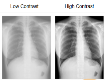
Human perceptions
Difference in perceptions can vary due to skill
Desirable characteristics of radiation detectors
Characteristics important for ay kinds of radiation detector
Absorption efficiency (% x-ray absorbed)
Conversion efficiency (% x-ray converted to electronic signal)
Capture efficiency (% of area of the detector that is ‘active’ detector)
Dose efficiency (how much incident dose on the detector contributes to image)
Desirable characteristics of radiation detectors
Temporal response
Timing of phosphorescence or afterglow
Wide dynamic range
High reporductibility abd stability
Detective Quantum Efficiency (DQE)
Measures how well incident x-ray is transferred into useful information (signal)
IDEAL DQE is 1 (100%)
A DQE of 0.5 means only 50% of incident x-ray on the detector are used for producing image.
DQE is affected by changes in input signal (mAs, kV) and patient
Evolution of Radiographic Detectors
Film → screen film → Computed Radiography (CR) → Digital Radiography (DR)
Film Radiography
X-rays interact directly with silver halide crystals
Advantage: high spatial resolution
Disadvantage: low efficiency (higher dose required)
Screen Film
Phosphor screen convert x-rays to visible light
advantage (higher efficiency)
Limitation (lower resolution vs film radiography due to light spread)
Intensifiers increase efficiency more (even lower dosage)
CR machine
Laser scanning releases stored energy as light
Advantage: wide exposure latitude
L: Moderate resolution (2.4 - 5lp/mm)
DR machine
Direct electronic conversion to image
A: fast workflow, wide dynamic range
L: costs
Clinical considerations
Higher DQE → better signal to noise ratio → lower dose
Digital systems reduce repeats
Risk of dose creep in digital systems due to wide dynamic range
Gas filled detectors
enclosed volume of detection medium (gas)
Carged electrodes
As radiation passes through, ionisation results from interactions
Number of ion paris produced depends on the LET of the radiation, High LET - more ionising
Ionisation chambers
X-rays interact in the chamber wall surrounding the air cavity
Electrons are generated which transverse the air in the cavity causing ionisations
Xenon gas detectors
Used in older CT scanners
Xenon gas molecules widely spaced in cavity so low absorption efficiency
Fast response