Physiology lecture 24
1/23
Earn XP
Description and Tags
Vision and the brain
Name | Mastery | Learn | Test | Matching | Spaced | Call with Kai |
|---|
No analytics yet
Send a link to your students to track their progress
24 Terms
LOs
• Describe the organisation of the retinal circuits
• Outline the responses of retinal ganglion cells to contrast and colour
• Explain what we mean by centre-surround receptive fields
• Describe where different parts of the visual field are represented in the brain
• Describe the visual pathway from retina to cortex
• Outline some of the responses of neurons in the visual cortex
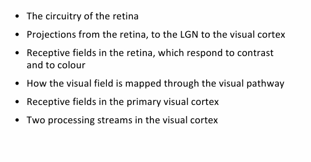
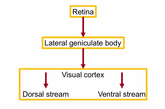
The visual pathway (a simplified view)
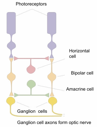
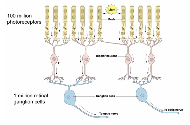
Retinal circuitry
involves vertical and horizontal connections
• Photoreceptors release the neurotransmitter glutamate in dark
• Glutamate excites some bipolar cells, inhibits others.
• Bipolar cells excite or inhibit retinal ganglion cells.
Retinal ganglion cells give rise to the optic nerve fibres.
• Horizontal and amacrine cells carry signals horizontally across retina.
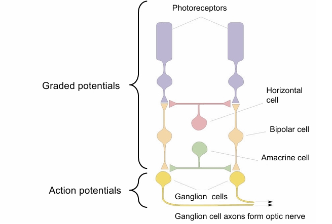
Retinal circuitry potential types
any potential that reaches the ganglion cells becomes an AP
what do many rod photoreceptors do?
Converge onto less bipolar cells
around 100 million photoreceptors into 1 million retinal ganglion cells
Retinal processing in light
• In response to light, photoreceptors reduce the amount of neurotransmitter (glutamate) released.
• This can lead to excitation or inhibition of bipolar cells
• Bipolar cells excite or inhibit retinal ganglion cells.
• Horizontal and amacrine cells for lateral collections between bipolar and ganglion cells.
• Photoreceptors, bipolar cells, horizontal cells and amacrine cells all respond with graded potentials. Action potentials first appear in ganglion cells.
• Bipolar cells connect to many photoreceptor cells and ganglion cells connect to multiple bipolar cells. Convergence of these signals means that information is conveyed by a smaller number of cells at each step.
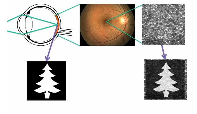
Importance of contrast
can allow differentiation to form the images between the contrast in the dark and light of the image
Difference in brightness consequence of enhanced contrast with surroundings when seeing things in contrast
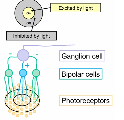
How is contrast detected?
• Photoreceptors respond to any light falling on them
• Ganglion cells respond best to particular patterns of light.
• The ganglion cell’s receptive field is the small small patch of retina where light excites that ganglion cell to fire impulses
• Ganglion cells have centre surround organisation because of the horizontal connections
○ On centre / off surround
○ Off centre / on surround
On centre type
Centre surround responses of rod ganglion cells
If light falls on centre, ganglions are excited
If off centre, it is inhibited
on centre and off centre types
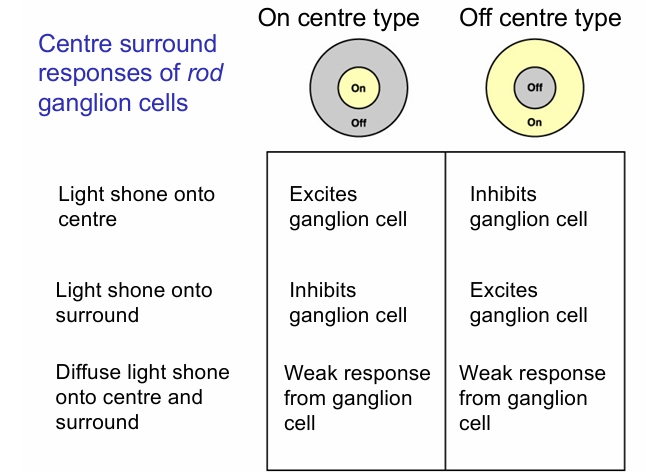
Response of contrast sensitive rod ganglion cell
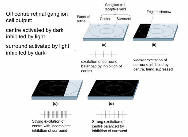
Benefits of centre surround organisation of rod ganglion cells
• Helps to emphasise contrast (differences in brightness) at edges of visual objects
• Important for identifying shapes, separating objects from background etc.
• Form of surround inhibition
• Another example of the nervous system extracting information about where a stimulus changes
• Basis for some visual illusions
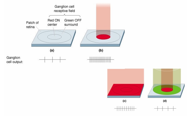
Cone ganglion cells
• Cone ganglion cells also have centre surround organisation
• Centre and surround fed by different cone types
• Two sorts of cone ganglion cell centre surround organisation
– Red (L) – Green (M) (one excites; other inhibits)
– Blue – Yellow (yellow = red + green)
• White light (includes both red and green) gives weak firing – excitation and inhibition balanced
• Illumination of part of field or with one colour excites or inhibits the cell
what properties do cone ganglion cells have?
colour opponent properties
Staring at red patch fatigues long wavelength (red) cones. When look at white surface – green component in white light dominates?
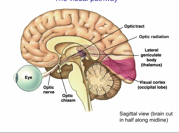
The visual pathway
LGN in thalamus
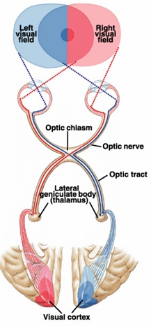
Visual field mapping and visual pathway
• Fibres from each half retina (nasal, temporal) in each eye take different routes to generate left and right visual fields
• Fibres from the nasal retina of each eye cross at the optic chiasm
• Fibres from the temporal retina do not cross, they stay on the same side as the eye they came from
• Left eye temporal and right eye nasal (red) go to left LGN and cortex - represent right visual field
• Right eye temporal and left eye nasal (blue) go to right LGN and cortex – represent left visual field
The lateral geniculate body (or lateral geniculate nucleus; LGN)
• There are two – one for each side of the visual field. The LGN for the left visual field is found in the right hemisphere of the brain, and vice–versa.
• Projections from each eye are kept separate in contralateral layers and ipsilateral layers.
• Contains three different types of cells, corresponding to three different types of layers.
• Maintains a topographic mapping of the visual world – neighbouring cells have receptive fields that are nearby in the world.
• The receptive fields of the cells here are similar to those of retinal ganglion cells – round with a centre-surround organisation.
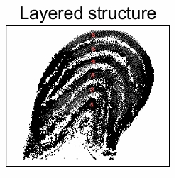
Visual cortex
• The visual cortex has five areas – V1 (primary visual cortex) to V5 (also referred to as MT), each of which has a particular role in vision.
• There are forward projections from V1 to ‘higher’ cortical areas V2, V3, V4, and V5 (MT). But there are also connections that run in the other direction.
Location of human primary visual cortex in the occipital lobe (at back) of the brain
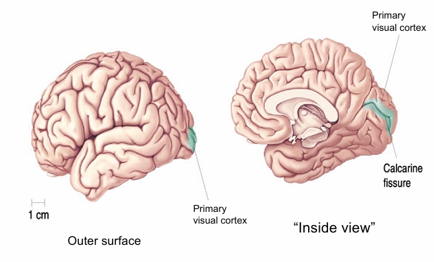
(Primary) visual cortex
• Maintains the topographic mapping of the visual world (how you interpret surroundings)
• More cortical area is devoted to the centre of vision (as is also the case in the lateral geniculate body)
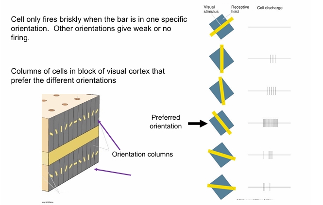
• Neurons in visual cortex have more selective responses than ganglion cells and lateral geniculate neurons:
– Prefer bars and edges - not spots
– Prefer specific position of stimulus in receptive field or
– bright bar in a specific orientation
– prefer stimulus moving in a specific direction
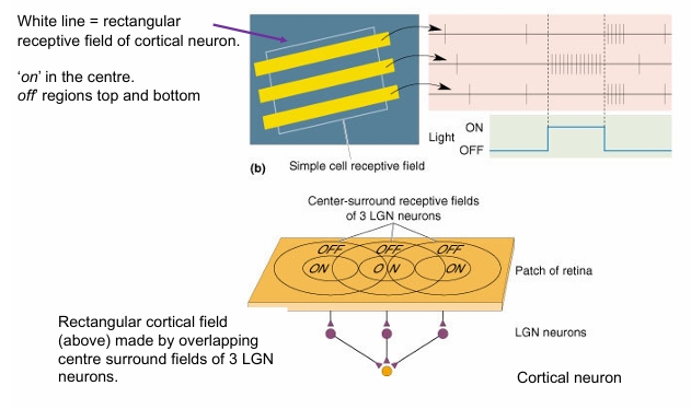
Responses of ‘simple’ cell and its possible construction from overlapping inputs from LGN neurons
what areb‘Complex’ neurons in cortex sensitive to?
specific orientation of stimulus
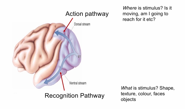
Several different visual areas in the cortex
Concept of ventral and dorsal processing streams
Summary