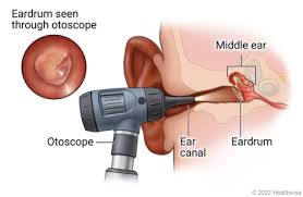
Anatomy of the Outer and Middle Ear
 Call Kai
 Learn
Learn Practice Test
 Spaced Repetition
 Match
Match Flashcards
 Knowt Play
1/58

There's no tags or description
Looks like no tags are added yet.
Name | Mastery | Learn | Test | Matching | Spaced | Call with Kai |
|---|
No analytics yet
Send a link to your students to track their progress
59 Terms
What are the 3 main components of the ear
outer ear
middle ear
inner ear
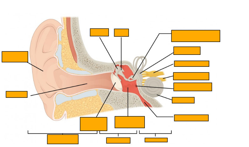
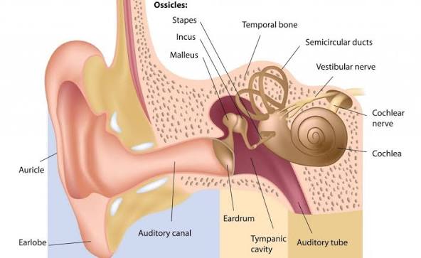
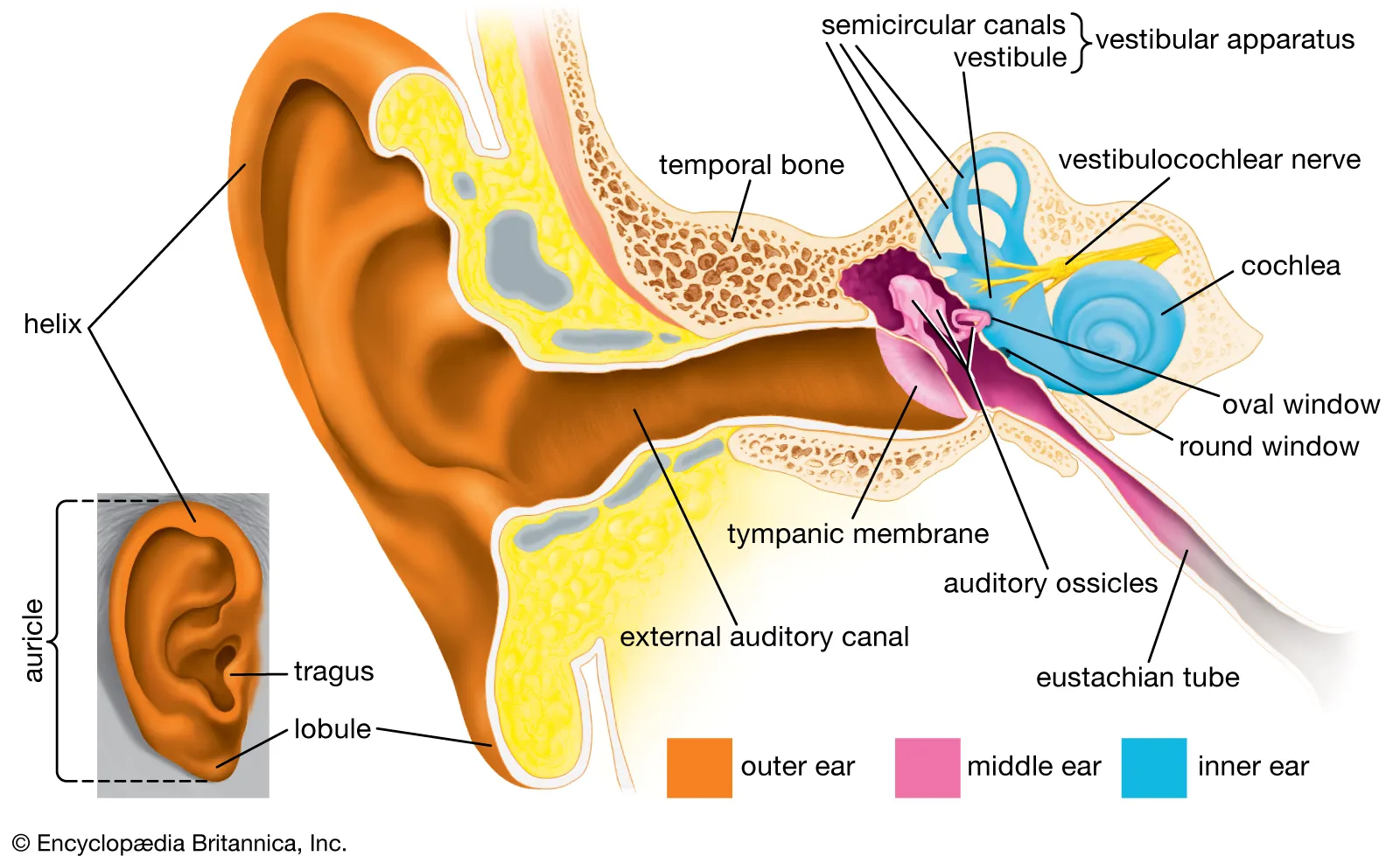
Label this diagram
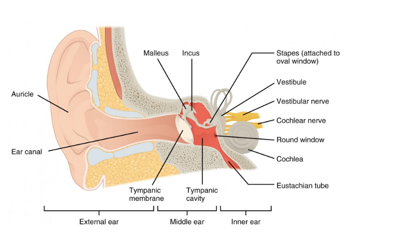
Which structure marks the boundary between the outer ear and the middle ear
the tympanic membrane/ eardrum
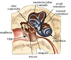
Which structures mark the boundary between the middle ear and the inner ear
round window and oval window
General function fo the outer ear
collects sound from auditory space
funnels sound towards the middle ear
General function of the middle ear
transduces sound energy (pressure) into movements of joints of the ossicle chain
General function of the inner ear
converts movements of the ossicle chain into electrical impulses of hearing
General function of the brain in relation to the ear
interprest signals from the ear into a meaningful special sense
Explain the process of hearing
ear canal (external acoustic meatus) catch sound waves and funnel them to the tympanic membrane
sound waves cause the eardrum to vibrate causing movement of the ossicles which amplify the vibrations
the stapes pushes on the oval window transmitting the vibrations to the cochlear, creating waves in the cochlear fluid
this causes hair cells in the cochlear to move and convert the mechanical energy into electrical signals
the auditory nerve carries these signals to the brain
What is another factor excluding function that explains why we can separate the ear into those 3 categories
they have disorders or pathologies unique to each division
they have different embryological derivations
Consequence of diseases to the outer ear
conductive deafness- usually occurs when sound waves are physically blocked from reaching the eardrum
What is the general definition of conductive deafness
soundwaves are blocked from reaching the inner ear due to issues in the outer or middle ear
Consequences of diseases to the middle ear
conductive deafness- usually caused by the middle ear becoming filled with fluid
Consequences of disorders of the inner ear
sensorineural deafness- eaither sensorial (problem with the transduction of mechanical to electrical energy by the heair cells) or neural (damage to the actual nerves themselves)
Embryological origin of the outer ear
first pharyngeal cleft (ectoderm)
Embryological origin of the middle ear
first pharyngeal pouch (endoderm)
Embryological origin of the mucosa lining the pharynx and eustachian tube
first and second pharyngeal pouch
Neural innervation of the outer ear
-sensory dupply shared between the trigeminal and facial nerves (1st and 2nd pharyngeal arch nerves)
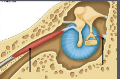
What are the 2 muscles found in the middle ear
tensor tympani- attached to the malleus
stapedius- attatched to the stapes
both protect the inner ear from damage caused by loud noises (e.g. chewing, shouting)
Motor supply to the tensor tympani
develops formt he 1st pharyngeal arch so its motor supply comes from the motor division of the trigeminal nerve (mandibular division of the trigeminal)
Motor supply to the stapedius
-develops from the 2nd pharyngeal arch so its motor supply comes from the motor division of the facial nerve
What is the outer ear also known as
the external ear
What is the outer ear further divisible into
the pinna/ auricle and the external auditory canal
What is the auricle/pinna of the era
external visible part of the ear
What is the structure of the pinna
-its skeleton is made form elastic cartilage and it is covered by skin
What is the main function of the pinna
to collect sound from the auditory space
What causes cauliflower ear
blunt force trauma/ repeated friction to the outer ear which causes a blood clot to form between the skin and cartilage cutting off nutrients to the cartilage causes it to harden, scar and deform
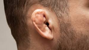
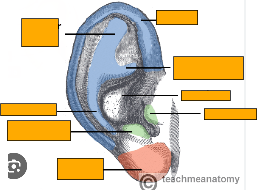
Label this image
Auricle/pinna of the ear
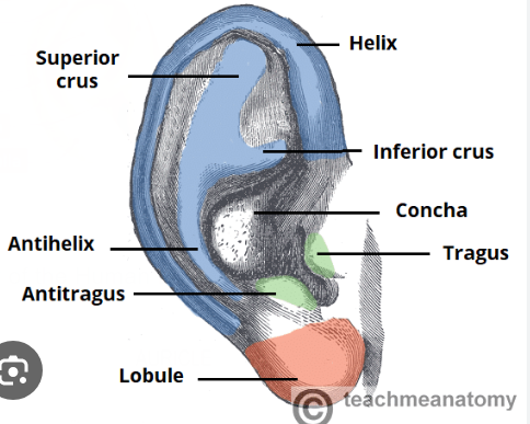
Which nerves provide sensory innervation to the skin covering the ear
lesser occipital nerve- innervates the skin of the auricle
greater auricular nerve
auricotemporal nerve (branch of CNV3)- innervates skind of auricle and external auditory meatus
auricular branch of the facial nerve- innervates deeer aspect of auricle and EAM
auricular branch of vagus nerve- innervates deeer aspect of auricle and EAM
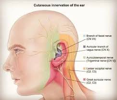
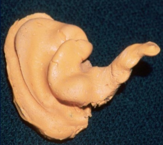
Shape of the external auditory canal
sinuous S-shaped canal ensures that the velocity of the air slows so bacteria/dust etc doesn’t travel further into the ear to the tympanic membrane
what are the 2 main parts of the external auditory canal
outer 1/3- made of elastic cartilage
inner 2/3- made of the tympanic plate of the temporal bone
What lines the EAC
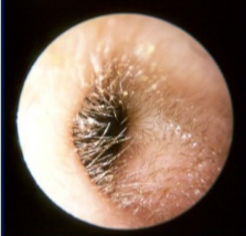
lined by skin throughout which then also covers the outer plate of the tympanic membrane
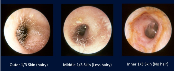
Features of the skin lining the outer 1/3 of the external auditory canal
-many hairs (trpas dust etc)
-has ceruminous glands that secrete bactericidal wax (prevents bacteria developing)
-has sabaceous glands
How do the features of the skin change as you go deeper into the external auditory canal
fewer hairs as you travel deeper into the canal
What is the ear-cough reflex
triggered by activation of the auricular branch of the vagus nerve which triggers the cough reflex mechanism
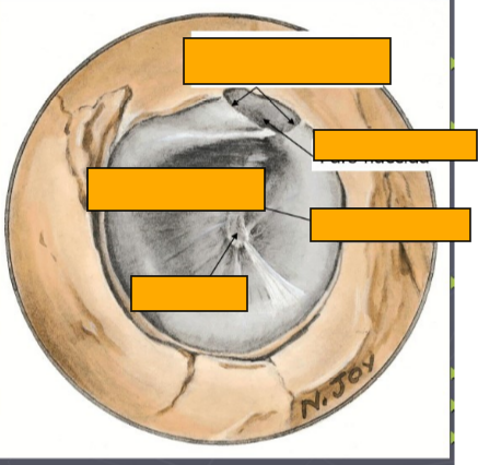
Label this image, which structure is shown
tympanic membrane
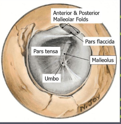
What are the 3 layers of tissues of the tympanic membrane
outer/lateral plate layer- lined by skin (keratinised stratified squamous cells
middle plate layer- fibrous layer
inner plate layer- respiratory epithelium (low columnar epithelium)
Which bone does the tympanic membrane adhere to
malleus
Function of the ossicular chain
-act as an impedence matching device
-sound has to travel from air into the cochlear fluid and most of it would bounce back
-however the ossicles are able to increase pressure at the oval window by acting as a lever and due to the oval window having a much smaller surface area than the eardrum
-this makes the sound ensuring it is strong enough tot move the fuid in the cochlea properly
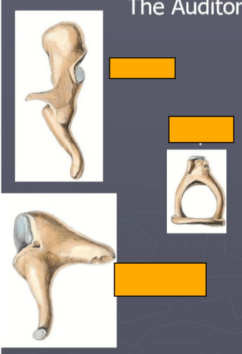
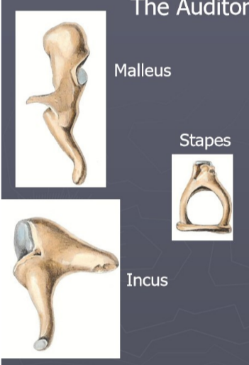
Sensory nerve supply to the external surface of the tympanic membrane
auriculotemporal nerve (branch ov CNV3)
small branch of the vagus nerve
Sensory nerve supply of the internal surface of the tympanic membrane
glossopharyngeal nerve
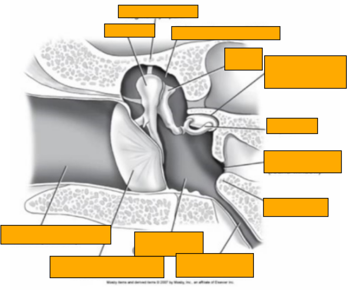
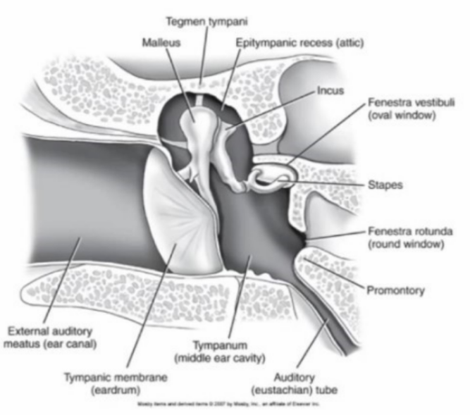
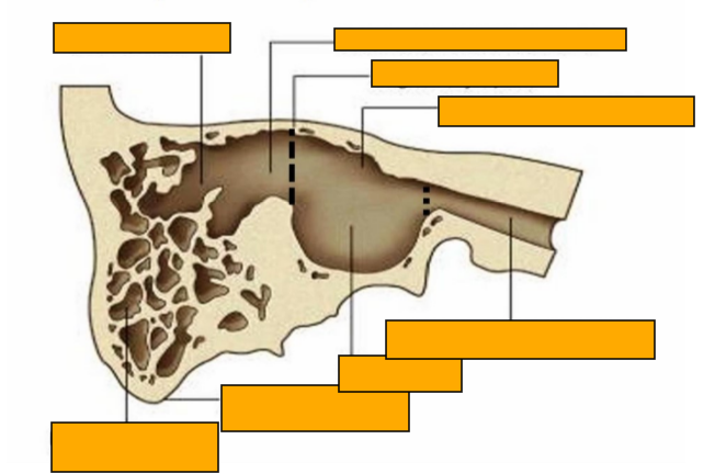
Label this image
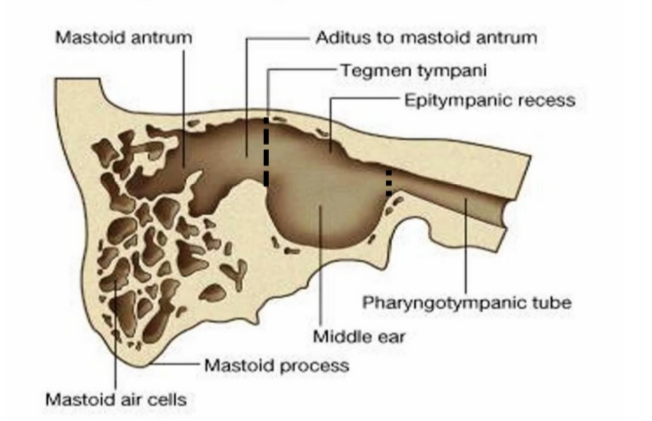
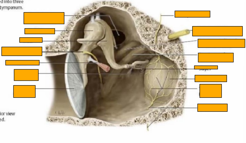
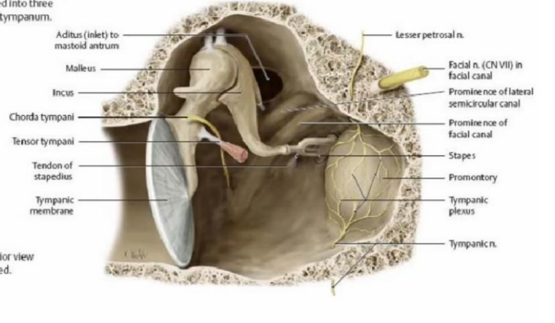
Features of the middle ear
bi concave compartment
aroudn 15mm diamete
normally filled with air
has 6 walls
Lateral wall of the middle ear
inner plate of the tympanic membrane
Medial wall of the middle ear
outer plate of the oval window
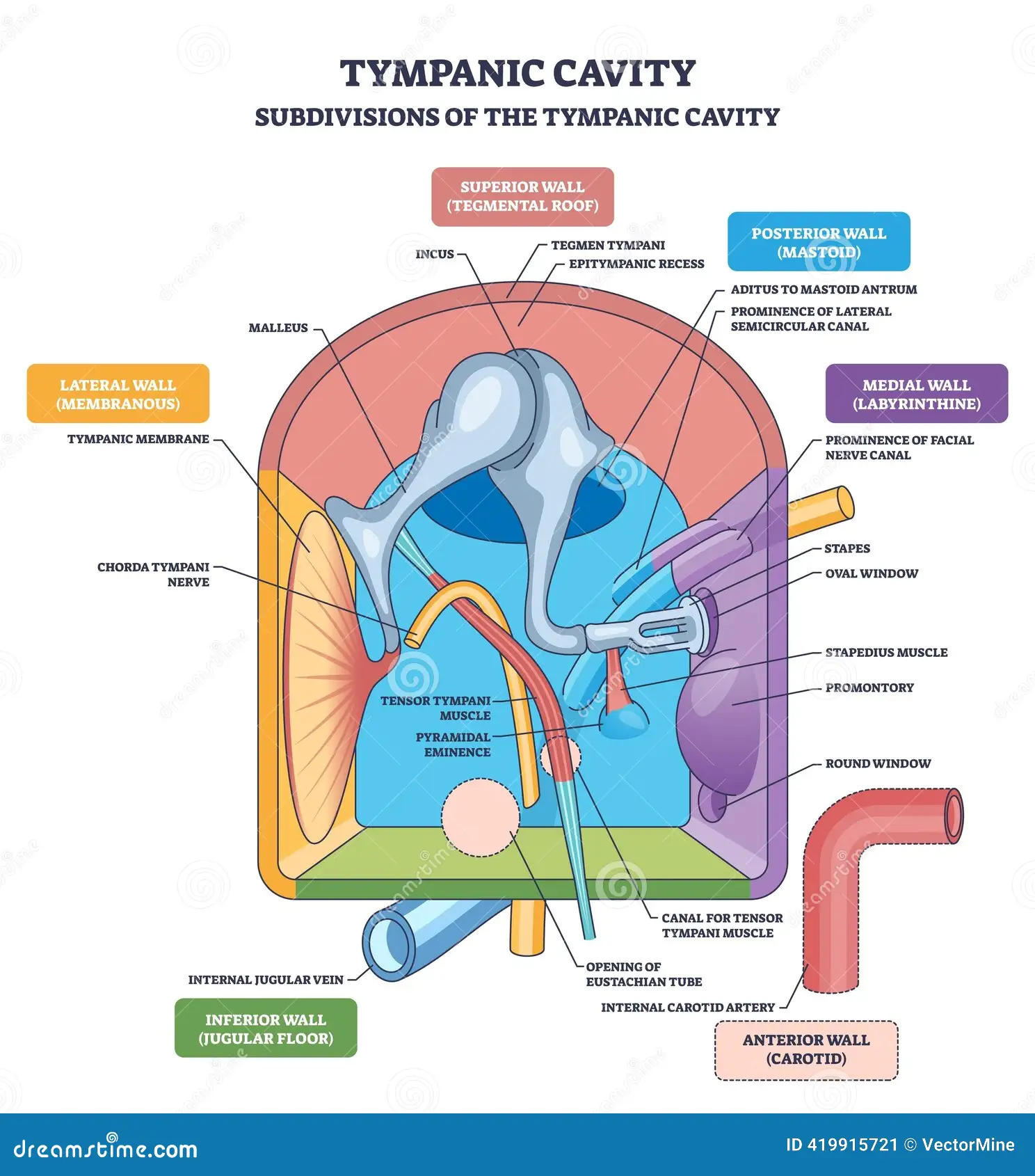
Roof of the middle ear
tegmen tympani
Flood of the middle ear
jugular wall
Posterior surface of the middle ear
mastoid wall
Anterior usrface of the middle ear
carotid wall
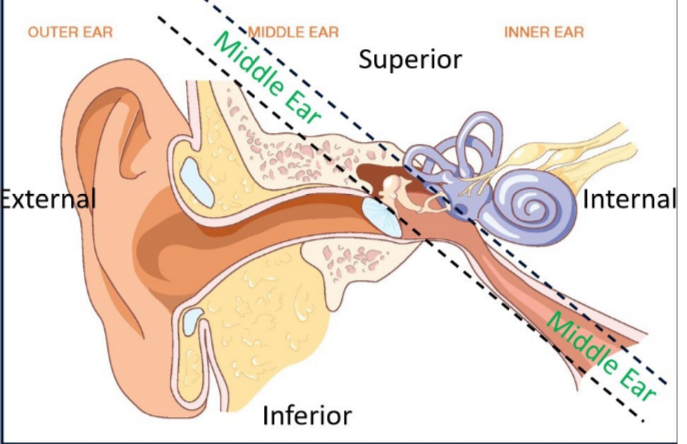
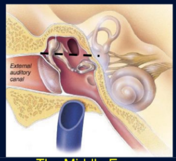
What are the 2 compartments that the middle ear is separated into by this line
epitympanic recess- above the line
tympanic cavity- below the line
How does the middle ear communicate with the nasopharynx
via the auditory/eustachian tube
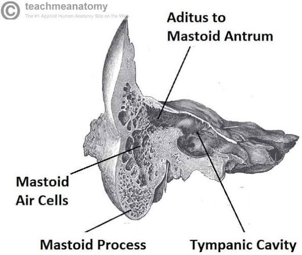
How does the middle ear communciate with the mastoid air cells
via the epitympanic recess and mastoid antrum
Contents of the middle ear
tympanic membrane
auditory ossicles
auditory tube
muscles of the ossicles
branches of facial and glossopharyngeal nerves
epitympanic recess
mastoid air cells
upper respiratory epithelium
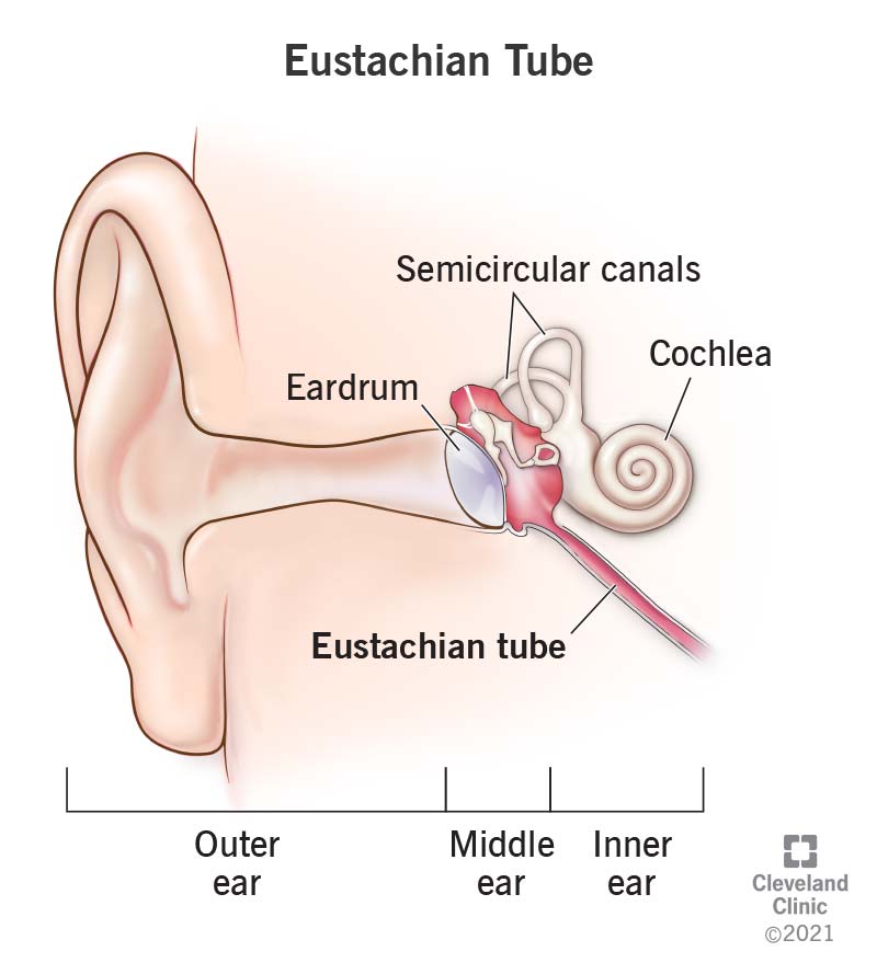
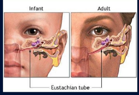
Function of the eustachian/pharyngotympanic tube
passes between the tympanic cavity and the nasopharynx and equalizes pressure across the membrane (keeps air pressure on both sides of the eardrum the same)
drains contents of middle ear cavity (epithelium secretes miucus)
Consequences of eustachian tube dysfunction
failure to drain mucuous secretions of the middle ear leading to congestion
What is an otoscopy
medical examination using an otoscope (light magnifying device to inspect the ear canal, tympanic membrane and outer ear