A&P Last Exam not including crainial nerves (Ch 11-13)
1/145
Earn XP
Description and Tags
Ch11-13 rn - numbers indicate which prompt it lines up to in the SG
Name | Mastery | Learn | Test | Matching | Spaced | Call with Kai |
|---|
No analytics yet
Send a link to your students to track their progress
146 Terms
sympathetic nervous system or Thoracolumbar division
fight or flight system,
parasympathetic nervous system - Craniosacral division
rest and Digest system,
peripheral nerves
Many axons bound together innervate most structures of the body, mixed nerves
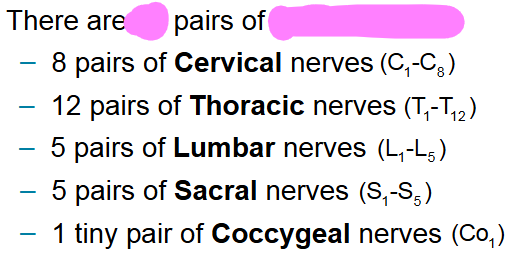
14 spinal nerves -
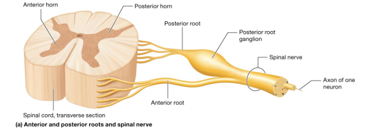
31 pairs, Branch from the spinal cord Axons are housed within posterior and anterior root
anterior root
side of the spinal cord, houses axons of motor neurons

posterior root
side of the spinal cord, houses axons of sensory neurons
cranial nerves
There are 12 that we are concerned with, they attached to the brain and innervate the structures of the head and neck
Epineurium
1- Sheath surrounding each spinal nerve

Fascicles
3- group of axons surrounded by perineurium
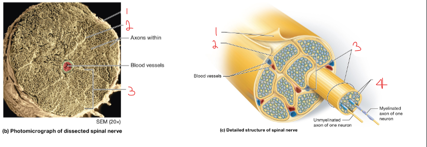
Perineurium
2- sheath that surrounds each fascicle

Endoneurium
4- sheath surrounding each Axon

anterior (orange) and posterior (pink) ramus - Spinal nerves leaving the vertebral cavity split into two major nerves the posterior and anterior ramus
Posterior Ramus—Travels to posterior side of the body
Anterior Ramus—Travels to anterior side of the body and/or
the upper and lower limbs
pink and orange
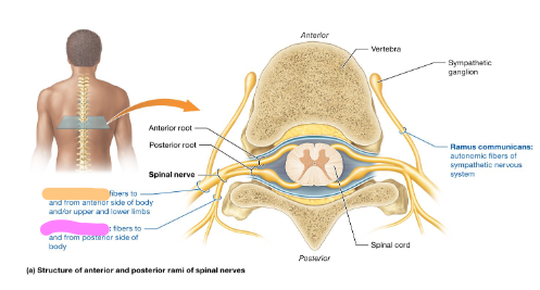
anterior ramus
orange
nerve plexuses
highly branched set of nerves the first exit from the spinal nerve Highway

cervical plexuses
In the neck area

Brachial plexuses
in the break of region just below the cervical
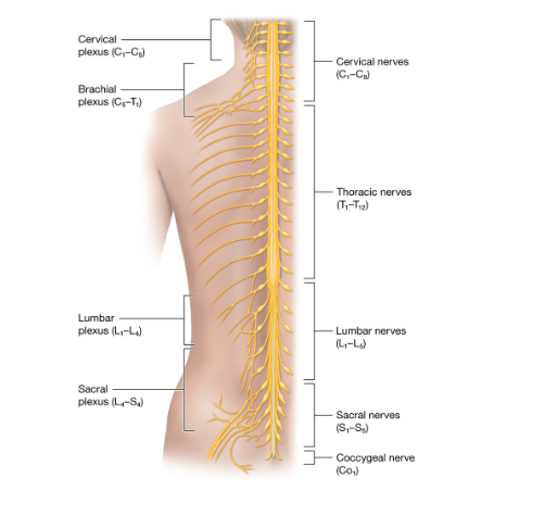
lumbar plexuses
in the lower back area above the sacral

sacral plexuses
lowest back area just before the end of the spinal nerves

Visceral reflex arcs
The steps of reactions in the ANS
sensory signals from organs are sent by aference Sensory neurons to brain or spinal cord
stimuli are integrated by CNS
motor impulses from CNS are sent via efferent motor neurons and cranial and spinal nerves to autonomic ganglia
autonomic ganglia send impulses via other efferent motor neurons to Target organs where they trigger motor response in Targeted cells
Autonomic ganglia
Connection point between pre and post ganglionic nerves in efferent neurons
Preganglionic neuron
Part of efferent nerve that connects to the CNS before the ganglia, uses acetylcholine only
Postganglionic neuron
part of nerve which connects to target cell, uses more than just acetylcholine as a neurotransmitter
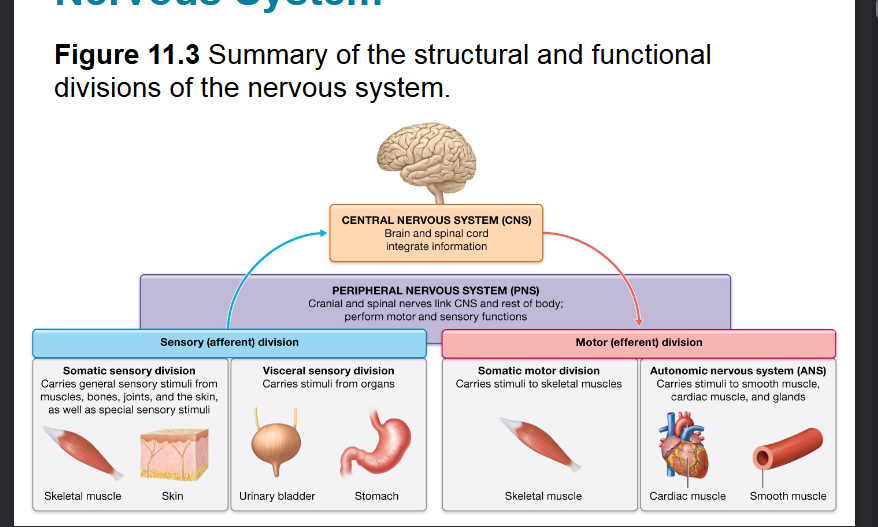
Central Nervous System (CNS)
Functions for integrative interpreting sensory info from PNS and tells PNS how to respond
includes brain and spinal cord
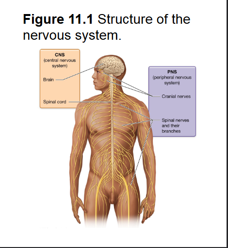
Peripheral Nervous System (PNS)
functions for sensory input sends to CNS which assess the info and tells PNS how to respond with motor responses
Has cranial nerves - shoot off of brain with 12 pairs
and Spinal nerves branch off of spinal cord 31 pairs

Nerves
carry signals to and from CNS made of Bundles of long neurons or arms packed with Blood vessels surrounded by CT sheaths
Also the name for bundles of axons in The PNS
12 pairs of cranial
31 pairs of spinal

1 Sensory (afferent) division - Sensory stimuli are detected by sensory receptors (varies from small tips of neurons to complex receptors)
contains somatosensory Division orange - external sensory - Neurons carry signals from skeletal muscles, bones, joints and skin including Sensory neurons from organs for vision hearing taste and smell ( special sensory)
And visceral sensory division purple - internal sensory- neurons carry signals from visceral / organs such as heart lungs stomach intestines Etc
Includes the somatic and visceral sensory division, deals with Sensations from Sensory neurons
light blue and its 2 sub divisions

Sensory receptors
Detect sensory stimuli- varies from small tips of neurons to complex receptors
1 Somatic sensory division - external sensory- Neurons carry signals from skeletal muscles, bones, joints and skin including Sensory neurons from organs for vision hearing taste and smell ( special sensory)
orange

1 Visceral sensory division –Internal sensory- neurons carry signals from visceral / organs such as heart lungs stomach intestines Etc
Sensory neurons carry signals from organs to CNS
1 Motor (efferent) division -
Carry out motor functions such as muscle contraction and gland secretion, organs that carry out the effects are effecters
contains somatic motor division yellow- voluntary movement - neurons transmit signals to skeletal muscles
and visceral motor division green (autonomic nervous system ANS) - involuntary movement - neurons carry signals to thoracic and abdominal viscera regulating secretion, controls smooth and cardiac muscle
Light pink
contains what 2 sub divisions

Effectors
organs that carry out the effects of the motor division
1 Somatic motor division - voluntary movement, motor neurons carry signals to skeletal muscles - voluntary movement - neurons transmit signals to skeletal muscles
yellow

1 Autonomic nervous system (ANS) or visceral motor division- involuntary movement, motor neurons carry signals to the cardiac, smooth muscles and glands
green

Neurons
Excitable cell type, Send and receive signals called Action potentials - responsible for sensory integrative and motor functions of nervous system, long living cells generally amitotic, very in size, have cell body one or more dendrites (receives signal) and one Axon (sends signal from cell)

Cell body
most metabolically Active part of neuron, manufacturers proteins, contains a lot of the same organelles as regular cells although the rough ER appears in clusters called nissl bodies

Neurofibril
Filaments of the neuronal cytoskeleton of the neurons cell body are called neurofibrils these extend into dendrites and axon microtubules

Dendrites
Short and forked, receive input from other neurons transmit it in the form of electrical pulses to cell body

Axon
Can generate and conduct Action potentials sending messages to other neurons contains axon hillock, axon collaterals, telodendria, axon terminals / synaptic knobs, Axolemma, axoplasm

Axon Hillock
Area of cell body where the axon begins to Branch off

Axon collaterals
branches of the Axon

Telodendria
small segments of axon that Branch off and form the end for connections it also has collaterals
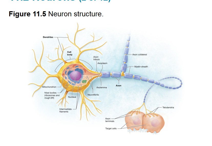
Axon terminals / Synaptic Knobs -
-the very end of the telodendria these communicate with target cells

Axolemma
Plasma membrane of Axon

Axoplasm
cytoplasm of Axon,
contains mitochondria intermediate filaments vesicles and lysosomes but no protein making organelles

multipolar neurons
Over 99% of neurons, single axon with highly branched dendrites wide variety of shapes and sizes
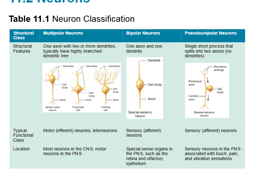
bipolar neuron-
one axon and one dendrite most are sensory, found in retina of eye and olfactory epithelium

Pseudounipolar neuron
single axon with one part that brings sensory stimuli to cell body and one part that carries stimuli spinal cord, detect touch Pleasure and Pain

Sensory/afferent Neurons
Carry signals towards CNS (pseudounipolar or bipolar) detect input from internal and external environments and facilitate motor coordination
Motor / efferent neurons
carry signals away from cell bodies in CNS to muscles and glands, most are multipolar
Nuclei
Clusters of cell bodies in the CNS
Ganglia
clusters of cell bodies in the PNS
Tracts
Bundles of axons in the CNS
Neuroglia / neuroglial cells
Part of nervous tissue hold neurons together, maintain the environment around neurons, protect and assist neurons and function. can undergo mitosis and fuel in gaps when neurons die
there's six different types four in the CNS (Astrocytes, Oligodendrocytes , microglia, Ependymal cells) and two in The PNS (Neurolemmocytes and satellite cells)
Astrocytes
CNS Star-shaped,
anchor neurons and blood vessels in place,
facilitate nutrient transport, regulate extracellular environment and brain,
remove unneeded stuff,
Assist information of blood-brain barrier,
repair damaged brain tissue ( divide rapidly but may impede the growth of neurons leading to more damage)
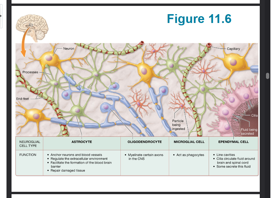
Oligodendrocytes
CNS Wrap around part of axons in certain neurons of the CNS, form layers of plasma membrane called myelin repeating segments of myelin form the myelin sheath which insulates an increases speed of nerve impulses
One cell has branches which wrap different parts of the axon so that one cell can make multiple wrappings

Myelin
cells wrap around axon of neurons insulating and allowing for increased nerve impulse conduction (Oligodendrocytes and Neurolemmocytes)

Microglia
CNS Tiny branching cells that are activated by brain injury, become phagocytes that in just disease causing organisms, did neurons and cell debris also stimulate inflammation

Ependymal cells
CNS Ciliated cells, main function is circulating cerebral fluid moved with cilia

Internodes
segments of axon covered by myelin sheath
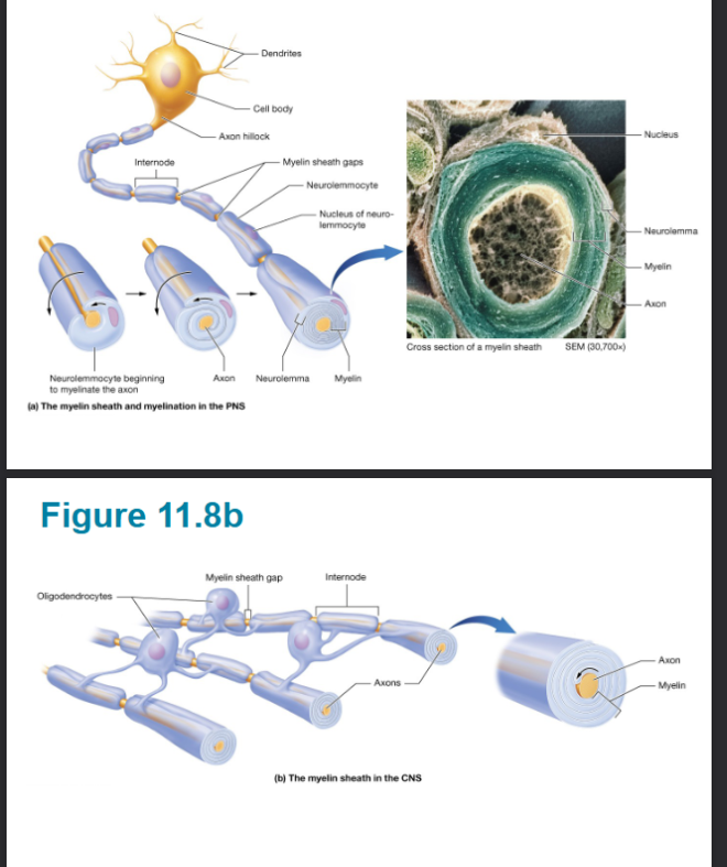
Gap/node of Ranvier
spaces in between internodes of myelin

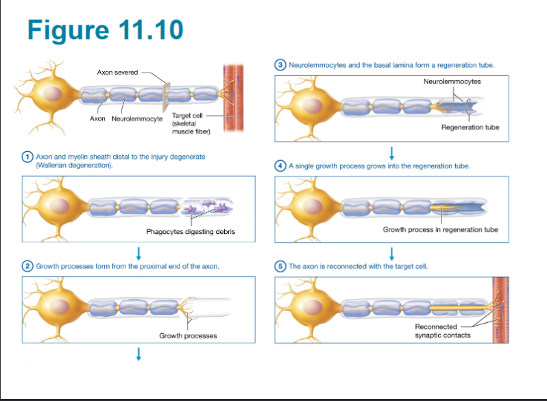
Regeneration
CNS- dendrites and axons Almost Never regenerate all good danger sites inhibit neural growth, astrocytes create scar tissue
PNS - capable of regeneration if the cell body remains intact and conditions are ideal
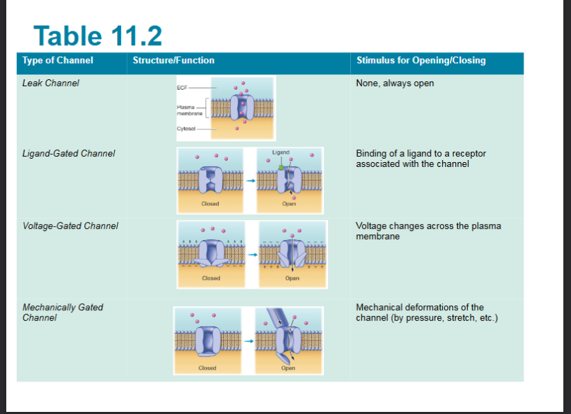
Ligand gated channels
Open and response to chemical ( ligand) binding with Channel or receptor associated with Channel

Ligand
chemical, binds to Channel or receptor to open ligand gated channels

Voltage gated channels
open or close and response to change in cells membrane potential

Mechanically gated channels
open or close and response to Mechanical stimuli, (stretch pressure or vibration)
Sodium-potassium pump (Na/K ATPase)
Moves ions against their gradient helps balance back to normal after hyperpolarization, moves two potassium onto cytosol and three sodium ions into extracellular fluid
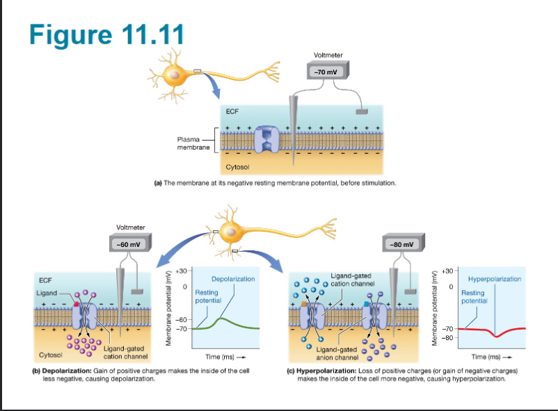
Depolarization
Opening a ligand-gated cat on Channel causing an influx of catons such as sodium, making membrane potential less negative
Repolarization
when a cell returns to its arresting membrane potential
Hyperpolarization
when a cell becomes more negative than it is at rest, loss of positive charges or gain of negative charges

Local potential -
Travel short distances, reversible, when a charge does not reach high enough to set off action potential
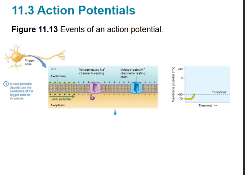
Action potential
irreversible, travels entire length of axon, Begins at -55mV, ends at +30mV includes depolarization, repolarization and hyperrepolarization phases as well as refractory periods

Depolarization phase
First step of action potential, voltage-gated sodium ion channels open at threshold of -55mV causing rapid increase in cell membrane charge

Repolarization phase
once cell membrane potential hits positive 30mV potassium channels open allowing cell electronegativity to decrease back to the normal negative 70mV

Hyperpolarization phase
potassium channels close slowly meaning that sometimes too many of them get out dropping cell membrane potential below the typical threshold until it can return to normal (Na+/K+ pump helps with this)

Threshold
negative 55mV, Is the cell membrane charge required to open sodium channels which begins in action potential and the depolarization phase

Refractory period
Brief time after a neuron has produced action potential in which the membrane cannot be stimulated to fire another one it has two phases
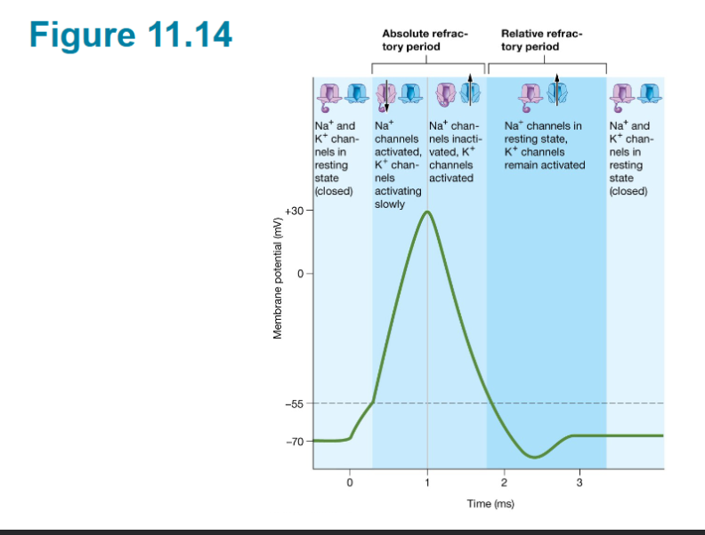
absolute refractory period -
during depolarization and repolarization no stimulus will be able to create an action potential

relative refractory.
a stronger than normal stimulus is required to overcome the fact that the membrane is repolarizing

All-or-none principle
If the neuron does not depolarize to threshold an action potential does not occur.
Propagated
Action potentials must act as a method of long distance signaling so they can be conducted or propagated down the length of the axon, they are self propagating one sets off another all the way down the axon like a chain reaction
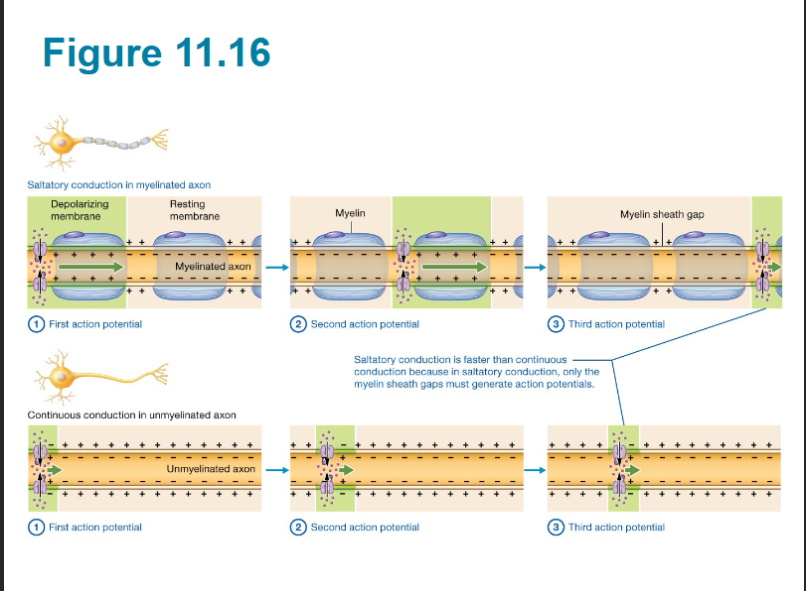
Conduction speed
The rate at which propagation occurs, determines how rapid signaling can occur within the nervous system, this is influenced by the diameter of the axon, and the presence or lack of my lawn sheath
if there is no myelin sheath continuous conduction occurs where the action potential must be created all the way down the Axon
if there is a myelin sheath then saltatory conduction happens where Action potentials only occur in the gaps between the sheath making things quicker
Saltatory conduction
if there is a myelin sheath then saltatory conduction happens where Action potentials only occur in the gaps between the sheath making things quicker

Continuous conduction
if there is no myelin sheath continuous conduction occurs where the action potential must be created all the way down the Axon

Presynaptic neuron
The neuron that is sending the message from its axon terminal
Postsynaptic neuron
the dendrite, cell body, or axon that is receiving the message from the presynaptic neuron
Synaptic transmission
the transfer of chemical or electrical signals between neurons at a synapse
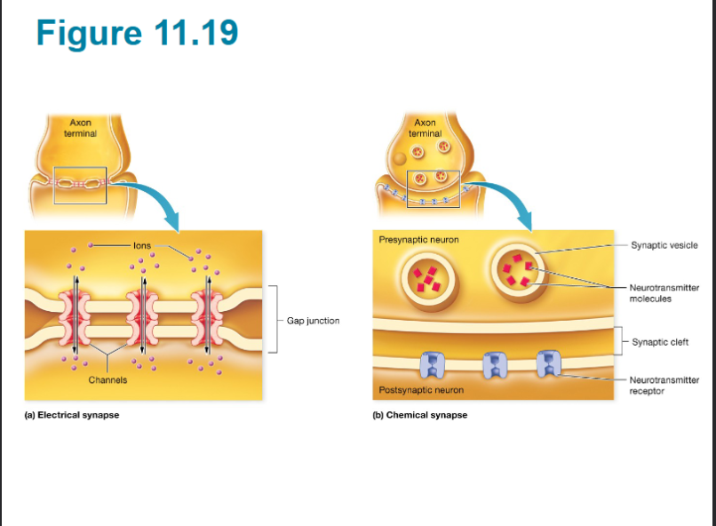
Chemical synapse
Very in size unlike electrical synapses, slower than electrical, use various neurotransmitters and receptors for different effects
Steps
Action potential in presynaptic neuron triggers calcium ion channels in axon terminal to open
calcium ion influx causes synaptic vesicles diffuse with the membrane of the presynaptic neuron and release neurotransmitters into synaptic cleft
neurotransmitters bind to the receptors on the postsynaptic neuron
ion channels open, leading to local potential and possibly an action potential

Neurolemmocytes
mylenates axons of PNS

Satellite Cell
surround and support cell bodies of PNS

Cerebrum
only portion with conscious / voluntary control
Diencephalon - small brain in brain center has 4 parts
Thalamus - Lg central, “main entry point / mail center” into the cerebrum for sensory input except for smell, sends sensory input to where it needs to go in the brain to be decoded
Hypothalamus- inferior, receives sensory input, boss of autonomic nerves system (ANS), involuntary functions, connects to Pituitary gland controls temp. BG, BP, sleep and thirst NOT the HR (controlled by brainstem), has Mammillary bodies that regulate sleep and wake signals that communicate with pineal gland to produce melatonin.
Epithalamus - Posterior and superior, has pineal gland which secretes melatonin
Subthalamus - has role in mvmt
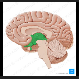
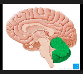
Cerebellum
it has the Arbor Vitae (tree of life) white matter being folded into tree shape, function- reduces motor error, helps with precise / complex movements, sits in the back base by brain stem.
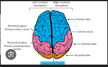
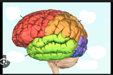
Cerebral hemispheres
the two sides of the brain has 4 lobe types plus the insulas
Frontal Lobes
frontmost lobe, posterior boundary is the central sulcus, Precentral gyrus (plural for gyri which is the peak in between folds of brain (the folds/creases)) is anterior to the central sulcus
- front most lobe has premotor cortex with planning, movement and coordination front eye fields, has broca’s area with movement making language, lobe of cerebral hemisphere
Function movement and reasoning
red
Spinal cord
contains neurons, connect with brain at foramen magnum, brain bod communication
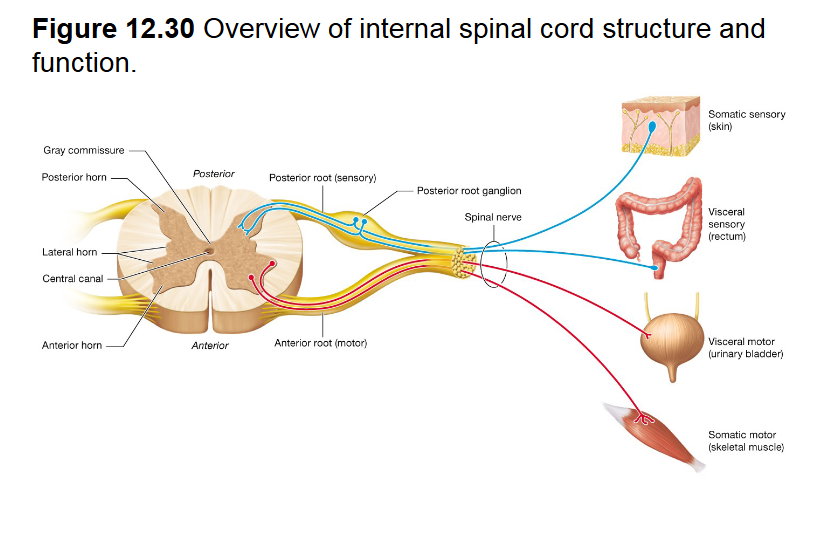
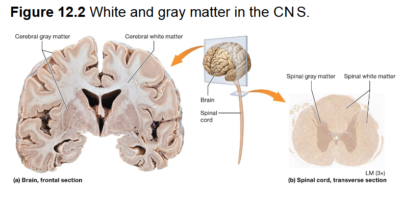
White matter -
Unmyelinated, On inside of spinal cord and outside of brain tissue

Grey matter
Myelinated, on outside of spinal cord and inside of brain tissue
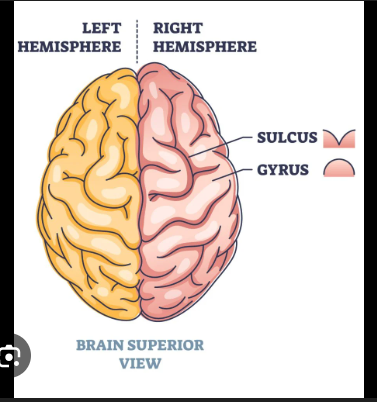
Sulci
shallow grooves, deep grooves are called fissures

Longitudinal fissure
separates the two halves of the brain