Pathology Final Exam Review
1/58
There's no tags or description
Looks like no tags are added yet.
Name | Mastery | Learn | Test | Matching | Spaced | Call with Kai |
|---|
No analytics yet
Send a link to your students to track their progress
59 Terms
ARDS
life-threatening pathology, leading to leakage of fluid into the interstitial space
increase technique
Asthma
widespread narrowing of airways due to stimuli; makes expiration difficult
decrease technique
Atelectasis
collapse of lung tissue
increase technique
COPD
encompasses several conditions which lead to ineffective exchange of respiratory gases
increase technique
Hyaline membrane disease
respiratory distress of newborn; airless alveoli and rapid respirations
increase technique
TB
chronic infection of lungs caused by rod-shaped bacterium
increase technique
Pneumoconiosis
caused by inhaling irritating particulates from environment or workplace
increase technique
Pneumonia
inflammation of lungs caused by bacteria or viruses
increase technique
Pneumothorax
air in pleural space that causes lungs to collapse
decrease technique
Chronic bronchitis
chronic inflammation of bronchi leading to increase of mucus production
increase technique
Croup
viral, pediatric infection that causes inflammation of the subglottic area
Cystic fibrosis
inherited condition; increased mucus secretion in lungs & bronchi
increase technique
Pleural effusion
accumulation of fluid in intra-pleural space
increase technique
Empyema
pus in pleural space
increase technique
Greenstick fracture
incomplete fracture with portion of cortex intact
Osgood Schlatter’s Disease
pediatric disease causing inflammation around the area of tibial tuberosity
Bone cyst
fluid-filled sacs within fibrous bone tissue
decrease technique
Bursitis
inflammation of bursa
increase technique
Ewing’s sarcoma
malignant; destructive bone tumor
decrease technique
Colle’s fracture
transverse fracture through distal radius
Compression fracture
compaction of the bone
subluxation
partial loss of continuity of joint
Spina bifida/meningocele
failure of vertebral arches to form properly, with associated soft tissue mass
no technique change
Congenital hip dysplasia
incomplete acetabulum formation
no change in technique
osteomyelitis
inflammation of bone & marrow caused by variety of infectious organisms
decrease technique
osteomalacia
abnormal softening of bones in adults
decrease technique
Paget’s disease
osteitis deformans; disruptive process
nonmetabolic bone disease causing bone destruction and unorganized bone repair
areas that are easier to penetrate are adjacent to areas that are harder making imaging difficult
Rheumatoid arthritis
inflammation & joint swelling
increase technique
Spondylolysis
defect in pars inter-articularis without displacement
Rickets
soft, pliable bones in pediatric patients
decrease technique
Comminuted fracture
multiple bone fragments
Osteoporosis
mass of bone per unit volume is decreased in amount but normal in composition (same structure, just less of it)
decrease technique
Osteopetrosis
increased bone density, inherited
increase technique
Multiple myeloma
widespread malignancy of plasma cells associated with bone destruction
decrease technique
Volvulus
twisting of bowel that may cause obstruction
increase due to contrast
Cancer of the esophagus
malignant neoplasm of esophagus, imaged during barium swallow study
increase due to contrast
Chron’s disease
inflammatory disorder usually involving the ileum
aka regional enteritis
increase due to contrast
Diverticulosis
presence of herniations through the wall of the colon
Esophageal varices
varicose veins at distal end of esophagus
increase due to contrast
Intussusception
prolapse of one segment of bowel into another section of bowel (telescoping)
increase due to contrast
Ileus
mechanical or adynamic; intestinal obstruction
increase due to contrast
Cholelithiasis
gallstones
increase due to contrast
Gastritis
inflammation of stomach mucosa
increase due to contrast
Hiatal hernia
portion of stomach protrudes through diaphragm
Pyloric stenosis
narrowing of pyloric sphincter
Ulcerative colitis
severe inflammation of colon and rectum; characterized by ulceration
increase due to contrast
Diverticulitis
inflammation of pouch-like herniations of colon wall
increase due to contrast
Annular carcinoma
“apple-core” pattern on BE study
increase due to contrast
Cystitis
inflammation of urinary bladder
polycystic kidney disease
enlarged kidneys containing numerous fluid-filled cysts
increase technique
Glomerulonephritis
inflammation of the glomerulus of the kidney
Pyelonephritis
inflammation of renal pelvis and parenchyma
Renal calculus
kidney stones
harder to penetrate stones, but overall no change in technique
Carcinoma of the bladder
seen as solid mass arising from bladder wall
increase technique
Renal carcinoma
solid mass cancer that causes renal bulging or enlargement
may need to increase technique
Renal cyst
fluid filled mass in kidney
increase technique
Wilm’s tumor
malignant cancer of kidney in children
increase technique
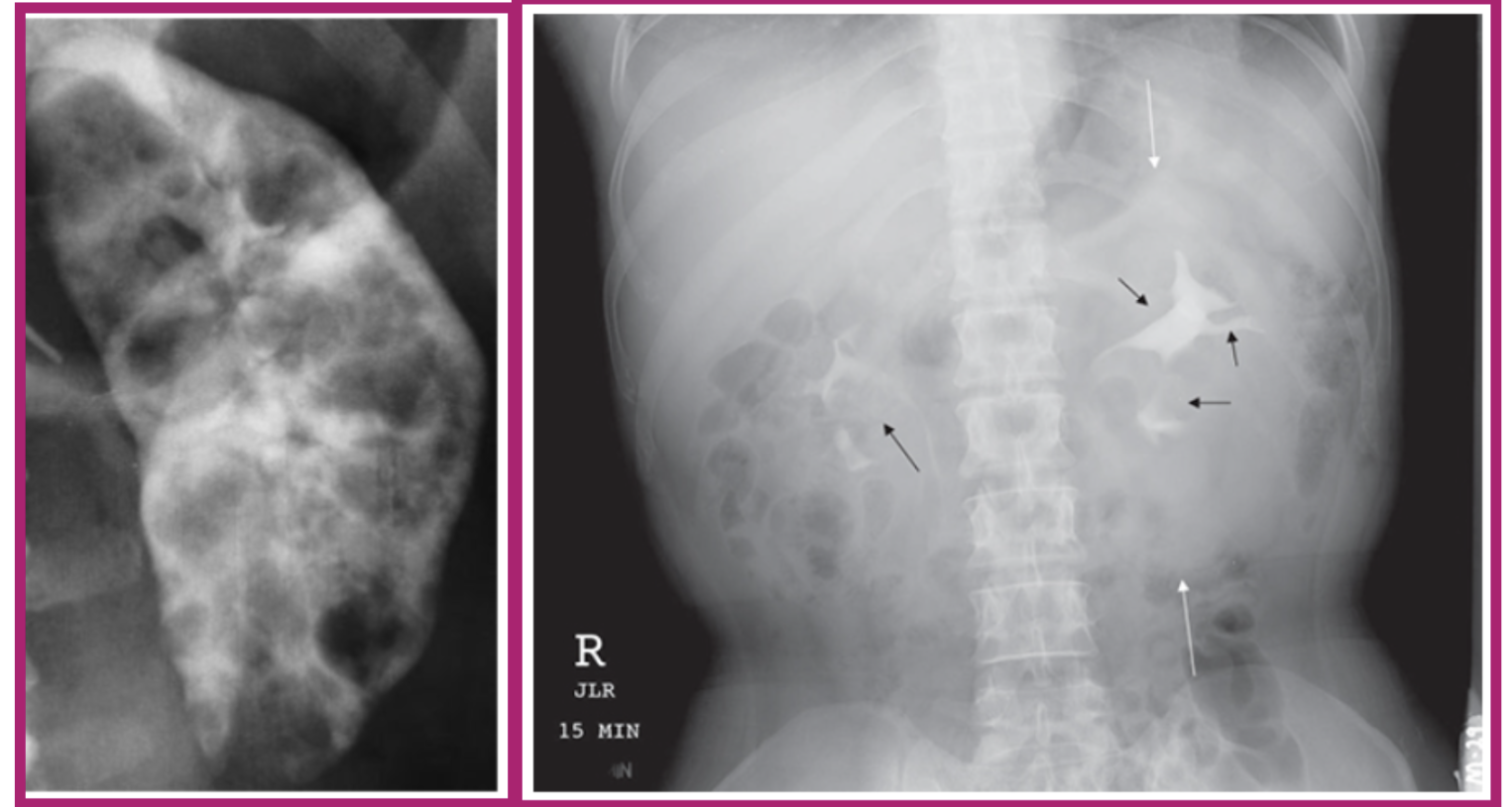
what pathology does this image show
polycystic kidney disease
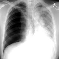
what pathology does this image show
pneumothorax