Transport in Mammals
1/18
There's no tags or description
Looks like no tags are added yet.
Name | Mastery | Learn | Test | Matching | Spaced | Call with Kai |
|---|
No analytics yet
Send a link to your students to track their progress
19 Terms
Why do mammals need a specialised transport system:
Greater the activities, higher demand of O2
Large and more complex
Cannot rely on diffusion like plants as there are cells deep within the body that are metabolically active
The circulatory system (pulmonary and systemic)
Pulmonary: From the heart to the lungs
Systemic: From the heart to the body

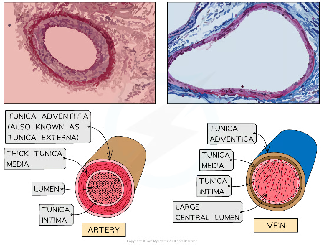
Structure of arteries and arterioles
Arteries:
Tunica externa: elastic fibres to allow wall to stretch as blood surges, collagen fibres and smooth muscles
Tunica media: same as tunica externa, thick to withstand high blood pressure surging
Tunica intima: endothelium cell lining to minimise friction, lumen small to maintain high pressure of blood for efficient delivery to tissues
Arterioles have the same proportion but contains more smooth muscle and a narrower lumen to slow down blood flow for efficient material exchange. This is due to having a nerve supply which receives impulses from the brain for vasoconstriction and dilation.
Structure of capillaries
Small vessel size takes blood as close as possible to all cells, allowing transfer of substances between cells and blood
Forms a network throughout every tissue except cornea and cartilage
Extremely thin wall made up of single layer of endothelial cell with tiny gaps between
Blood is able to squeeze as close as 1um to tissue cells to allow gaseous exchange
Veins and venules
Blood will then reach to larger vessels known as venules and join to form veins
Initial blood pressure when entering a vein is low, because tunica media is thinner and has lesser elastic fibres and muscle and has the largest lumen
Low pressure is solved by skeletal muscle contraction which raises b.p but it’s not enough, so semi-lunar valve adaptation prevents back-flow and pressure loss in veins
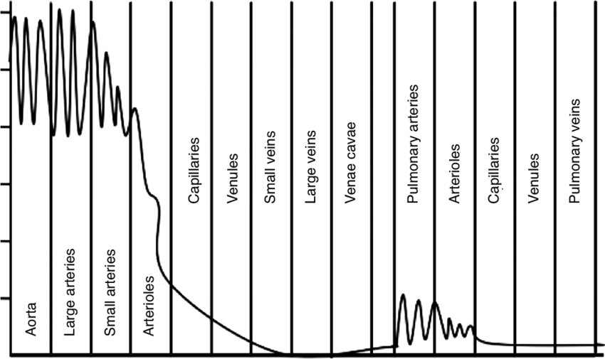
Pressure changes in circulatory system
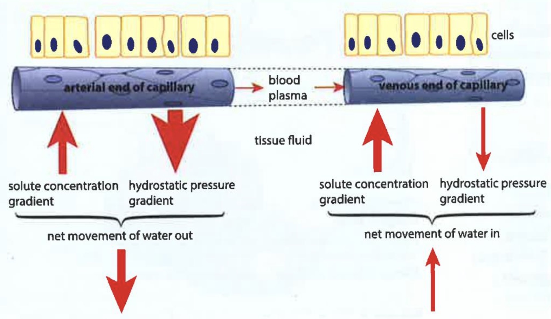
Blood plasma and capillaries
Some plasma leaks out the gap in capillary wall
Tissue fluid has the same properties as plasma but with less protein
At the atrial end, the pressure is enough to push plasma out of cells
And due to osmosis, water is drawn back to the capillaries
“How does blood from arteriole end differ from venule end?”
1) lower pressure at venule end
2) less oxygen at venule end
3) lower water potent. at venule end
4) higher [solute] at venule end
5) more urea and secretory waste
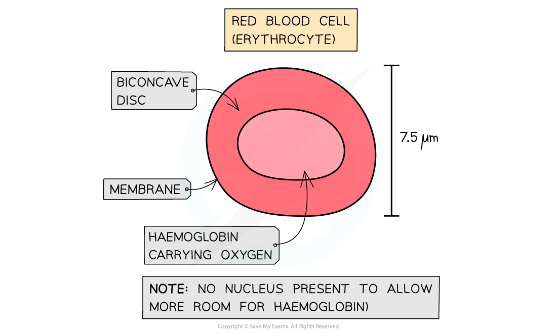
Characteristics of red blood cells (erythrocytes)
Globular protein that transports oxygen from lungs to respiratory tissues
Fragile and will burst if their membranes are in a tight spot
Small in size, 7um in dm and able to squeeze in capillaries
Biconcave shaped increase sa:v ratio for diffusion
Flexible, can deform and pass through capillaries due to their exoskeleton
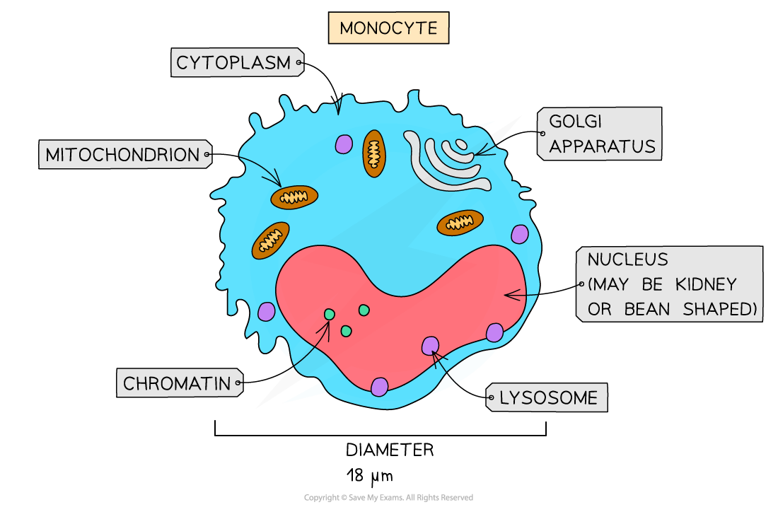
Characteristics of white blood cells (leucytes)
Larger than red blood cells and has a nucleus
Involved in fighting against infectious diseases
Phagocytes
Destroys foreign organisms using phagocytosis
Normally lobed nuclei and granular cytoplasm which are dark purple in colour
E.g: neutrophil, monocytes, and macrophages
Lymphocytes
Destroys microorganisms with the secretion of antibodies protein
Antibodies attach and destroy invading cells
Smaller than phagocytes, having a large round nucleus and small amount of cytoplasm
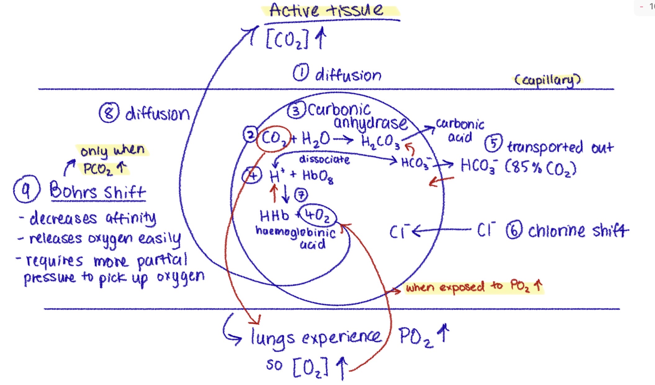
What happens when partial pressure of CO2 is high in active tissue (Bohrs shift):
1) High conc. of CO2 in tissue diffuses into haemoglobin
2) CO2 + H2O → H2CO3 (forms carbonic acid from the enzyme carbonic anhydrase)
2.5) H2CO3 → H+ + HCO3- (dissociates)
3) HCO3- goes into the capillary and Cl- shifts into the haemoglobin
4) H+ + HbO8 → Hhb (haemoglobonic acid) + 4O2
5) 4O2 diffuses into active tissue
6) “Bohrs shift”: decreases affinity which releases O2 easily and requires increased partial pressure to pick up O2
What happens when partial pressure of O2 is high in active tissue:
1) Hhb dissociates into H+
2) HCO3 shifts back into haemoglobin
3) Forms H2CO3 again to revert back into CO2 + H2O
4) CO2 goes back inside the lungs
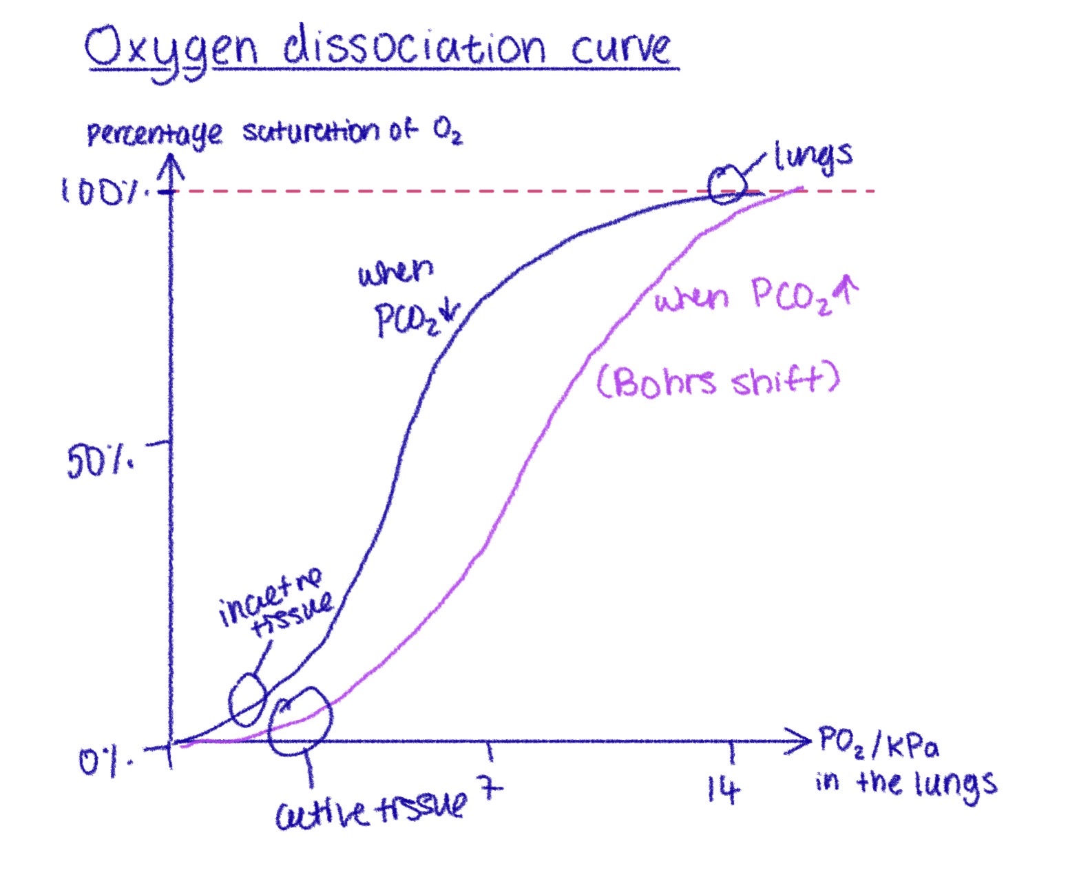
Why is it an s-shaped curve?
Behaviour of haemoglobin (Hb) in different partial pressures of O2
Each haem group distorts to fit more O2, 1st is hardest and last is easiest
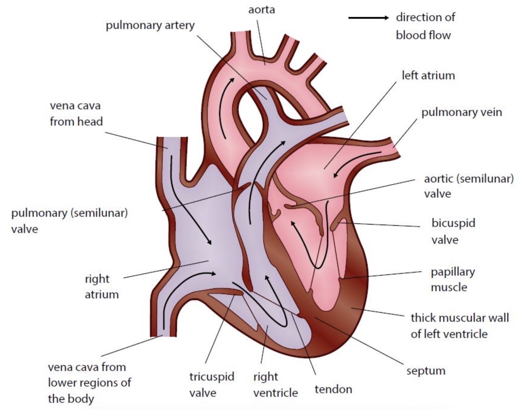
Diagram of the heart and adaptations
Left ventricle is thicker and has more muscle to produce a higher pressure so blood can be pumped through the whole body
Right ventricle pumps to the lungs which is nearer
Pulmonary artery flows at a lower pressure than aorta
Cardiac cycle simplfiied
1) Atrial systole: both atria contract. blood from atria flows into the ventricles. back flow is prevented by closure of valves
2) Ventricular systole: both ventricles contract. av valves are pushed shut by the pressurised blood in the ventricles. sl valves in aorta and pulmonary artery are pushed open. blood flows from ventricles to arteries
3) Ventricular diastole: atria and ventricles relax. sl valves in aorta and pulmonary artery are pushed shut. blood flows from the veins through the atria into ventricles
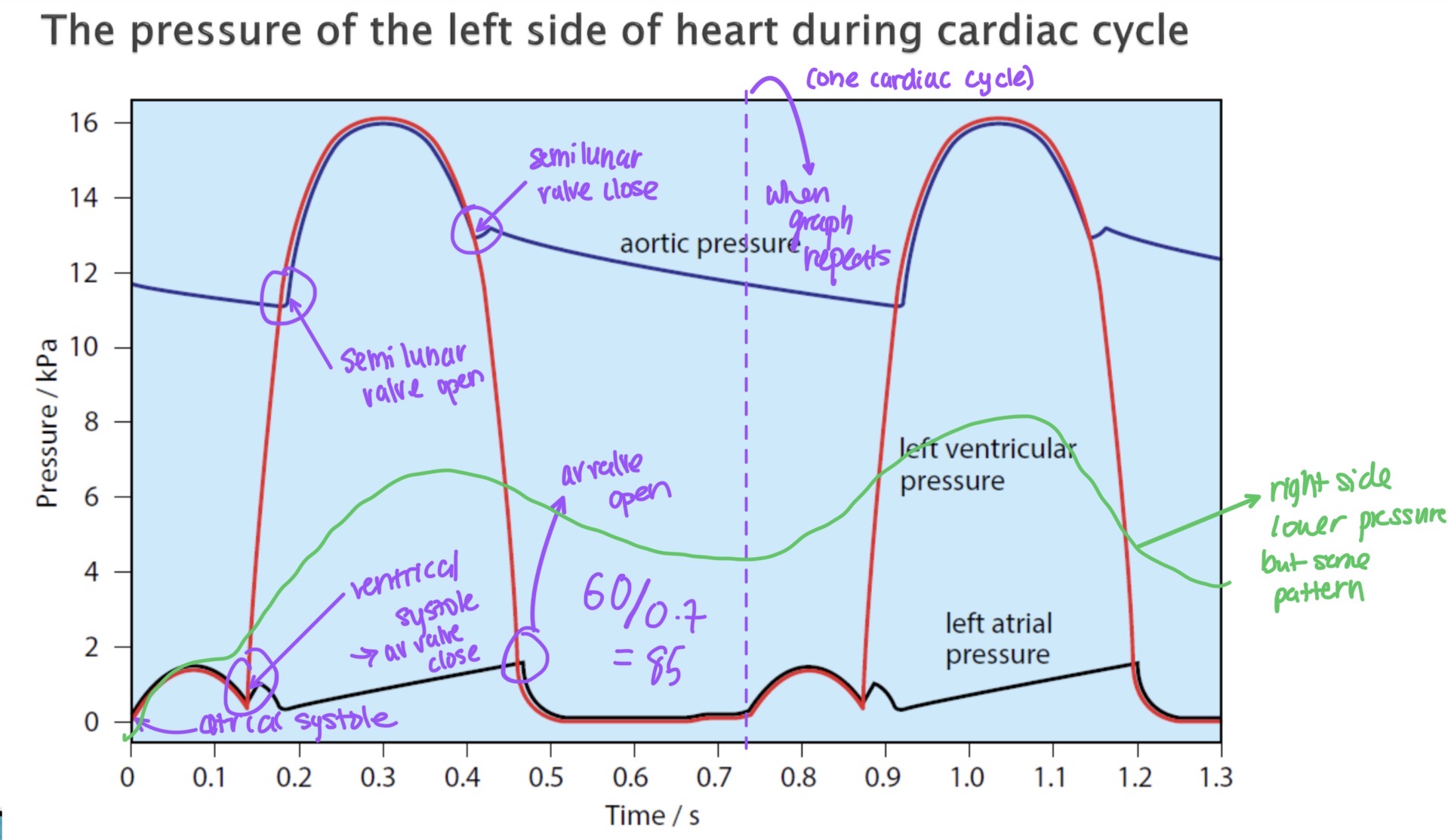
Pressure graph for the left side of the heart. Right side has the same shape, but with lower pressure
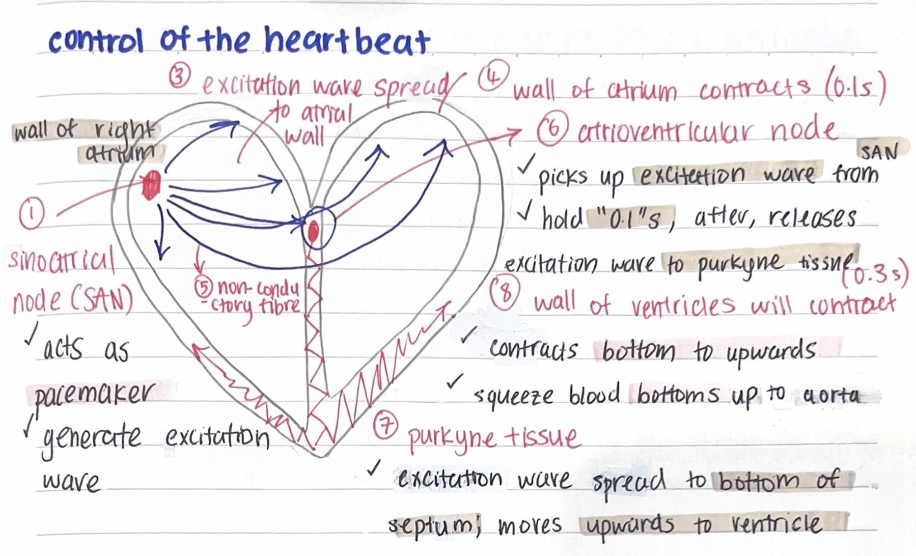
Control of the heartbeat:
1) Sinoatrial node act as a pacemaker which generates excitation wave
2) Excitation wave spreads to atrial wall
3) Wall of atrium contracts (0.1s)
4) Atrioventricular node picks up excitation wave from SAN and holds its for 0.1s before releasing it to purkyne tissue
5) Excitation wave from purkyne tissue spreads to the bottom of septum and moves upwards to ventricle
6) Wall of ventricles then contract bottom to up which squeezes blood upwards to aorta