BIO 330 - Neuroanatomy Lab 10
1/42
There's no tags or description
Looks like no tags are added yet.
Name | Mastery | Learn | Test | Matching | Spaced | Call with Kai |
|---|
No analytics yet
Send a link to your students to track their progress
43 Terms
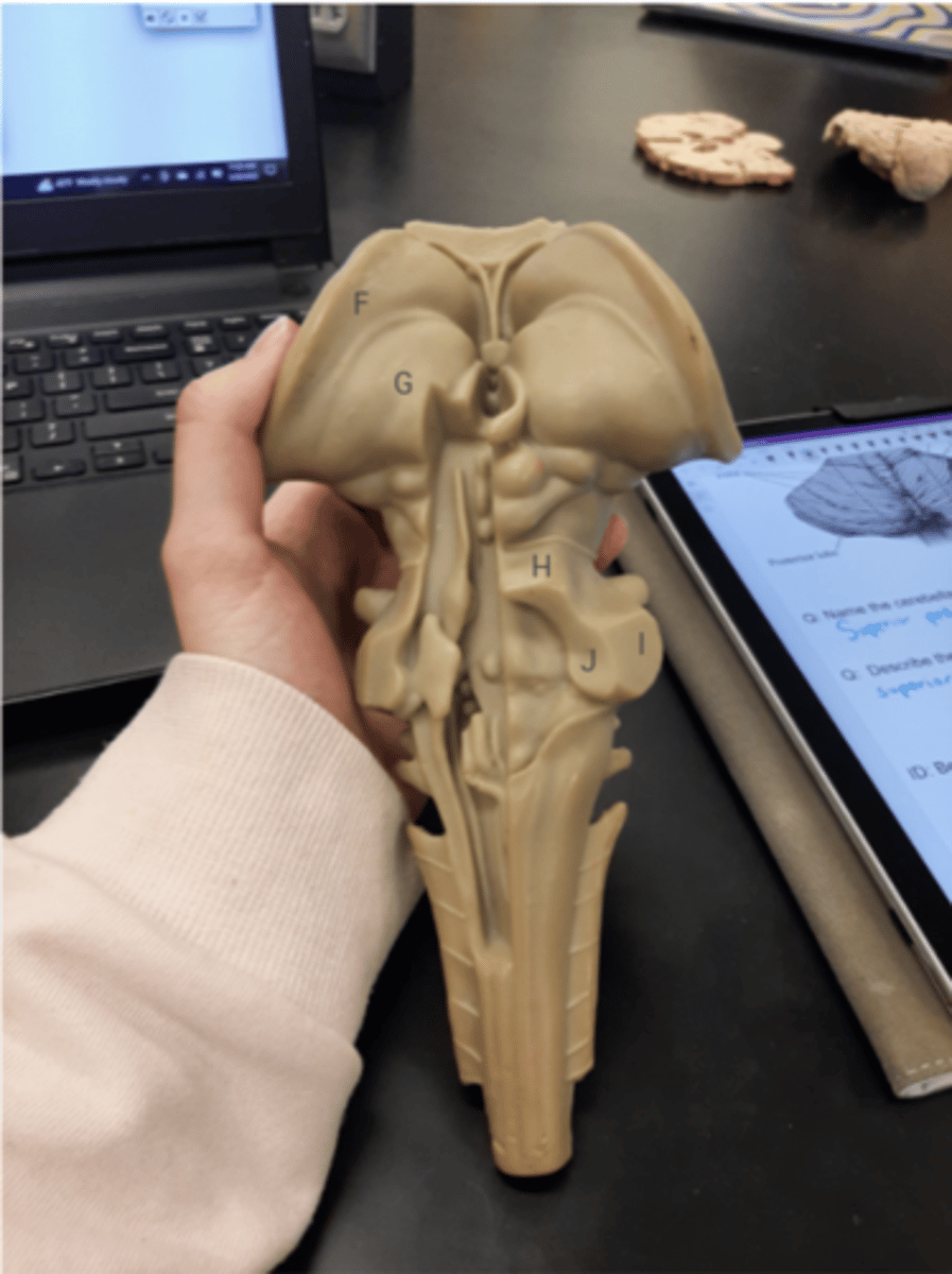
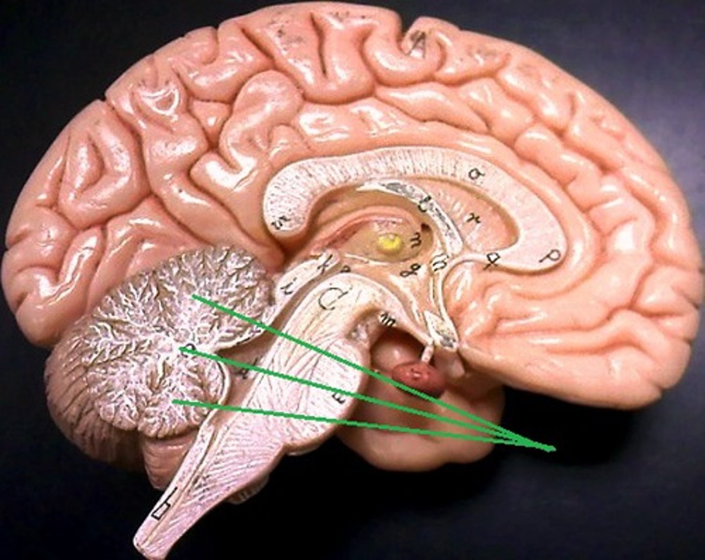
Label this image of the dorsal brainstem model : caudate nucleus, thalamus, 3 cerebellar peduncles
F = caudate nucleus
G = thalamus
H = superior cerebellar peduncle
I = middle cerebellar peduncle
J = inferior cerebellar peduncle
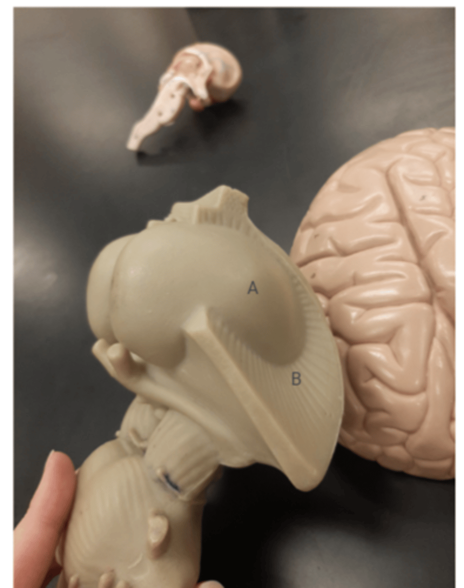
Label this image of the brainstem model
A = putamen
B = internal capsule
What are the 5 main basal ganglia?
caudate nucleus, putamen, globus pallidus, subthalamic nucleus, substantia nigra
What fiber tract separates the thalamus from the basal ganglia?
internal capsule
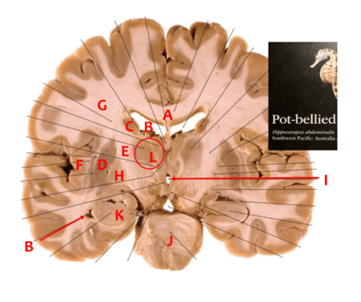
Label the following letters with their respective structures : A,B,C,D,E,F,G,H,I,J, K,L
A = corpus callosum
B = lateral ventricle
C = caudate nucleus
D = putamen
E = internal capsule
F = insula
G =
H = globus pallidus
I = 3rd ventricle
J =
K =
L = thalamus
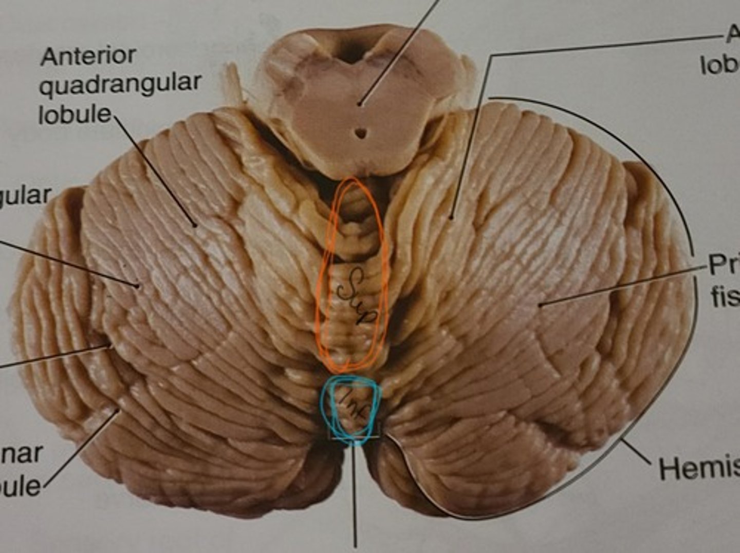
What is the orange outline of the cerebellum called?
vermis
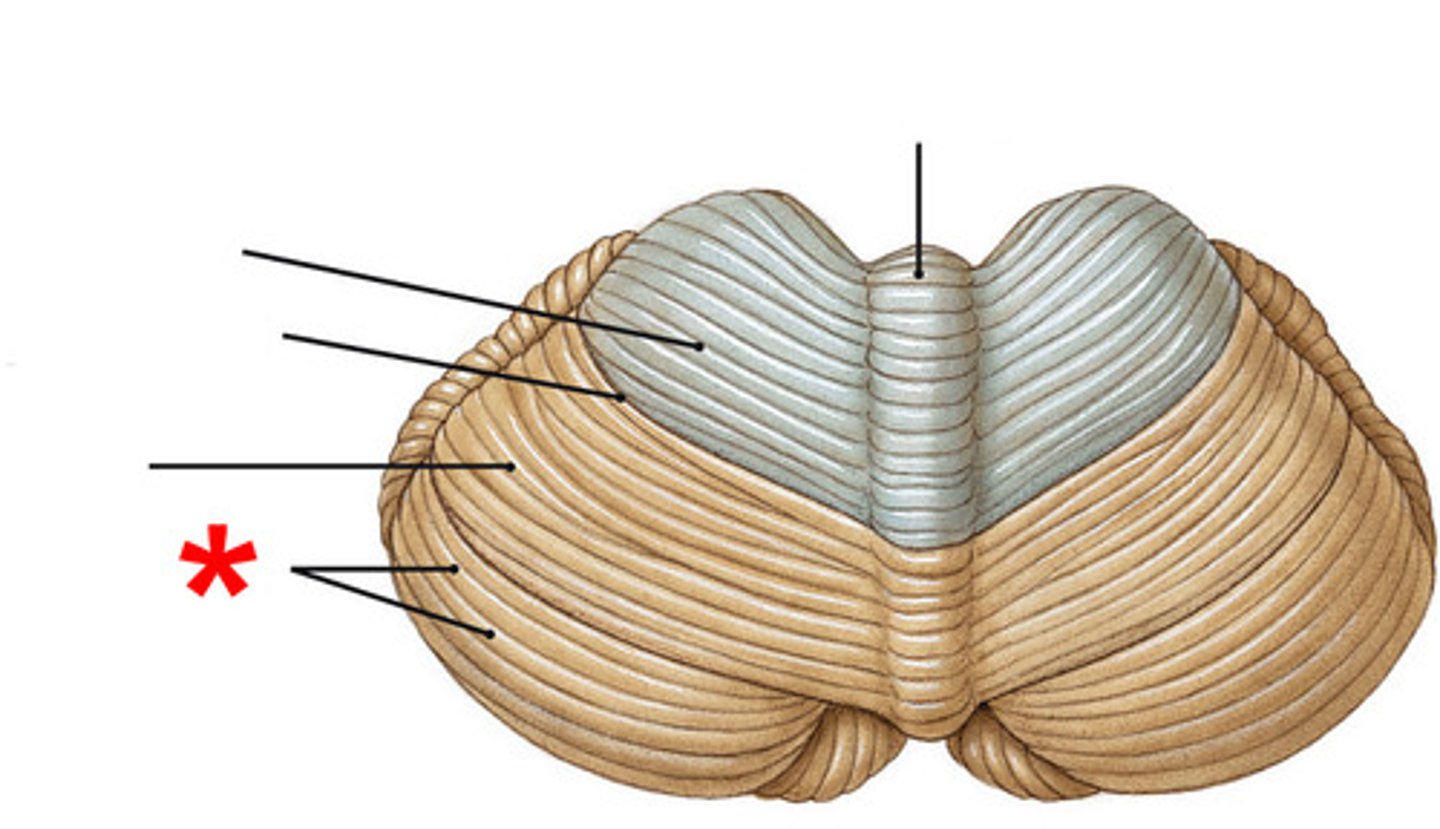
What are the bumps/folds on the cerebellum called?
folia
What is the white matter of the cerebellum called?
arborvitae
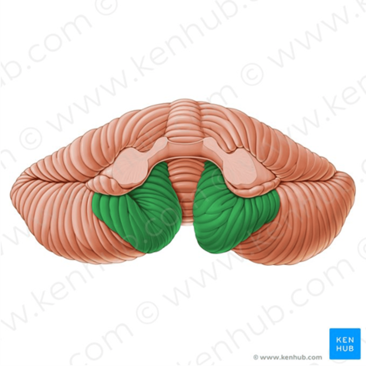
What are these green areas of the cerebellum called?
tonsils
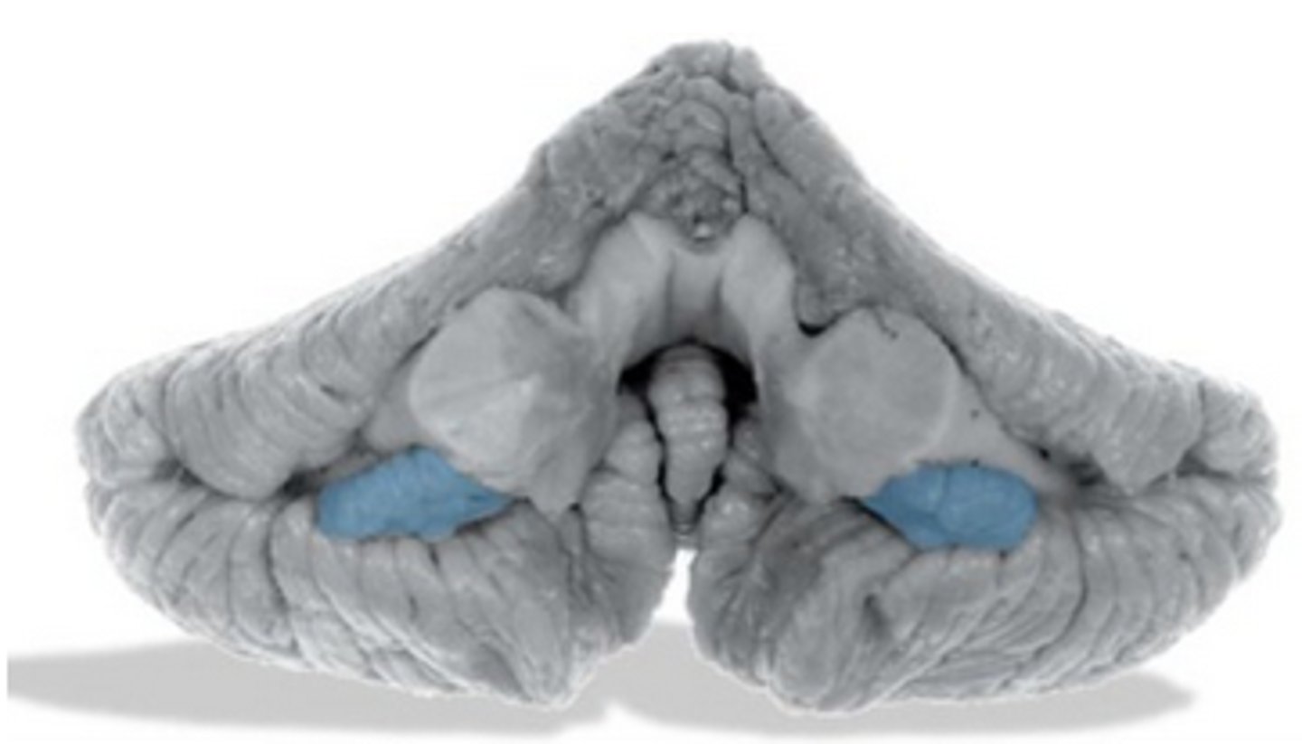
What are these blue areas of the cerebellum called?
flocculus
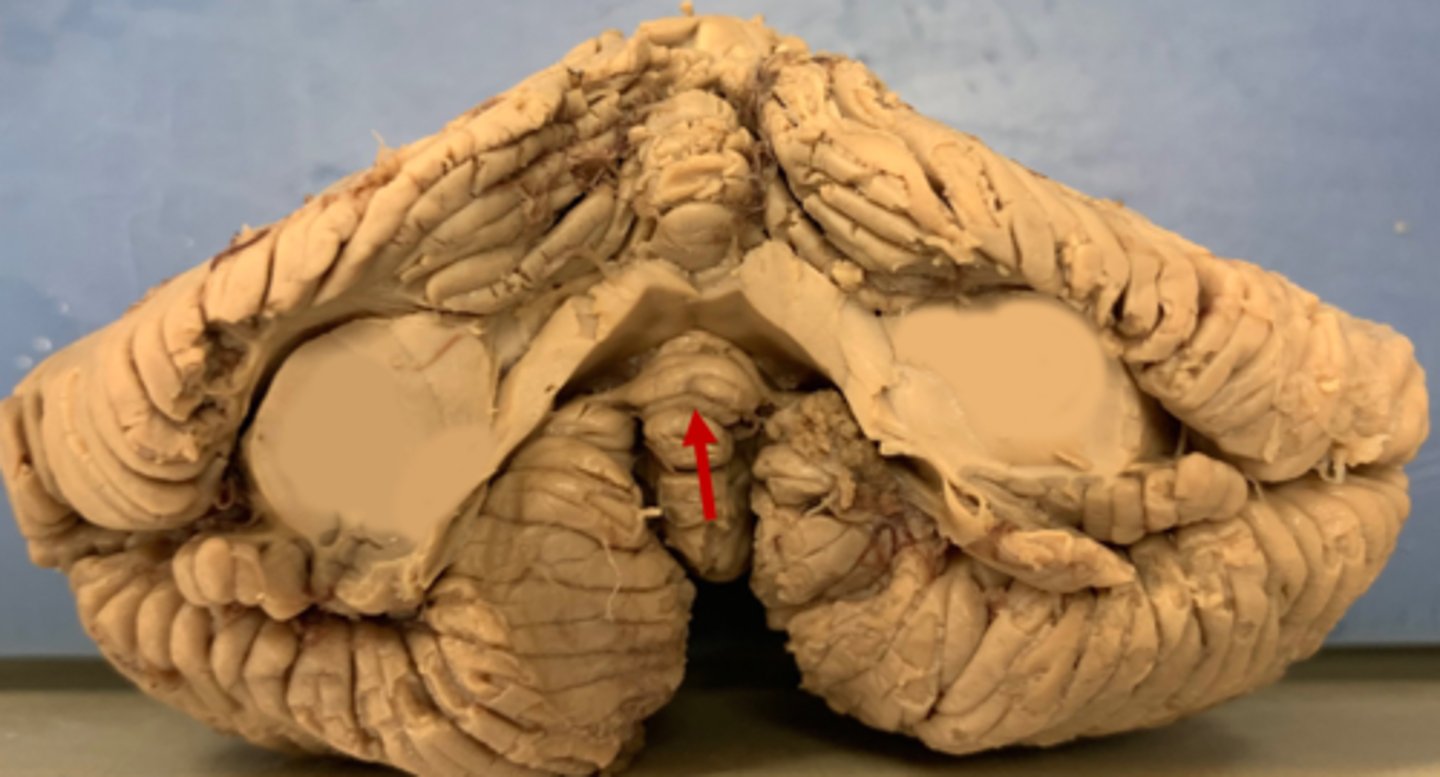
What is this bottom area of the vermis called? (red arrow)
nodulus
The nodulus and flocculus of the cerebellum make up what is called the
flocculonodular lobe
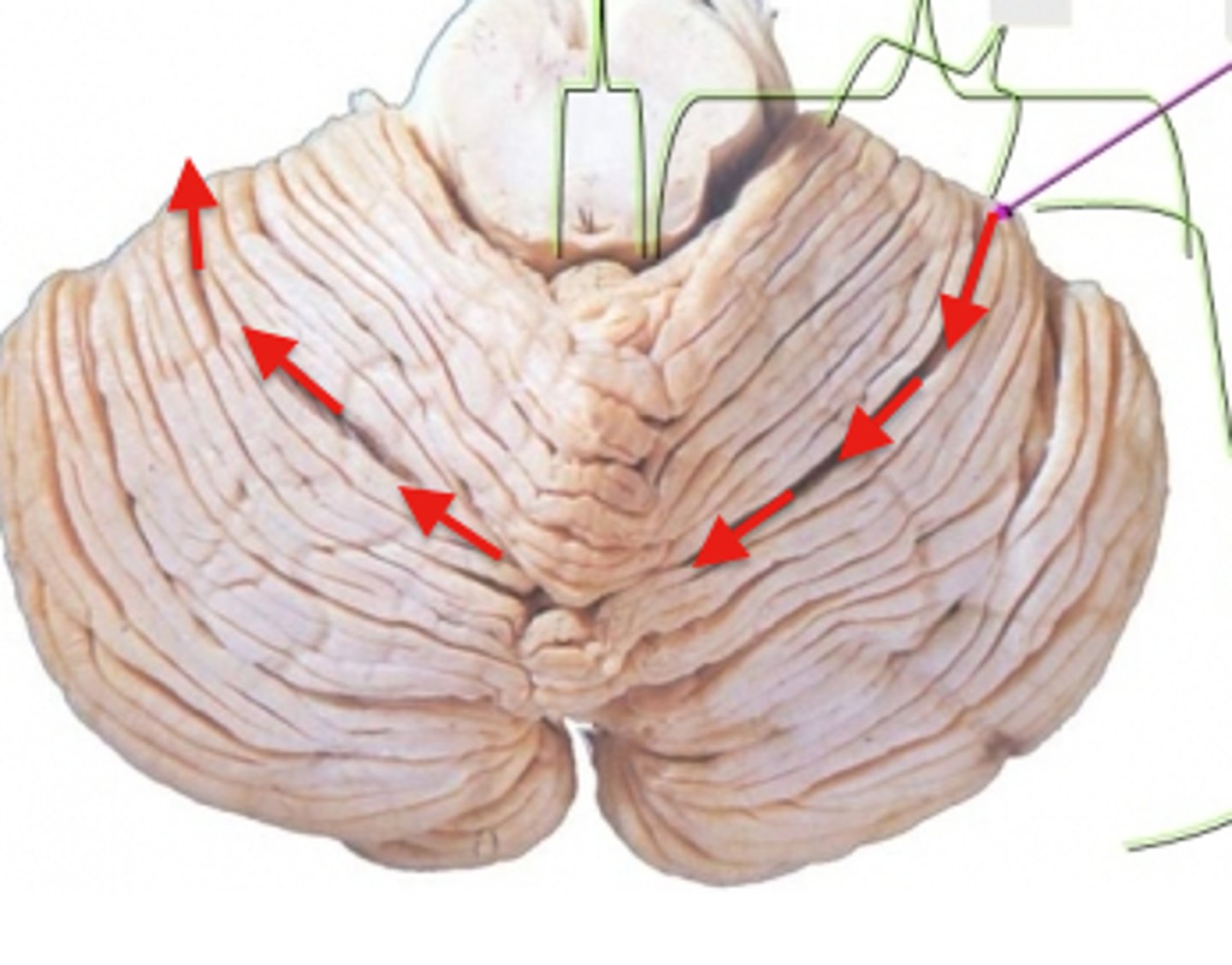
What is this dividing line that separates the Anterior and Posterior lobes of the cerebellum?
primary fissure
If the brain swells, what part of the cerebellum pushes on the brainstem? Specific brainstem structure?
tonsils push on medulla
If the brain swells and the cerebellum pushes on the brainstem, why is this bad?
phrenic nerves are being hit which causes disrupted respiratory control
If the brain swells and the cerebellum pushes on the brainstem, what foramen could the cerebellum herniate through?
Foramen magnum
Which 2 nuclei do the basal ganglia and cerebellum project to?
ventral anterior or ventral lateral nuclei of the thalamus
Which cerebellar peduncle allows the output of information out of the cerebellum?
superior cerebellar peduncle
Which cerebellar peduncle allows the input of information from the premotor and primary cortexes into the cerebellum?
middle cerebellar peduncle
Which cerebellar peduncle allows the input of information from lower brainstem structures and the spinal cord into the cerebellum?
inferior cerebellar peduncle
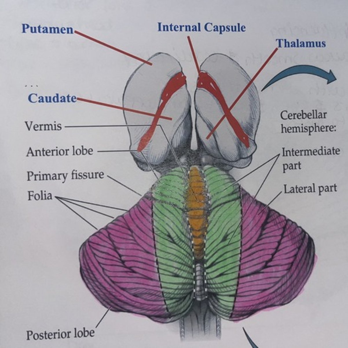
In this image of the cerebellum, pair the colors with their respective hemisphere regions or structural name if it is not a hemisphere
orange/yellow = vermis
green = intermediate hemisphere
pink = lateral hemispheres
The vermis of the cerebellum influences movement of
present/ongoing trunk muscles
When the vermis is active, what type of body movements are carried out after the vermis talks to the cerebral cortex? (think motor tract)
balance and gait movements/trunk muscles (ACT)
What type of movement does the flocculonodular lobe of the cerebellum influence?
coordination of eyes with movement (vestibulo-ocular reflexes)
The intermediate hemisphere of the cerebellum influences the movement of
present/ongoing limb movements
The lateral hemisphere of the cerebellum influences the movement of
future/planning of limb movements
If someone has damage to their intermediate hemisphere what is the result?
erratic/shaky limb movements
If someone has damage to their vermis what is the result?
truncal ataxia (unsteady gait)
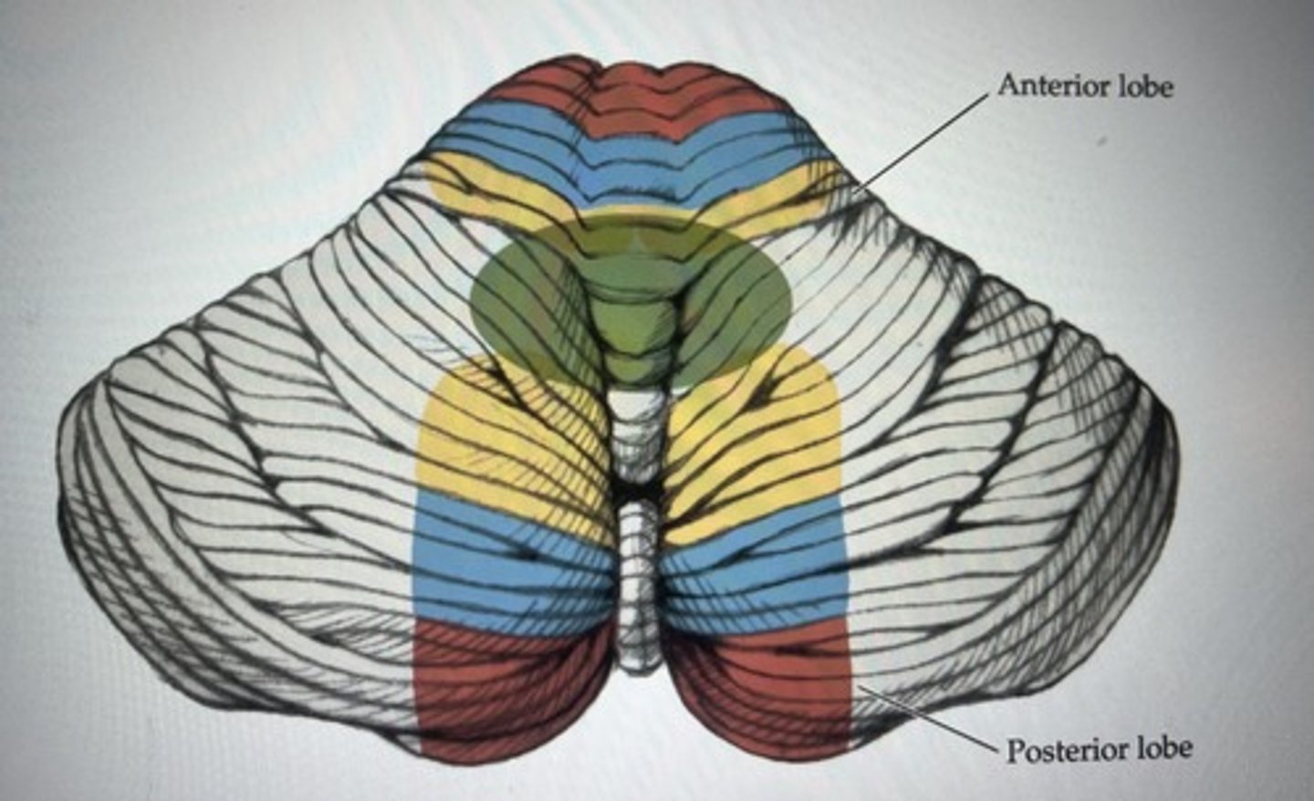
The cerebellum has 2 homunculi, in this image label what each color represents in respect to what part of the homunculus it is associated with
yellow = head
blue = arms
red = legs and feet
green = eyes
it is the same for both anterior and posterior lobe
If the LCT wants to perform a muscle movement, where would the cerebellar cortical neurons be active?
left hemisphere
left side of cerebellum in legs / feet area of both anterior and posterior lobes
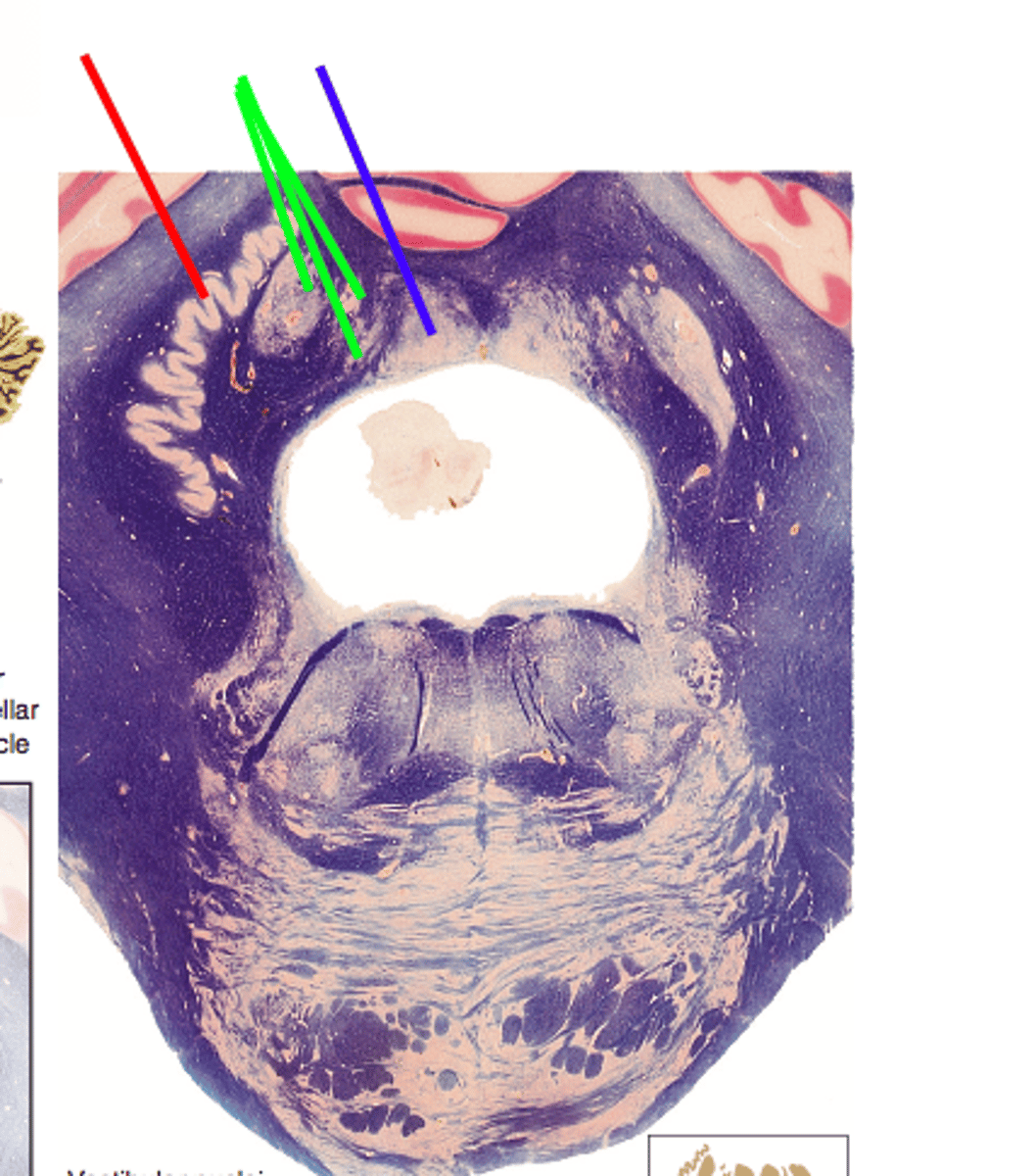
In this image of the pons / cerebellum the colored areas are deep cerebellar nuclei ; match the color to the respective deep cerebellar nuclei
red = dentate
green = emboliform & globose (emboliform is the one closest to dentate)
blue = fastigial
The present/ongoing movement of trunk muscles purkinje cells associated with the _______ of the cerebellum synapse with which deep cerebellar nuclei
vermis ; fastigial nuclei
The present/ongoing movement of limbs purkinje cells associated with the _______ of the cerebellum synapse with which deep cerebellar nuclei
intermediate hemisphere ; emboliform & globose (interposed nuclei)
The future/planning of limb movements purkinje cells associated with the _______ of the cerebellum synapse with which deep cerebellar nuclei
lateral hemisphere ; dentate nuclei
The future/planning of eye movements punkinje cells associated with the ________ of the cerebellum synapse with which deep cerebellar nuclei
flocculonodular lobe ; vestibular nuclei
Purkinje cells associated with the lateral hemisphere synapse with ________ deep cerebellar nuclei and leave cerebellum the __________ cerebellar peduncle
dentate ; superior peduncle
Purkinje cells associated with the intermediate hemisphere synapse with ________ deep cerebellar nuclei and leave cerebellum the __________ cerebellar peduncle
interposed (emboliform & globose) nuclei ; superior peduncle
Purkinje cells associated with the vermis hemisphere synapse with ________ deep cerebellar nuclei and leave cerebellum the __________ cerebellar peduncle
fastigial ; superior and inferior peduncles
Purkinje cells associated with flocculonodular lobe synapse with ________ nuclei and leave cerebellum by the ________
vestibular ; MLF
The cerebellum does have a decussation point, but it happens after leaving the superior cerebellar peduncle. Where is this decussation point located in the brainstem, and what is it called?
superior cerebellar decussation in caudal midbrain
In a finger-nose-finger test of the LEFT pointer finger, what part of the cerebellar cortex is active and what deep cerebellar nuclei are used?
left intermediate hemisphere ; interposed nuclei
Describe the cerebellar pathway used if you are currently moving an upper or lower limb
-interposed nuclei receive signal from purkinje cells in the intermediate hemisphere
-interposed axons travel up and leave through the sup. cerebellar peduncle
-interposed axons at the sup. cerebellar deussation
-interposed axons synapse with VA or VL of the thalamus
VA or VL axons go through internal capsule and corona radiate on contralateral side of the brain
-VA or VL axons synapse with UMN in the primary motor cortex
Describe the cerebellar pathway used if you are planning to throw a dart and how it will be performed
-dentate nuclei receive signals from pukinje cells in the lateral hemisphere
-dentate axons travel up and leave through the sup. cerebellar peduncle
-dentate axons decussate at the sup. cerebellar decussation
-dentate axons synapse with ventral anterior or ventral lateral nuclei of the thalamus
-VA or VL (thalamic) axons travel through internal capsule and corona radiata on contralateral side of the brain
-VA or VL axons synapse with UMN in premotor cortex, arm area