Anatomy - Gluteal Region and Thigh
1/76
There's no tags or description
Looks like no tags are added yet.
Name | Mastery | Learn | Test | Matching | Spaced | Call with Kai |
|---|
No analytics yet
Send a link to your students to track their progress
77 Terms
The superficial gluteal muscles includes the
- Glute max
- Glute med
- Glute min
- TFL
The glute max is the only superficial gluteal muscle that is innervated by the
Inferior gluteal nerve
What are the deep muscles of the gluteal region
- Piriformis
- Superior/inferior gemelli
- Obturator externus/internus
- Quadratus femoris
What is the medial border of the adductor canal
Adductor longus/magnus
What is the lateral border of the adductor canal
Vastus medialis
What is the anterior border of the adductor canal
Sartorius
What are the contents of the adductor canal
- Femoral artery
- Femoral vein
- Branches of femoral nerve
Saphenous nerve is a continuation of the
Femoral nerve
After the adductor canal the saphenous nerve continues
Medially to provide cutaneous sensation to the medial leg
After the adductor hiatus, the femoral artery and vein becomes the
Popliteal artery and vein
The gluteofemoral bursae separates the
Iliotibial tract from superior part of proximal attachment of vastus lateralis
What is the purpose of the gluteal bursae
It minimizes friction to allow the greatest ease of movement
The ischial bursae separates the
Inferior part of the glute max from the ischial tuberosity
The trochanteric bursa separates the
Superior fibers of the gluteus maximus from the greater trochanter
What muscles attach to the pes anserine
- Sartorius
- Gracilis
- Semitendinosus
The deep fascia of the thigh limits
Outwards expansion of contracting muscles
The deep fascia of the lower limb allows muscular contraction to
Be more efficient in compressing veins to push blood towards the heart
The TFl encloses the
Large thigh muscles, especially laterally
The 3 compartments of the thigh are composed of
The fascia lata and three fascial intermuscular septa
Of the 3 intermuscular septa of the thigh, the lateral one is _ and the other 2 are relatively _
Especially strong; relatively weak
The lateral intermuscular septum extends from the
IT band to the lateral lip of the linea aspera and lateral supracondylar line of femur
The saphenous opening is located
Inferior to the medial part of the inguinal ligament
The saphenous opening allows for the passage of
Lymphatic vessels from the superficial inguinal lymph and the great saphenous vein and its tributaries
After passing through the saphenous opening, the great saphenous vein will
Enter the femoral vein
The anterior compartment muscles of the thigh are
Knee extensors
The posterior compartment muscles of the thigh are
Knee flexors
All of the anterior compartment muscles of the thigh are innervated by the
Femoral nerve (L2-L4)
The quadriceps muscles can be how much stronger than the hamstring muscles?
3x
In gait, the quads are active during
The termination of the swing phase, preparing the knee to accept weight
What is the primary responsibility of the quads in gait
Absorb the shock of heel strike
In bent knee activities, the quads serve as
Fixators
During downhill walking or descending stairs, the quads work
Eccentrically
Which of the quads will work on the hip joint
Rectus femoris
Ther articularis genu is an anterior muscle that is
A derivative of the vastus intermedius
What does the articularis genu do
It pulls the synovial membrane of the knee superior during extension to prevent folding
When you walk, most of the work that the hamstrings do is
Eccentric
The workhorse of the knee is the
Biceps femoris
The Q-angle is formed from the
Line from the ASIS to the patella and line from the tibial tuberosity to the patella
The normal Q-angle in men is _ and in women is _
12 degrees; 17 degrees
Genu varum is associated with a decrease in
Q-angle
Coxa valga leads to _ which leads to _
Genu varum; Decreased Q-angle
Genu valgum is associated with an increase in
Q-angle
Coxa vara leads to _ which leads to _
Genu valgum; Increased Q-angle
Genu valgum is characterized by
Knock knees
Genu varum is characterized by
Bow legs
The medial and lateral condyles of the femur serve as proximal attachment sites for the
Medial and lateral collateral ligaments
The pes anserine is medial to the
Tibial tuberosity
Coxa vara and valga exist in which plane
Frontal plane
Q-angle
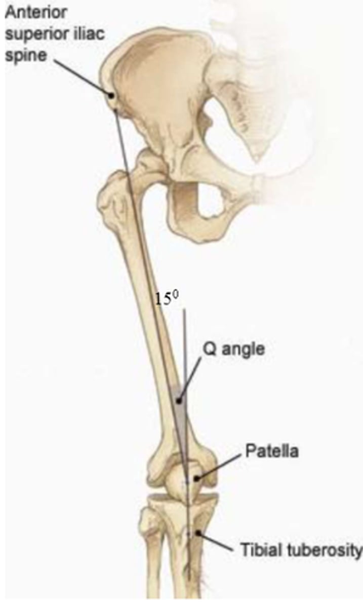
The knee naturally exists with slight
Valgus
The fascia lata is comprised of
Fat, cutaneous nerves, and superficial veins
Superiorly, the fascia lata attaches to and is continuous with the
Pubic region and abdominal wall fascia
Posteriorly and laterally, the fascia lata attaches to the
Iliac crect
Posteriorly and medially, the fascia lata attaches to
Inferiorly, the fascia lata attaches to
The fascia lata thickens laterally to form the
IT band
The shared aponeurosis of the _ and _ attach to the IT band
Glute max and TFL
Muscles of the anterior compartment of the thigh are innervated by the
Femoral nerve
The sartorius acts as the roof of the
Adductor canal
Concentric contraction of the quads is used to extend the knee against
Gravity
The rectus femoris is particularly efficient in movements where
Knee extension and hip flexion are combined (preparing to kick a ball)
Medial compartment muscles are primarily innervated by the
Obturator nerve
The most anterior adductor muscle is the
Adductor longus
The adductor brevis lies deep to the
Pectineus and adductor longus
The obturator splits which nerve into anteiror and posterior divisions
Obturator nerve
The most superficial adductor muscle is the
Gracilis
The adductor hiatus is formed between the
Distal attachment of the adductor part and hamstring part of the adductor magnus
The hamstrings are primarily innervated by the
Tibial division of the sciatic nerve
The two actions of the hamstrings cannot be
Performed maximally at the same time
The semimembranosus lies more _ to the semitendinosus
Medial
When the knee is flexed to 90 degrees, the tendons of the medial hamstrings pass to
The medial side of the tibia
The external iliac artery passes deep to the _ to become the _
Inguinal ligament; femoral artery
The femoral artery will branch into the
- Profunda femoral artery
- Lateral and medial circumflex femoral artery
- Perforating arteries
Veins have a one way valve to prevent
Backflow
What is a DVT and how can it be formed
Deep vein thrombosis; can be formed after surgery from blood clots in the veins by slow blood flow, vein damage, and increased clotting tendencies
The main superficial veins of the LE are
Greater and lesser saphenous veins
Deep veins of the LE are named according to their
Corresponding artery