Anatomy Lab Practical 2
1/169
There's no tags or description
Looks like no tags are added yet.
Name | Mastery | Learn | Test | Matching | Spaced | Call with Kai |
|---|
No analytics yet
Send a link to your students to track their progress
170 Terms
3 principal divisions of the brain
forebrain
hindbrain
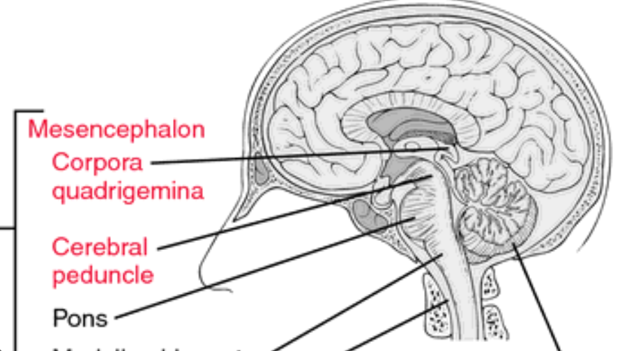
midbrain

parts of the forebrain
cerebrum
diecephalon
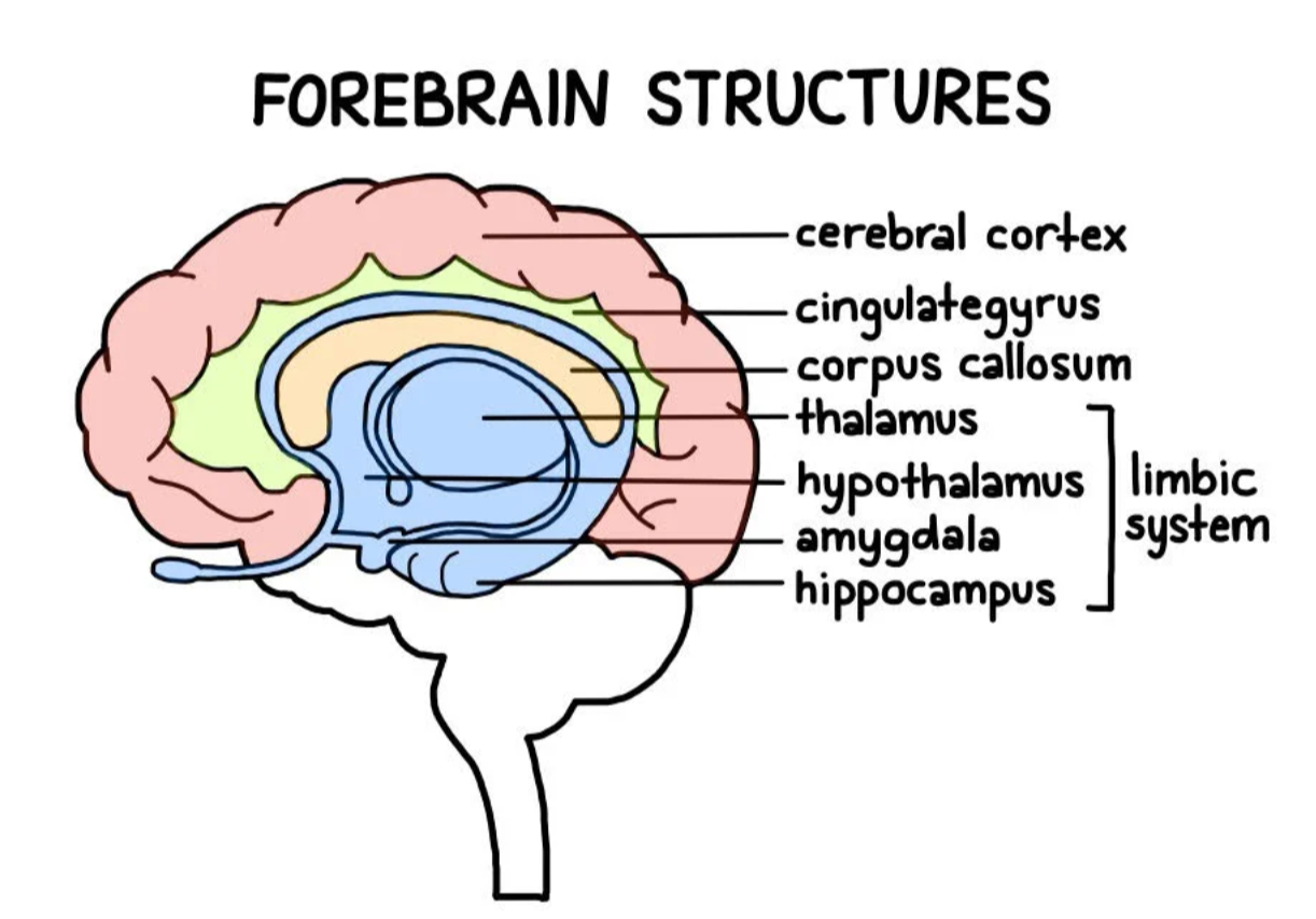
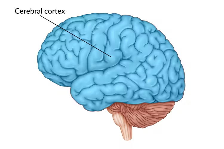
parts of the cerebrum
cerebral cortex
internal structures
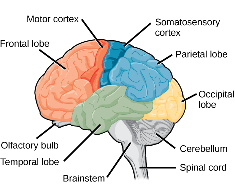
cerebrum → parts of the cerebral cortex
longitudinal fissure
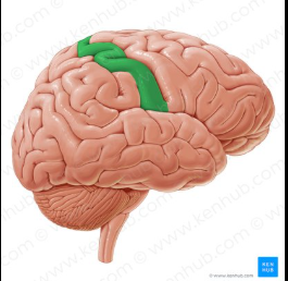
precentral gyrus
central gyrus
postcentral gyrus
lateral sulcus
frontal lobe
parietal lobe
temporal lobe
occipital lobe
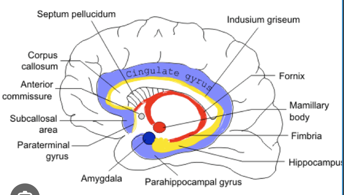
internal structures → parts of the cerebral cortex
corpus callosum
fornix
basal nuclei
septum pellucidum
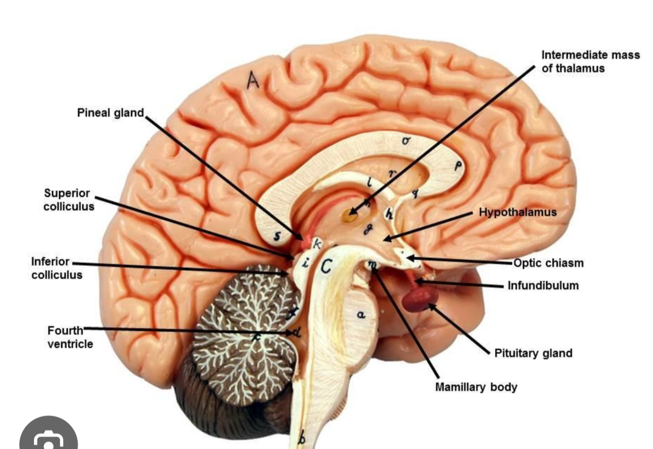
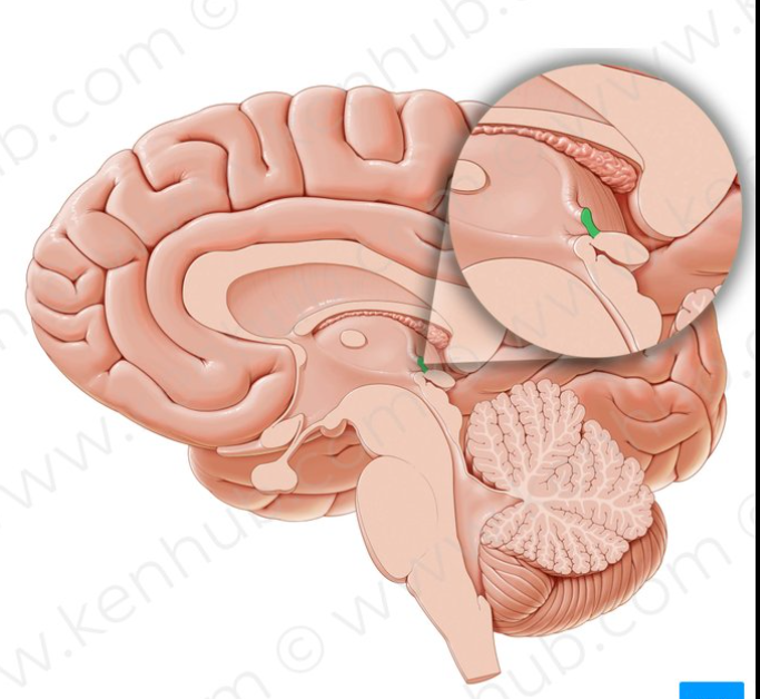
parts of the diecephalon
thalamus
hypothalamus
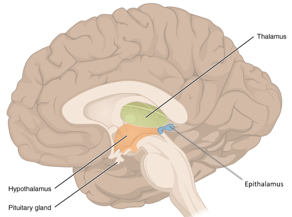
parts of hypothalmus & thalamus
intermediate mass
mammillary bodies
infundibulum
pituitary gland
parts of the midbrain
cerebral penduncles
corpora quadrigemina
sulcus
A shallow depression or groove on the surface of the cerebral cortex
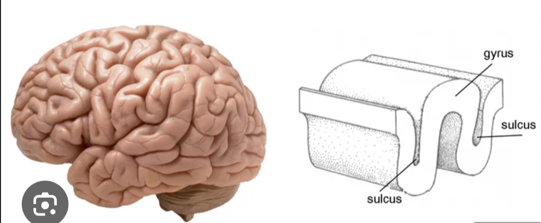
gyrus
The raised ridge or bump between two sulci
fissure
A considerably deeper, larger furrow or cleft than a sulcus
composition of grey matter
unmyelinated neuronal components
glial cells
network of blood vessels
gray matter location
throughout the central nervous system (CNS)
encompasses the brain and spinal cord
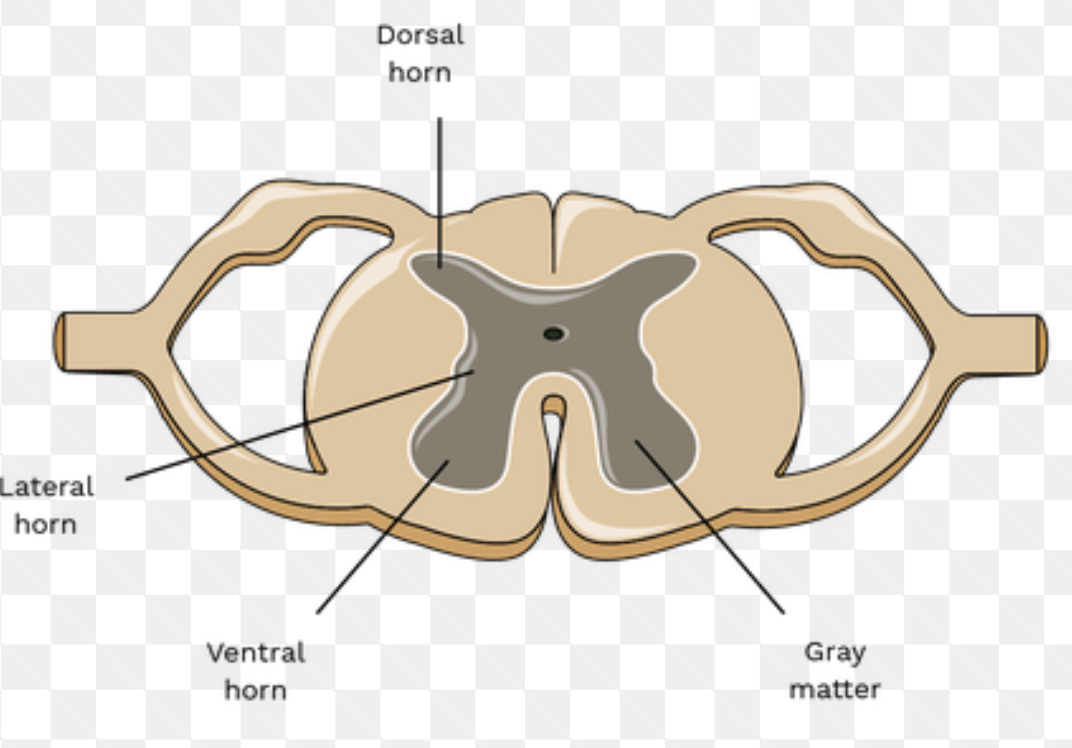
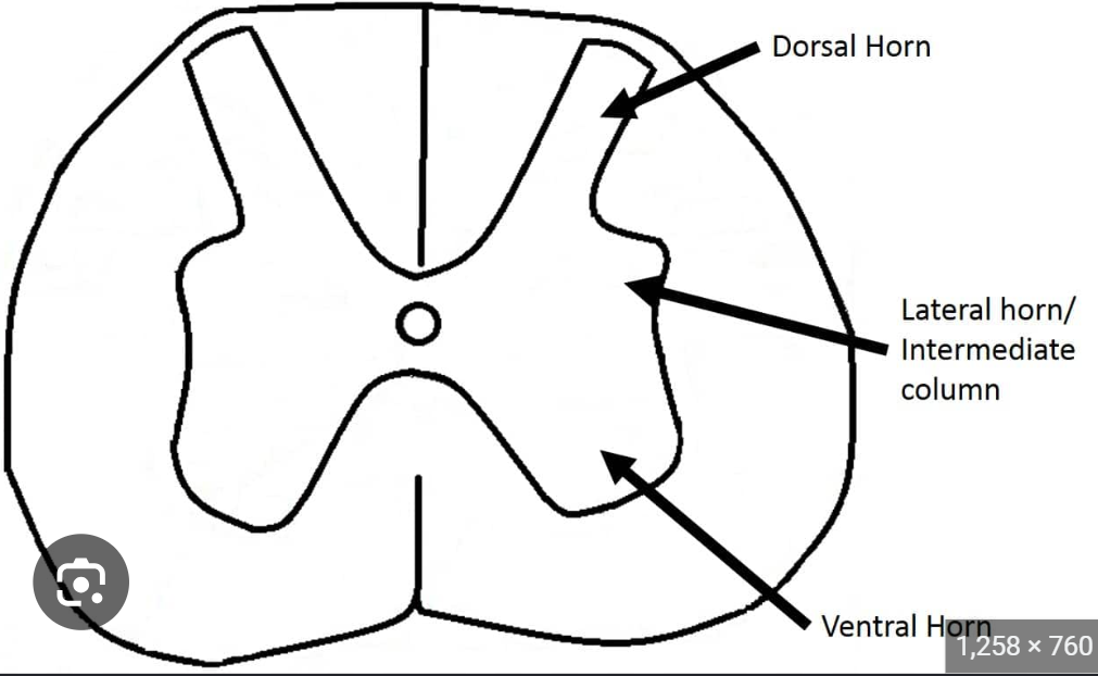
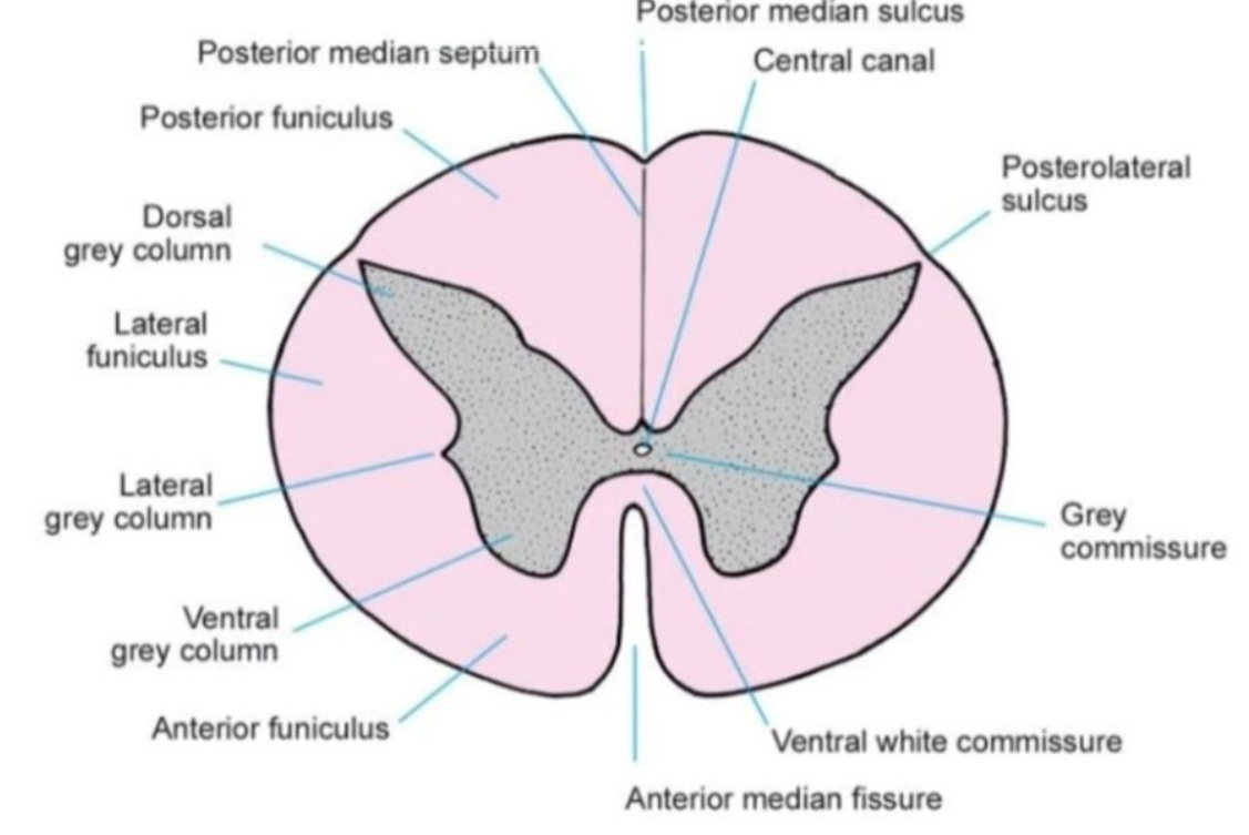
anatomical components of grey matter
central canal
ventral (anterior) horn
laternal horn
dorsal (posterior) horn
anatomical composition of spinal cord
spinal nerve (dorsal + ventral ramus)
dorsal root + ganglion
ventral root
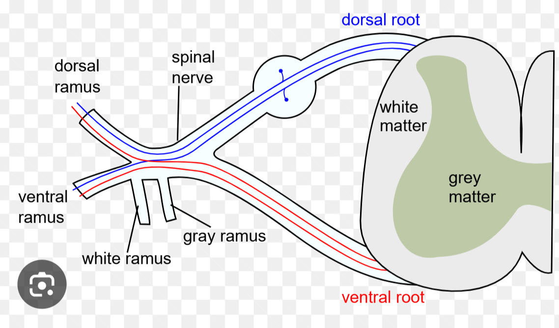
composition of white matter
myelinated bundles of axons
a fatty substance made of proteins and lipids
white matter location
throughout the central nervous system
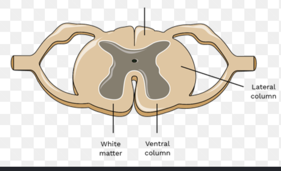
anatomical components of white matter
ventral (anterior) fissure
dorsal (posterior) sulcus
funiculus (ventral, dorsal, lateral)
white & grey matter in brain vs spinal cord
Brain = grey matter on outside & white matter on inside
Spinal Cord = grey matter on inside & white matter on outside
nerve vs tract
NERVE is a bundle of axons in the Peripheral Nervous System (PNS)
TRACT is a bundle of axons in the Central Nervous System (CNS)
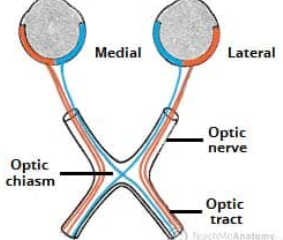
commissure
anatomical location where two structures or tissues join or meet
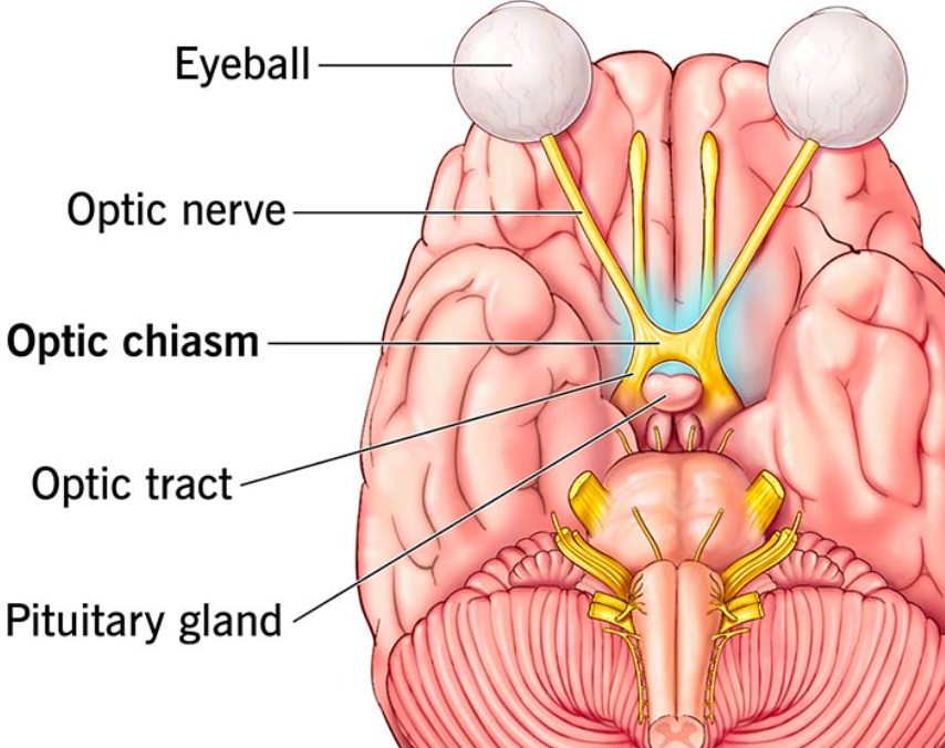
optic chiasm
an X-shaped neural structure where the optic nerves intersect
cranial meninges
three protective tissue membranes that envelop and support the brain within the skull
3 cranial meninges
dura mater
arachnoid mater
pia mater
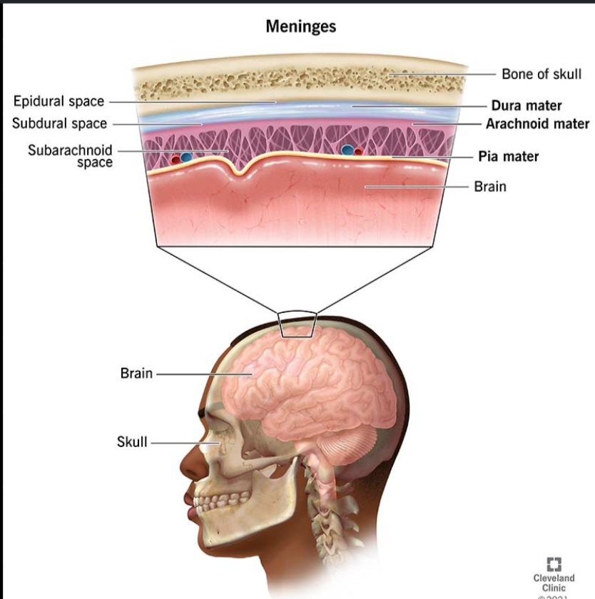
dura mater
the outermost, thickest, and most durable of the meninges
protect the brain and spinal cord

arachinoid mater
the middle, spiderweb-like layer of the meninges
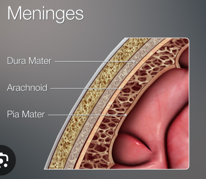
pia mater
the delicate, innermost layer of the meninges
acts as a protective barrier and blood vessel network for the CNS

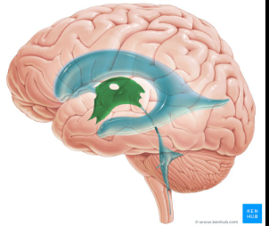
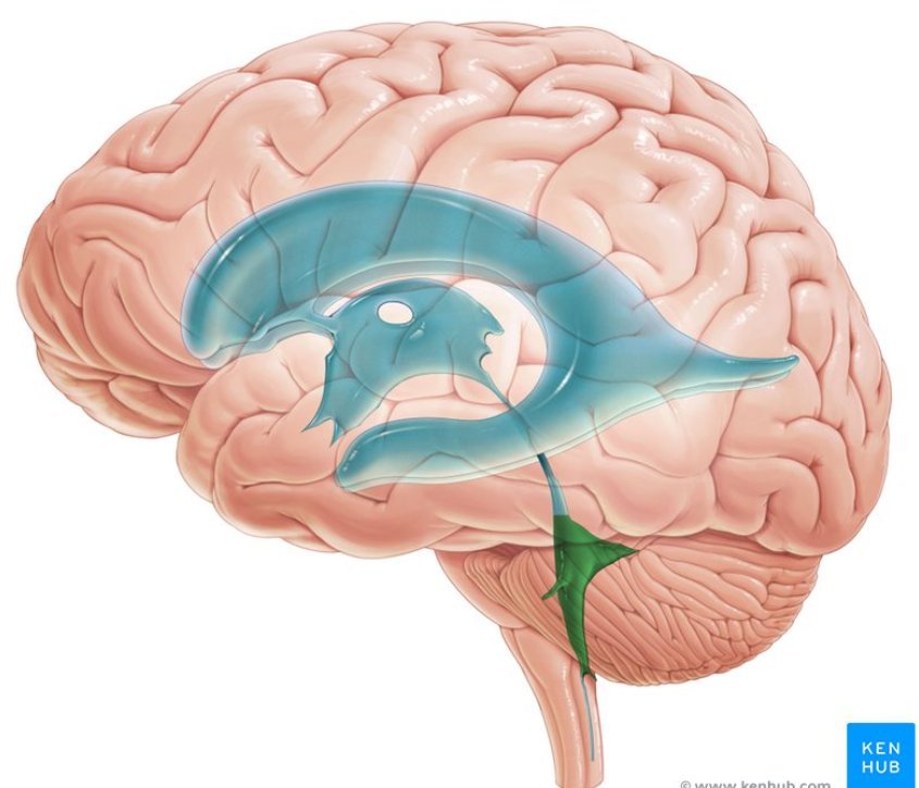
ventricles of the brain
lateral ventricle
choroid plexus
third ventricle
fourth ventricle
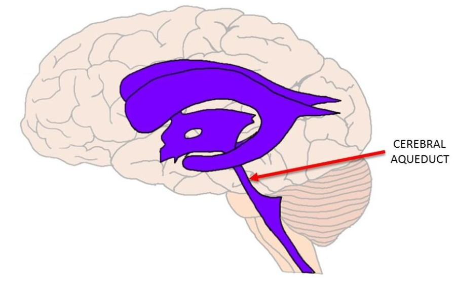
cerebral aqueduct (communicate btw 3rd ad 4th ventricles)
lateral ventricle
C-shaped cavities located within the cerebral hemispheres of the brain
responsible for production, circulation, and containment CSF to protect the brain
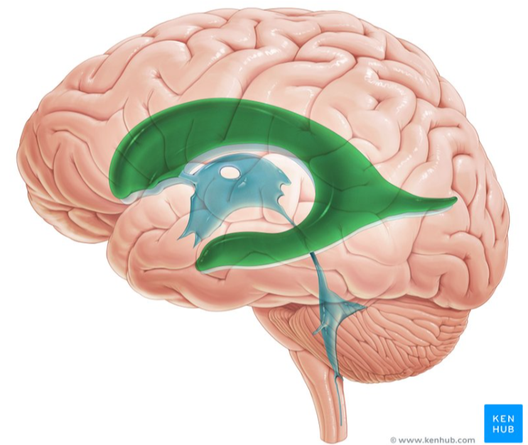
choroid plexus
network of specialized cells and blood vessels located within the brain's ventricles
produce cerebrospinal fluid (CSF) and form the blood-CSF barrier
third ventricle
situated between the right and left halves of the thalamus and hypothalamus
produces, cushions, and circulates cerebrospinal fluid (CSF)
fourth ventricle
located between the brainstem and the cerebellum
a crucial passageway that allows CSF to flow out brain's internal cavities and into the protective spaces around the CNS
cerebral acqueduct
a narrow, fluid-filled tube located in the midbrain
allows CSF to flow directly from the third ventricle to the fourth ventricle
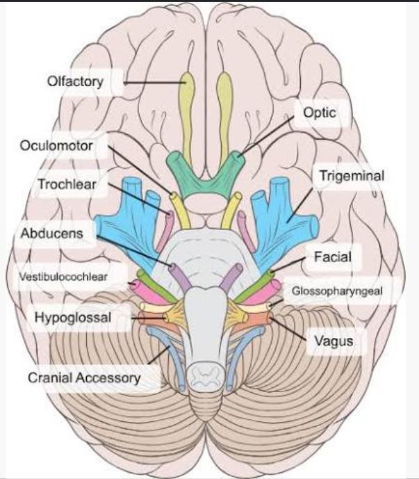
CN I
olfactory nerve
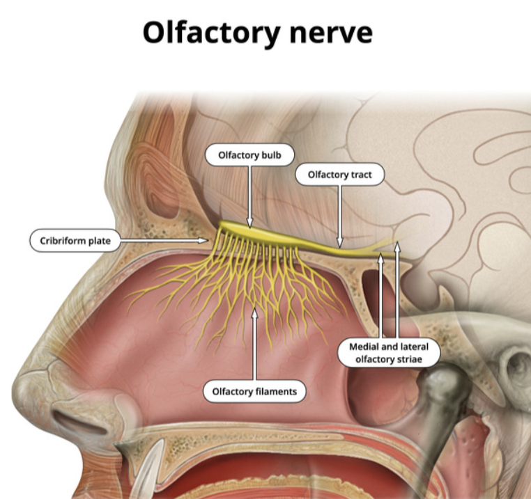
CN I function
transmit sensory info about smell and odors
CN II
optic nerve
CN II function
transmit sensory visual info from retina to brain
CN III
oculomotor nerve

CN III function
control eye movement, pupil size, and shape
CN IV
trochlear nerve
CN IV function
control superior oblique muscles of the eye
CN V
trigeminal nerve

CN V function
give sensory info to face, scalp, teeth and mouth for chewing
CN VI
abducens nerve

CN VI function
control lateral rectus muscles of the eye
CN VII
facial nerve

CN VII function
controlling motor, sensory and parasympethetic functions
2 branches of CN VIII
vestibular branch
cochlear branch

vestibular brach (CN VIII)
transmit vital spacial, gravitational, and movement signals from the inner ear to the brain
cochlear branch (CN VIII)
transmit auditory signals to the brain
CN IX
glossopharyngeal nerve

CN IX function
swallowing, taste, salivation, and gag reflex
CN X
vagus nerve

CN X function
heart rate, digestion, and immune reponses
CN XI
accessory nerve

CN XI function
control nexk and shoulder movement, and vocal cord function
CN XII
hypoglassal nerve

CN XII function
control all movement of the tongue
what cranial nerves DO NOT exit from the brainstem
CN I → olfactory nerve
CN II → optic nerve
what nerves provide parasympathetic innervation to a target
CN III → oculomotor nerve
CN VII → facial nerve
CN IX → glossopharyngeal nerve
CN X → vagus nerve
components of the CNS
brain
spinal cord
what system is a “nerve” a part of
the PNS
where does a nerve begin
as it enters through the intervertebral foramen
spinal nerve type
a MIXED nerve
contains both sensory & motor components
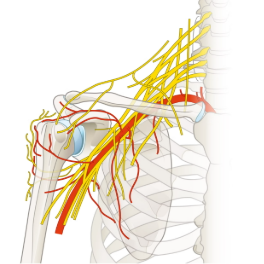
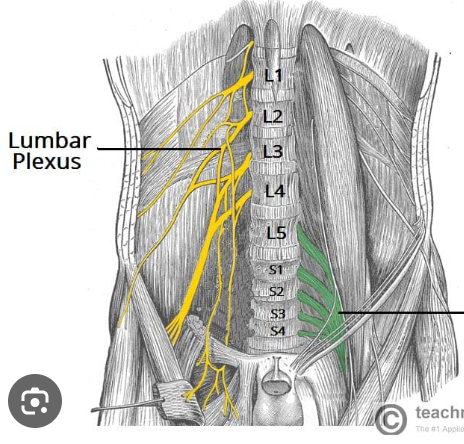
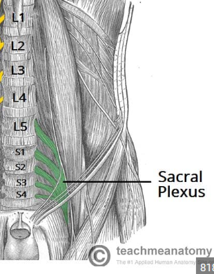
4 spinal plexuses
cervical plexus
brachial plexus
lumbar plexus
sacral plexus
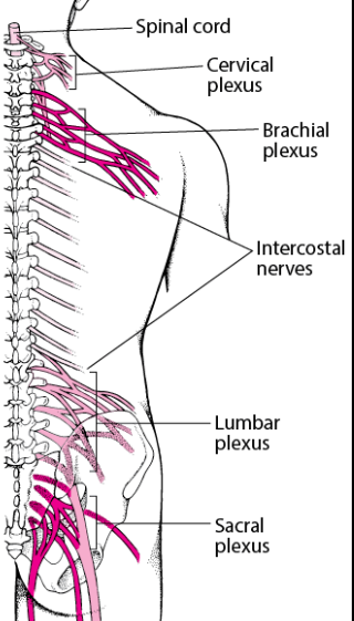
cervical plexus
innervates upper body (head, neck, shoulders, diaphragm)
major nerve = PHRENIC NERVE
brachial plexus
innervates the upper limbs
major nerve = MEDIAN NERVE
lumbar plexus
innervates the thigh
major nerve = FEMORAL NERVE
sacral plexus
innervates the lower limb and glutes
major nerve = SCIATIC NERVE
frog dissection → corneal reflex
still works even when forebrain removed
DOES NOT work when hindbrain is removed
DOES NOT work when spinal cord is cut
frog dissection → swimming movement
still works even when forebrain removed
DOES NOT work when hindbrain is removed
DOES NOT work when spinal cord is cut
frog dissection → posture
still works even when forebrain removed
DOES NOT work when hindbrain is removed
DOES NOT work when spinal cord is cut
frog dissection → breathing
still works even when forebrain removed
DOES NOT work when hindbrain is removed
DOES NOT work when spinal cord is cut
frog dissection → tongue muscle tone
still works even when forebrain removed
DOES NOT work when hindbrain is removed
DOES NOT work when spinal cord is cut
frog dissection → righting reflex
still works even when forebrain removed
DOES NOT work when hindbrain is removed
DOES NOT work when spinal cord is cut
frog dissection → acid reflex
still works even when forebrain removed
STILL WORKS even when hindbrain is removed
DOES NOT work when spinal cord is cut
frog dissection → withdrawl reflex
still works even when forebrain removed
STILL WORKS even when hindbrain is removed
DOES NOT work when spinal cord is cut
pupil constriction controlled by
parasympathetic nervous sytems
uses acetycholine
effector mediating pupil constriction
sphincter pupillae of the iris
pupil dilation controlled by
sympathetic nervous system
uses epi/norepi
effector mediating pupil dilation
dilator pupillae of the iris
types of spinal reflex tests
patellar (knee-jerk) reflex test
achilles tendon (ankle-jerk) reflex
what are spial reflexes also called
stretch reflexes
because the hammer tap stretches a muscle/tendon
concidered protective readjustments because the response prevents muscle/tendon overstretching
patellar (knee-jerk) reflex test
this test assesses spinal motor neurons in L2-L4
a rapid, involuntary stretch reflex
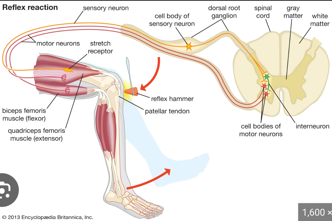
patellar (knee-jerk) reflex test COMPONENTS
stimulus → tap on patellar tendon with a hammer
receptor → muscle spindles in quadricep
effector → quads recieve signal from motor neurons to stretch
response → kicking/extending leg motion
hyporeflexia
less than normal response to patellar tendon tap
hyperreflexia
more than normal response to patellar tendon tap
achilles tendon (ankle-jerk) reflex
assesses spinal motor neurons at S1 level
deep tendon reflex
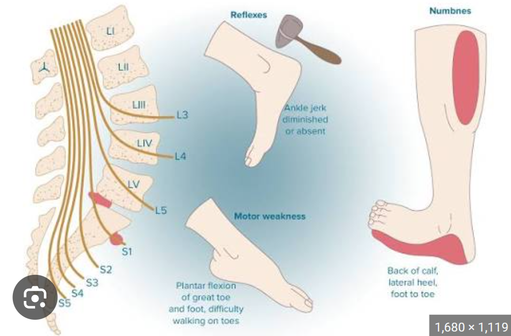
achilles tendon (ankle-jerk) reflex COMPONENTS
stimulus → tap to achilles tendon with hammer
receptor → muscle spindles in calf muscles detech sudden stretch and fire signals
effector → gastrocnemius + soleus muscles recieve signal and carry out movement
response → involuntary, rapid contraction of calf muscles which cause foot to jerk downward
general sensory tests
two-point descrimination test
adaption of tactile pressure receptors
adaptation of temperature (thermal) receptors
radiating pain
two-point descrimination test
assesses the sensitivity of different skin areas by asking subjects if they feel two objects or one, on different areas of the skin
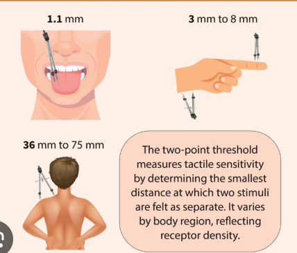
two-point descrimination test SENSORY PATHWAYS
ascending sensory afferent pathway
dorsal column-medial lemniscus pathway
1st, 2nd, 3rd order neurons
areas MOST SENSITIVE in two-point descrimination test
fingertips
lips
tongue
adaptation of tactile pressure receptors test
uses classic constant stimulus coin method
seeing how long it takes for the brain to ignore a constant, steady pressure
adaptation of tactile pressure receptors test MECHANISM
measures how quickly rapidly adapting receptors like Meissners and Pacinian corpuscles stop firing when a stimulus remains constant
slowly adapting receptors (like Merkel discs) continue to fire
cutaneous receptors activated at ONE COIN
uses superficial, rapidly adapting mechanoreceptors because the pressure is minimal (less time to adapt)
EX: meissners corpuscles
cutaneous receptors activated at FOUR COINS
increased pressure deforms deeper mechanoreceptors because its a new/stronger stimulus (longer duration to adapt)
EX: pacinian or ruffini corpuscles
sensory adaptation
an automatic process which our sensory receptors become less respnsive to constant, unchanging stimului
allows brain to filter inputs to focus attention to novel or threatening changes
adaptation of temperature (thermal) receptors test
shows how sensory nerves adjust to a constant stimulus
overtime the receptors reduce theie firing rate, causing hot and cold sensationg to fade
adaptation of temperature (thermal) receptors test RESULT
aone hand was submerged in hot water and the other in cold water for 2 min and then you put both in room-temp water
the hot water hand will feel COLD in room-temp water
the cold water hand will feel HOT in room-temp water