BIOL229: Lab Final
 Call Kai
 Learn
Learn Practice Test
 Spaced Repetition
 Match
Match Flashcards
 Knowt Play
1/345

There's no tags or description
Looks like no tags are added yet.
Name | Mastery | Learn | Test | Matching | Spaced | Call with Kai | Chat |
|---|
No analytics yet
Send a link to your students to track their progress
346 Terms
total magnification
objective lens magnification x ocular lens magnification
field of view
what you can see through the oculars at a given magnification
does increasing magnification increase or decrease the field of view?
decrease
working distance
space between objective lens and slide
coarse focus knob
moves stage large distances; only used on scanning lens
fine focus knob
used to bring an object back into focus after switching magnification
parfocalization
being able to switch magnification after focusing on an object and still remain nearly in focus
refraction
when light passes through a medium into another, light rays often bend
immersion oil
used with 1000x objective because the oil has same index of refraction as glass; improves resolution
prokaryotic cells
very simple and lack a nucleus or membrane bound organelles and are small in size
eukaryotic cells
more complicated; contain a nucleus and many specialized organelles
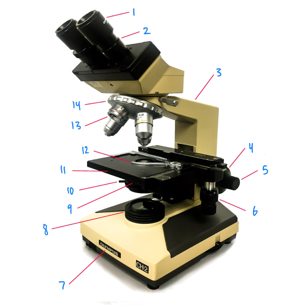
1
ocular eyepiece
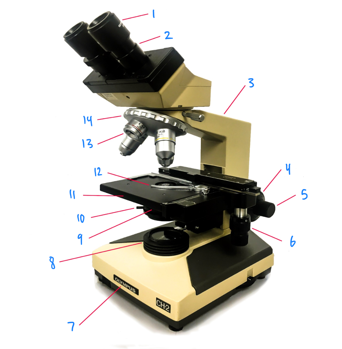
2
observation tube
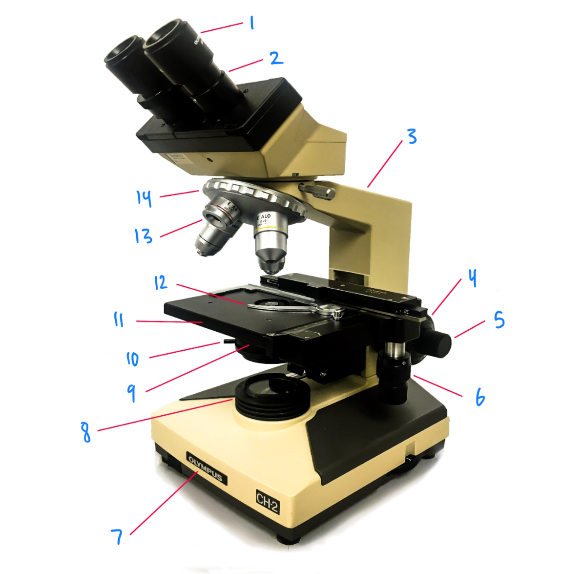
3
arm
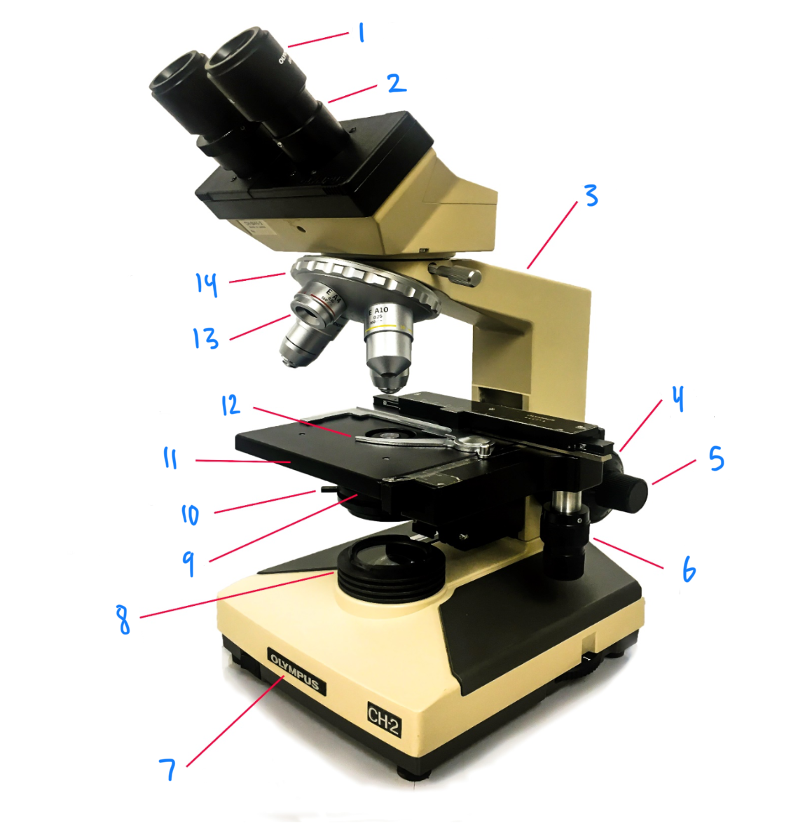
4
coarse focus knob
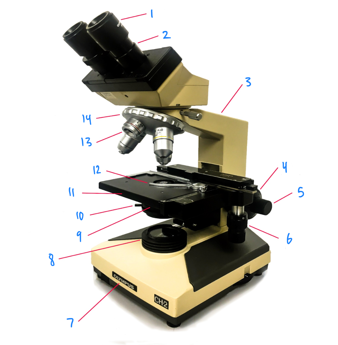
5
fine focus knob
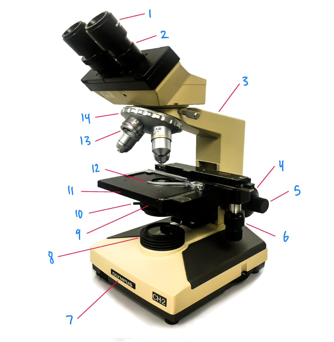
6
stage control knob
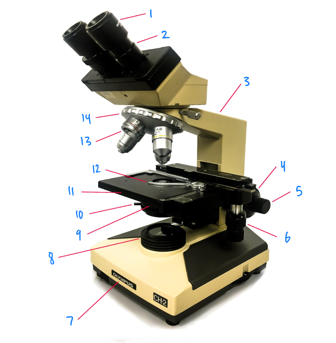
7
base
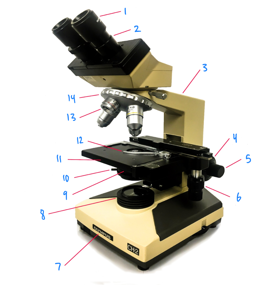
8
light source
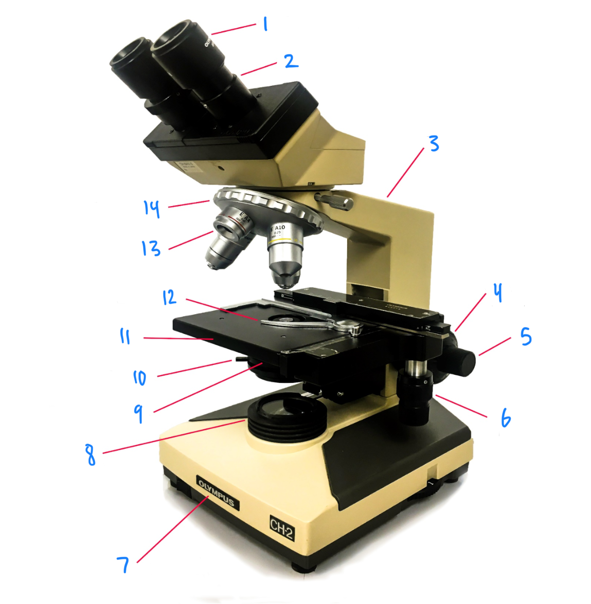
9
condenser lens
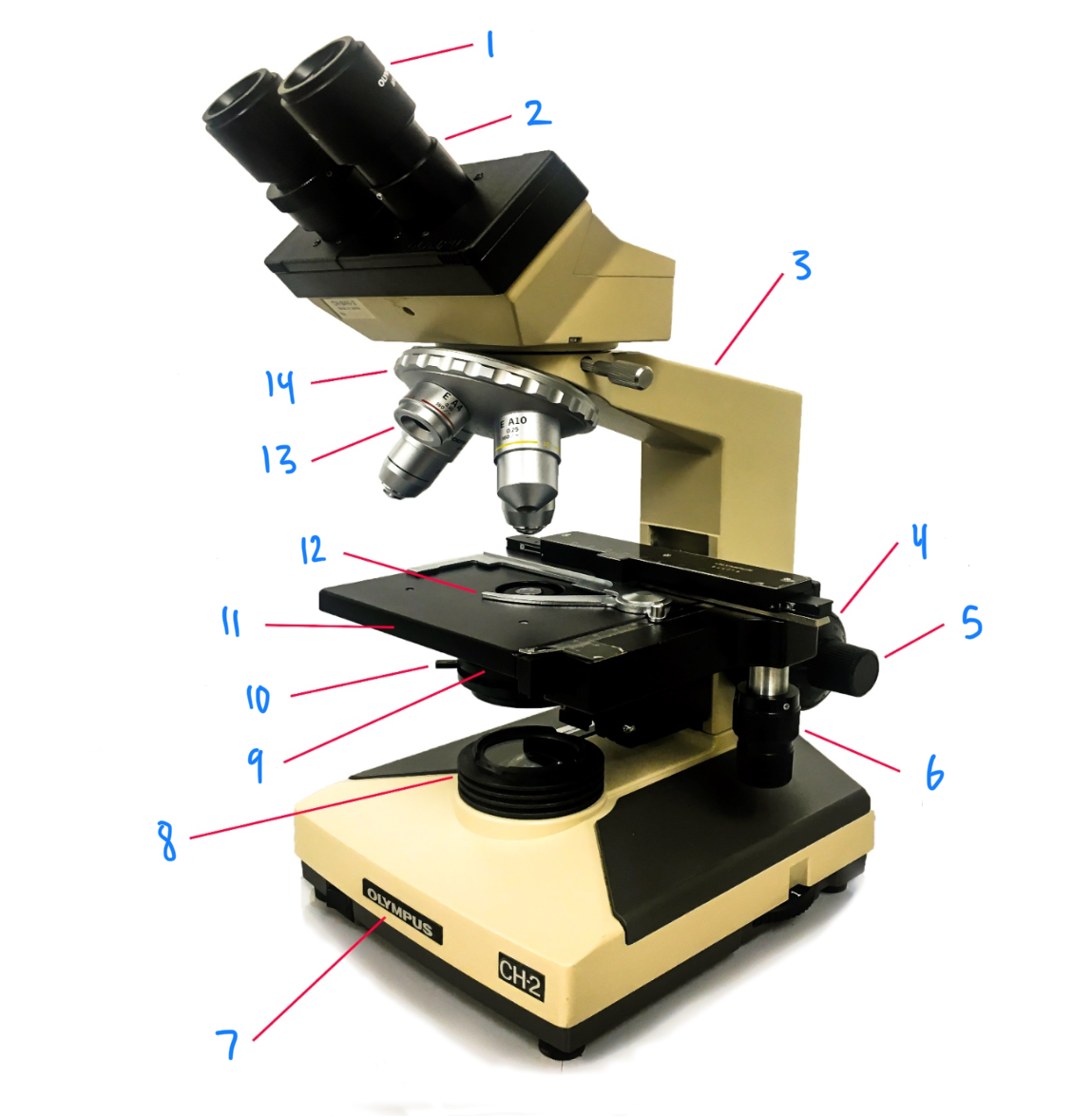
10
iris diaphragm
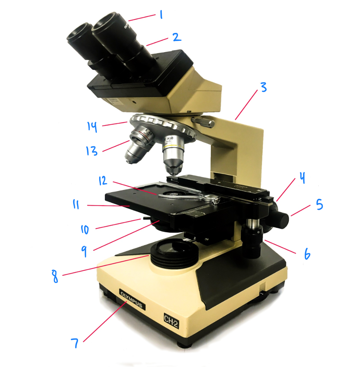
11
stage
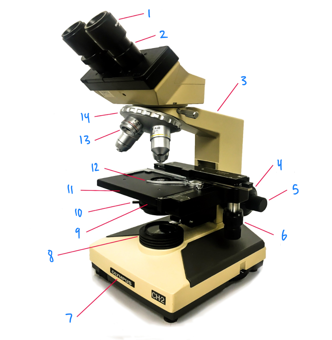
12
stage clip
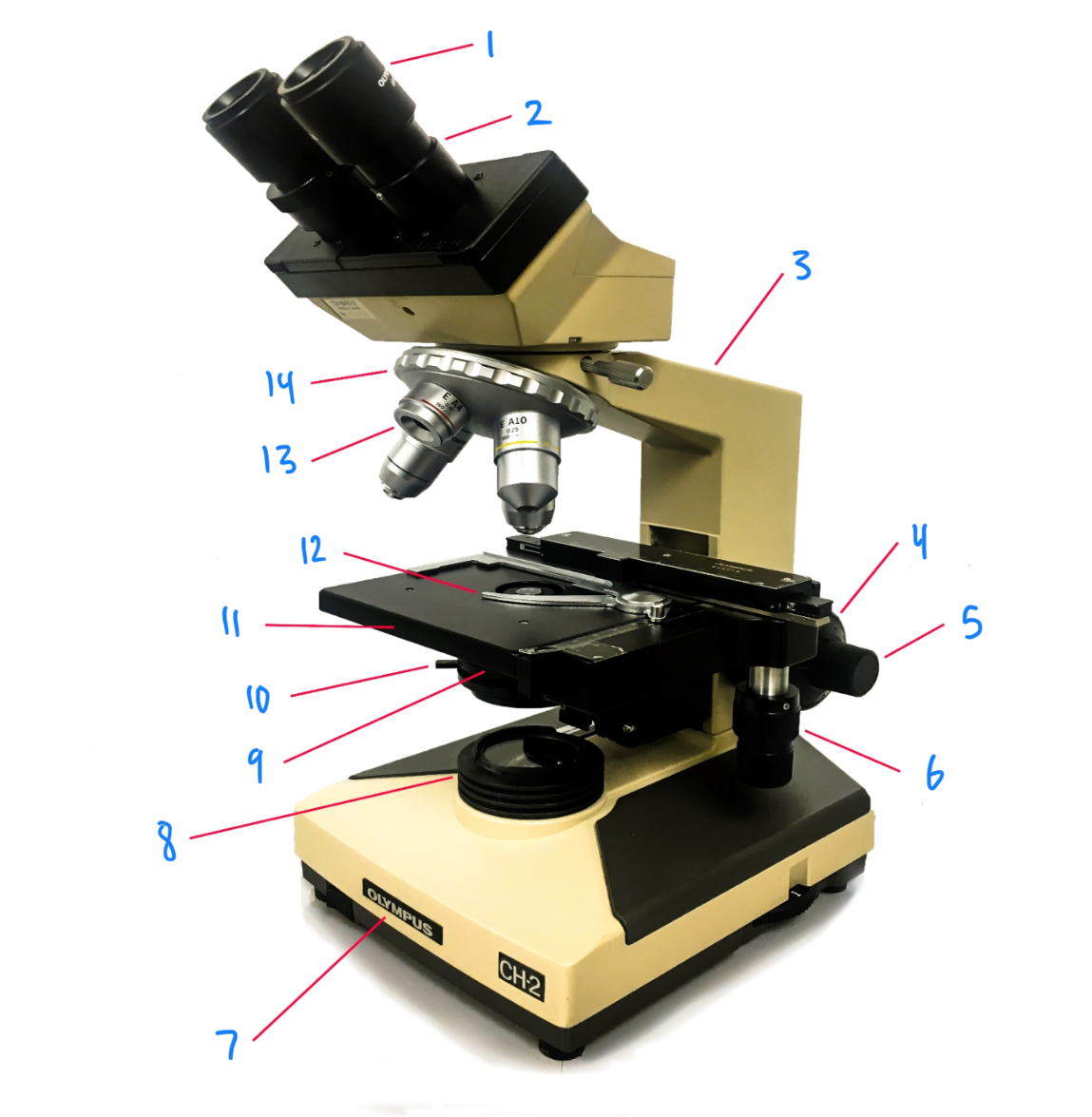
13
objective lens
14
nosepiece
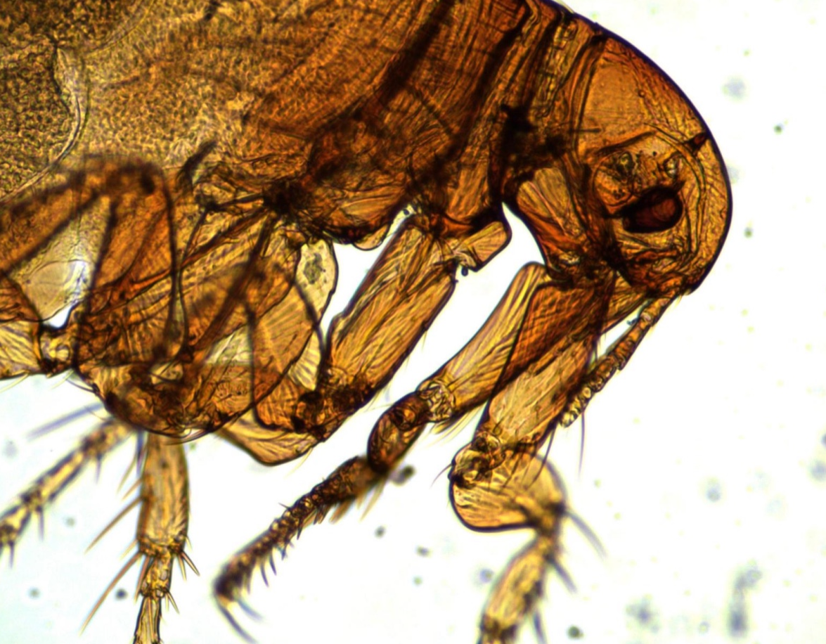
flea
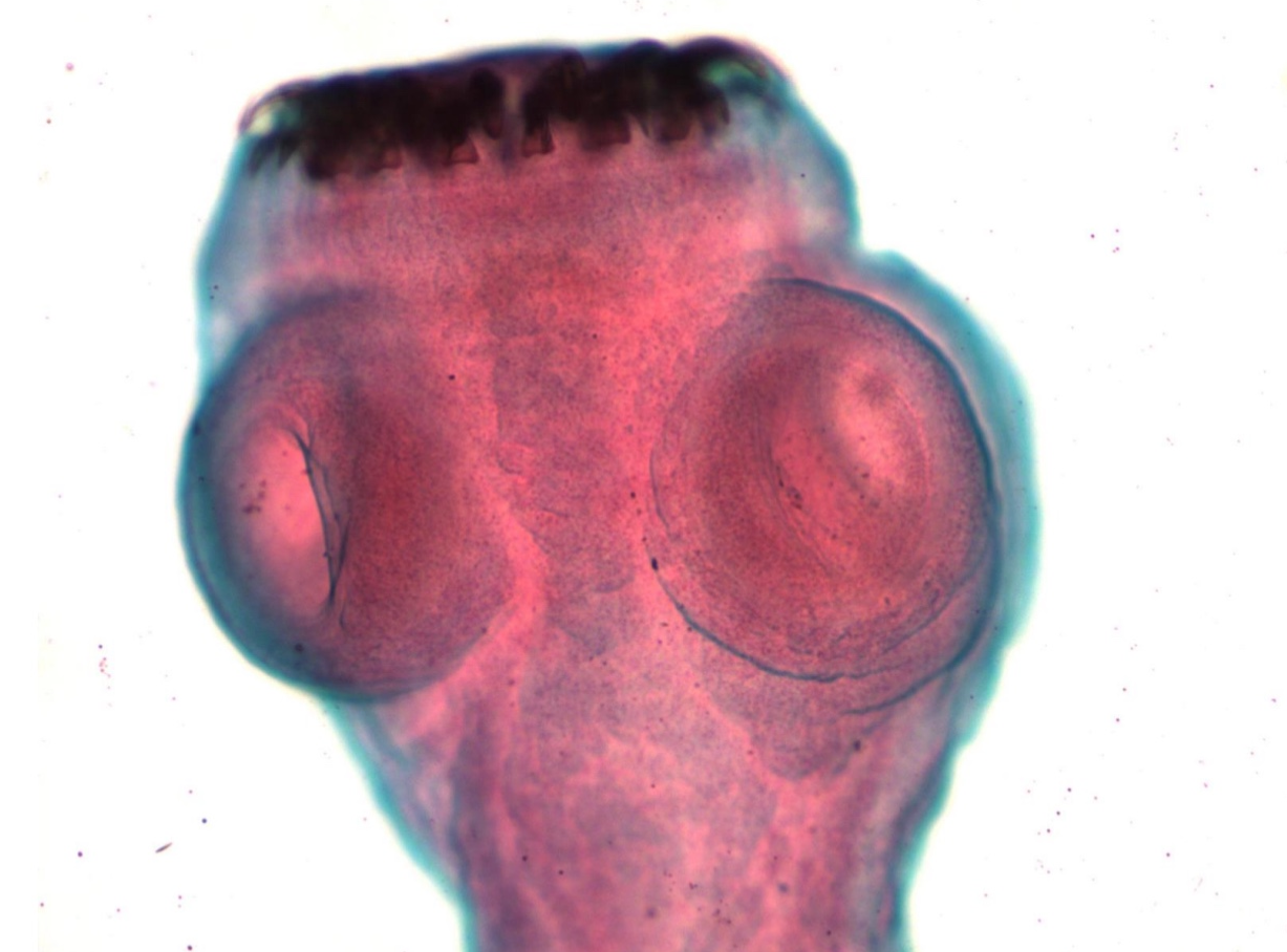
*note hooks
tapeworm scolex
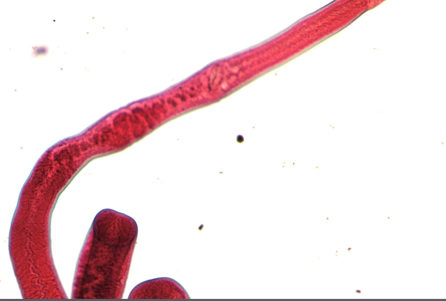
Schistosoma adult worm
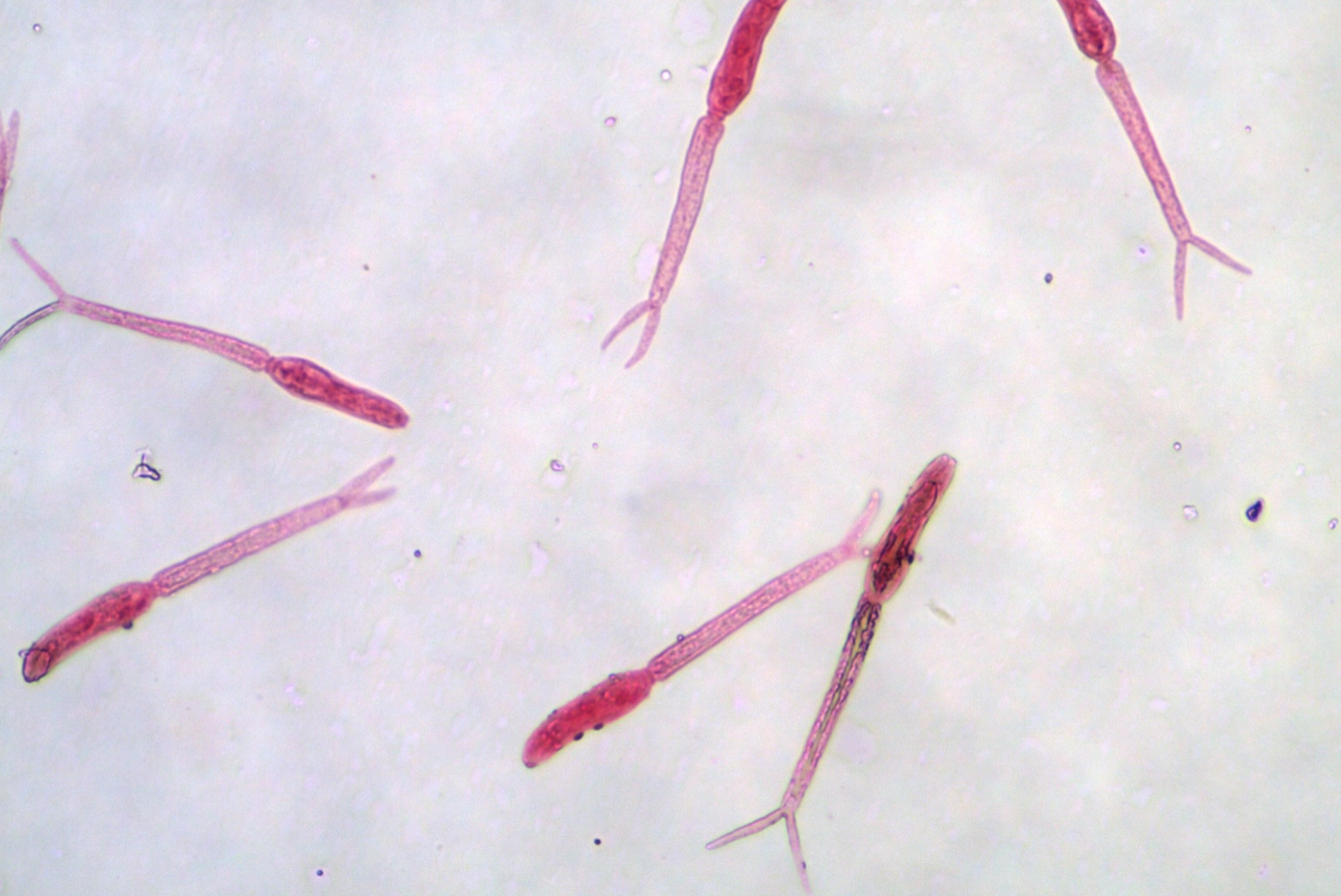
Schistosoma cercariae
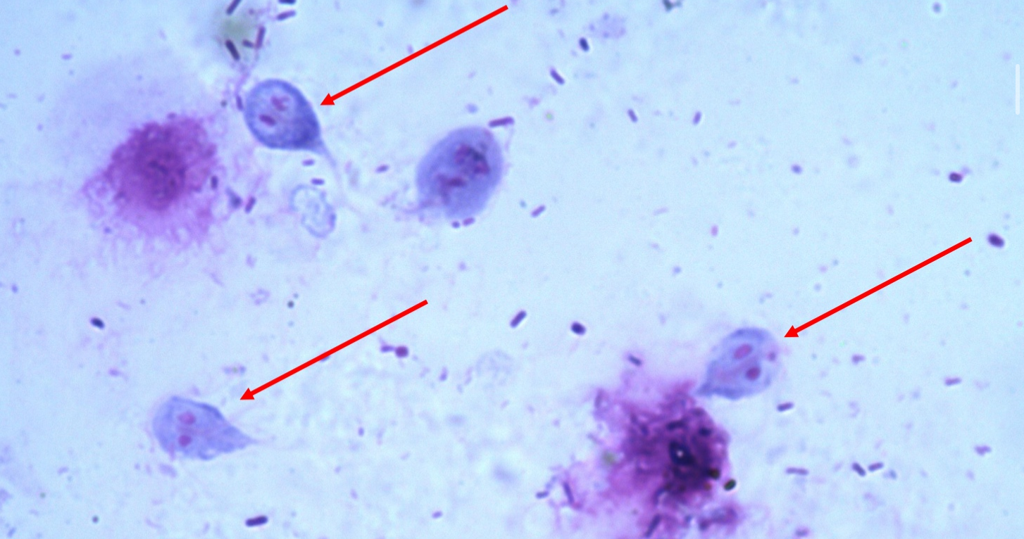
Giardia trophozoites
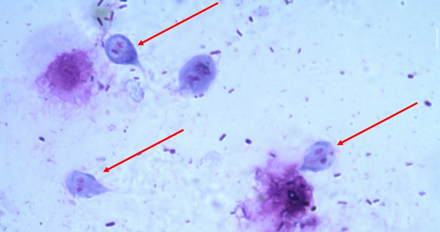
“smiley face” - 2 nuclei
trophozoites
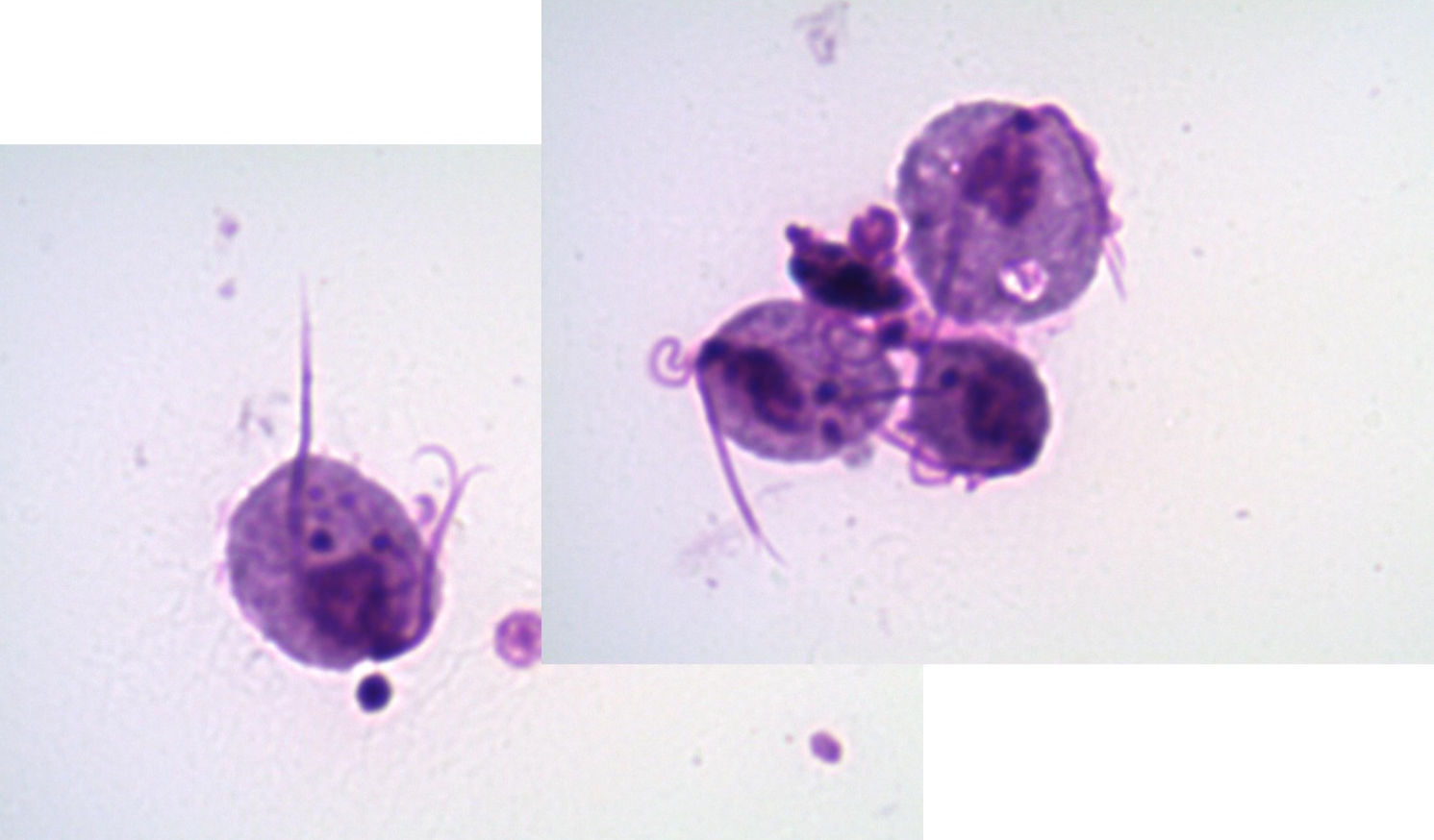
*note flagella, nucleus
Trichomonas vaginalis
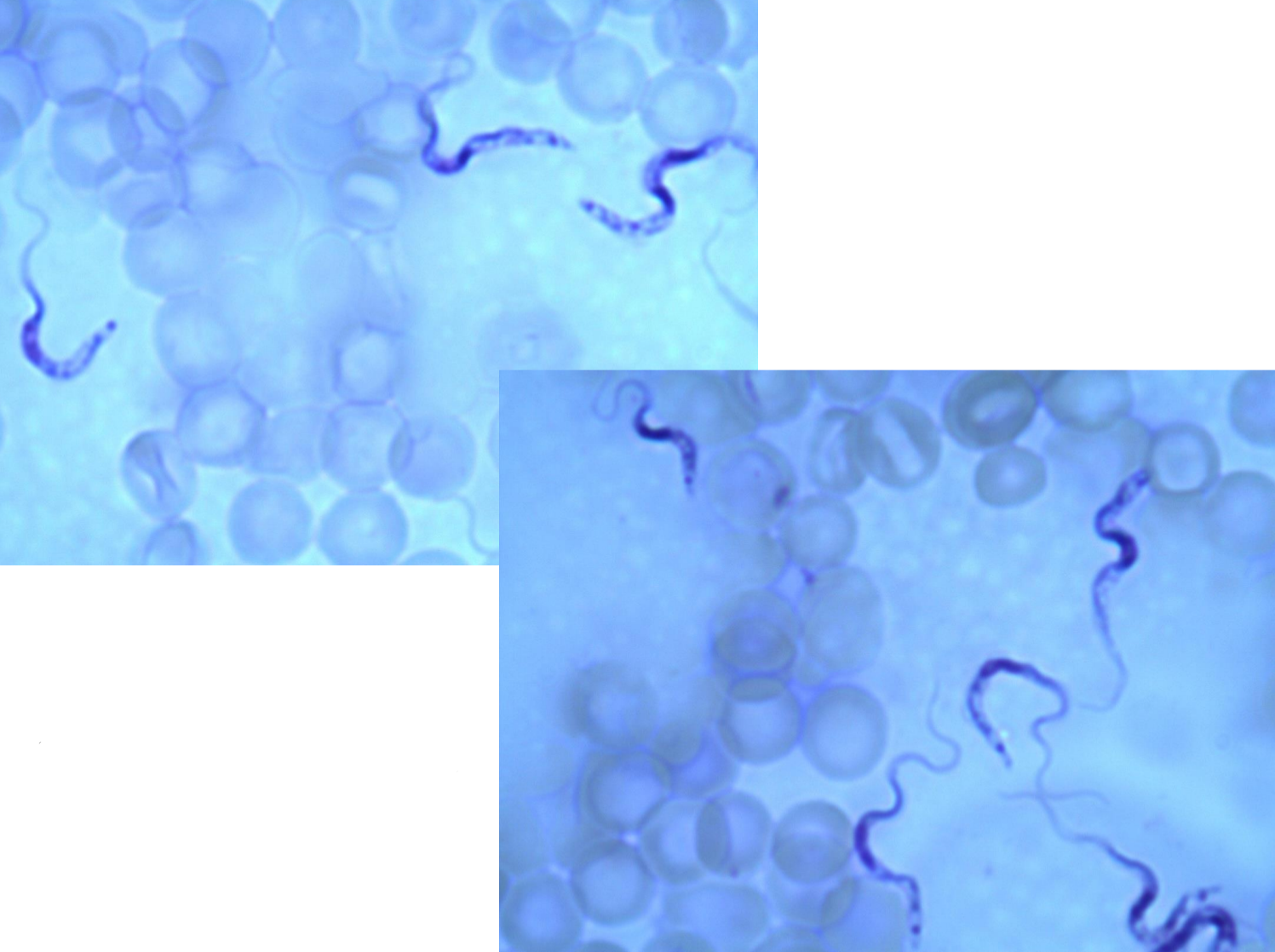
*note red blood cells, parasite, nucleus, flagella
Trypanosoma
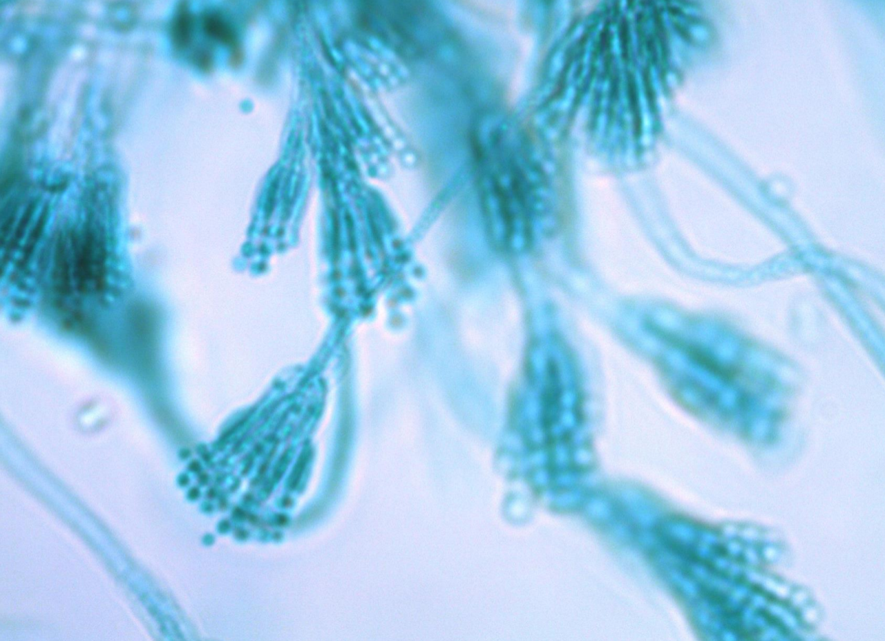
Penicillium
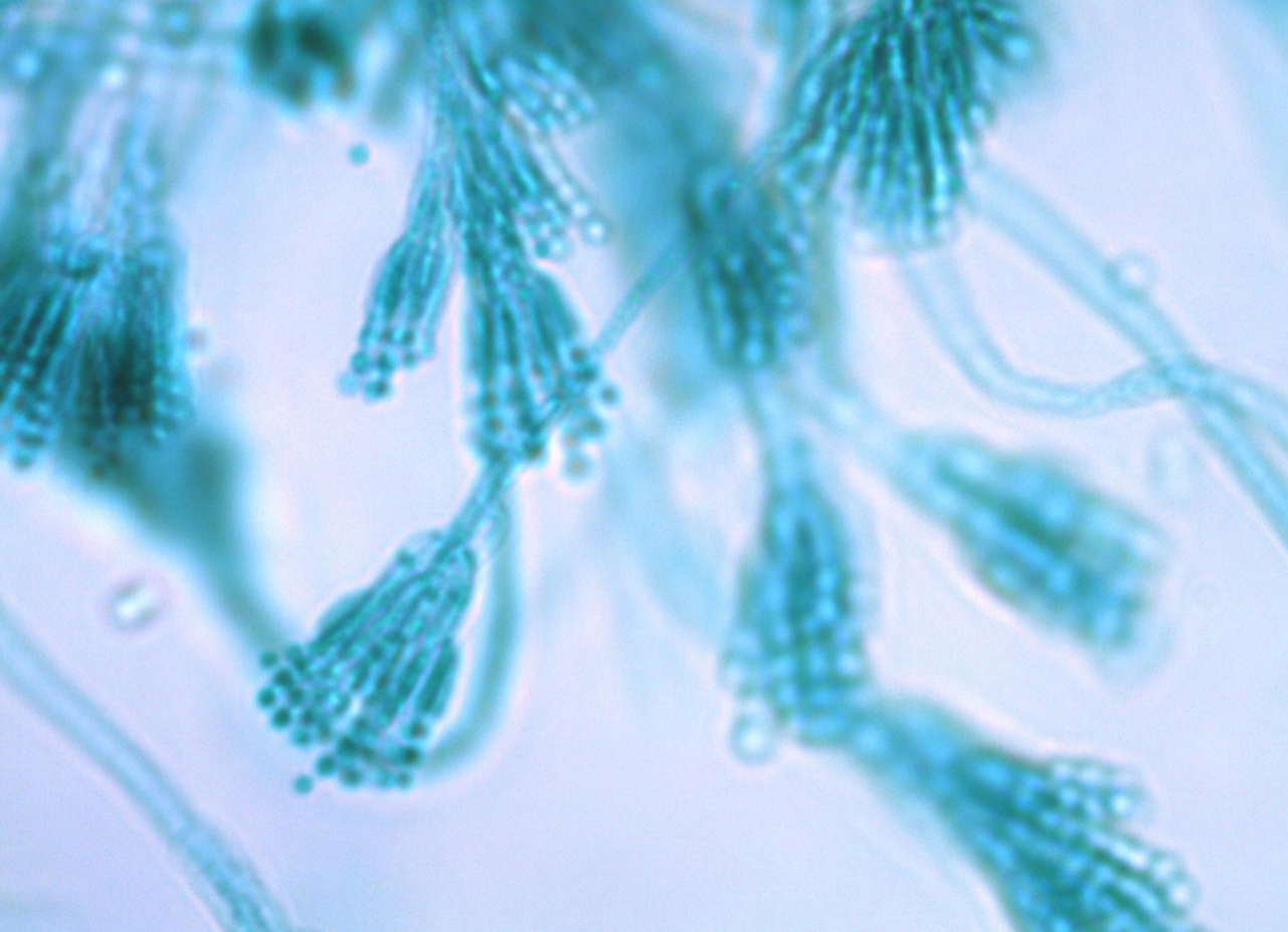
little circles/dots
conidia (spores)
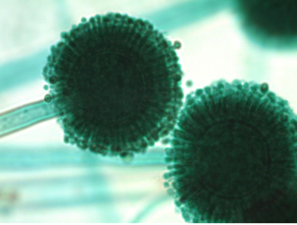
Aspergillus
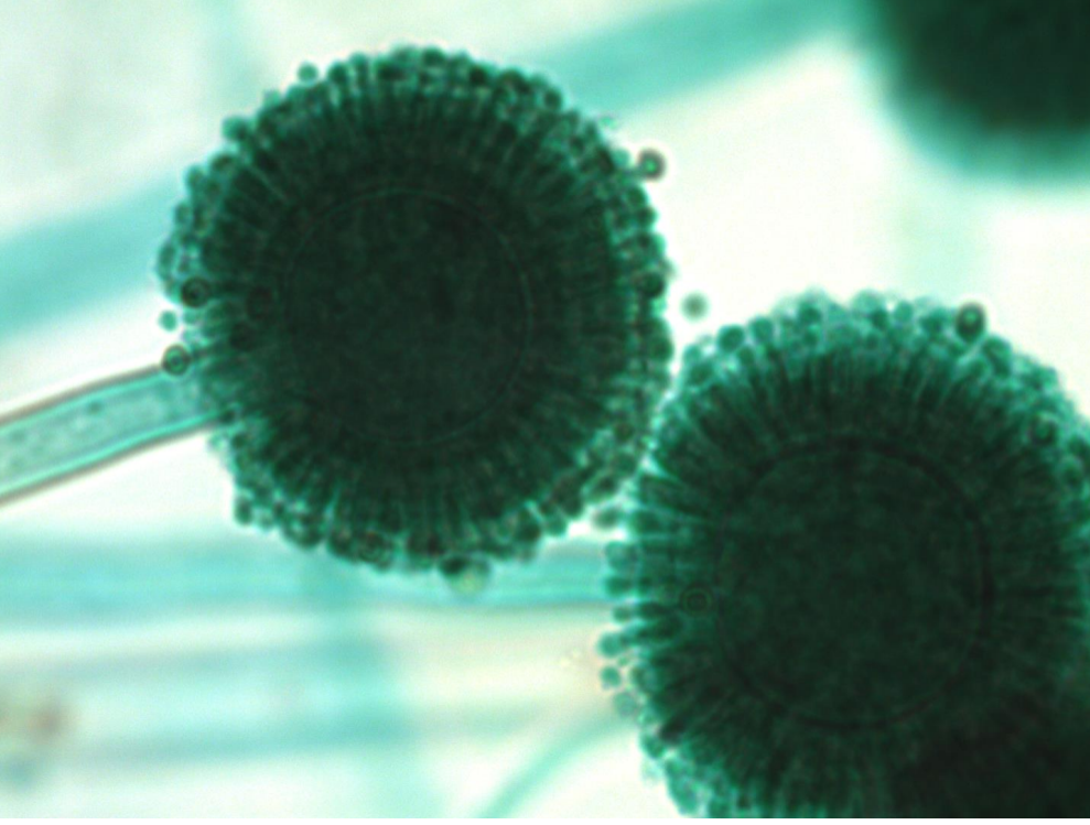
little circles/dots
conidia (spores)
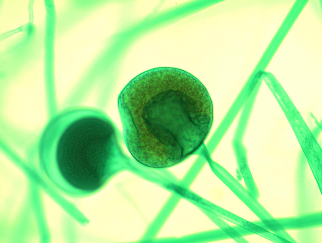
Rhizopus sporangia
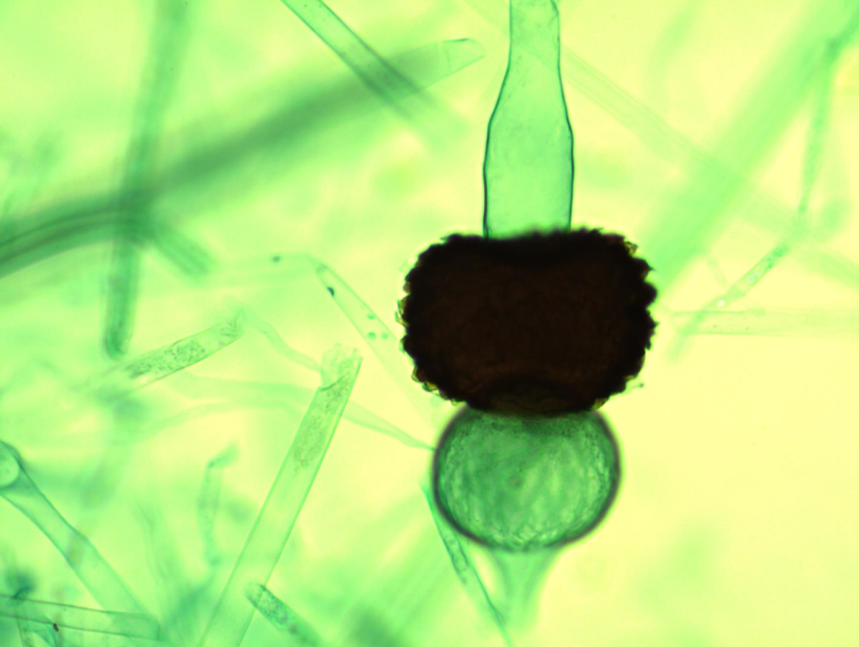
Rhizopus zygospore
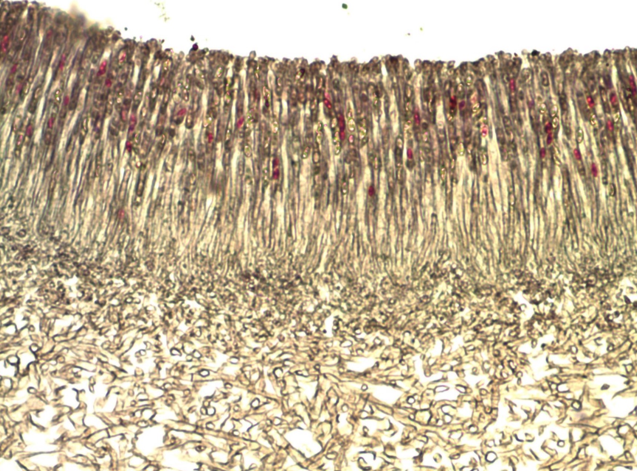
Peziza
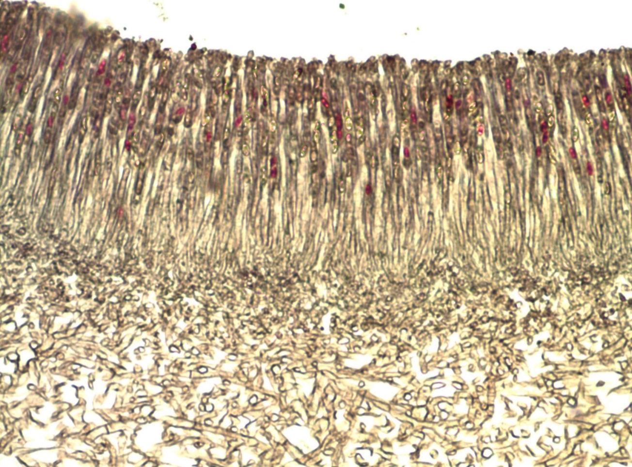
area surrounding red dots
ascus
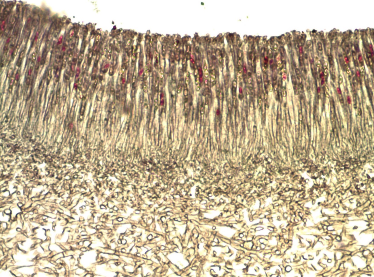
red dots
Ascospores
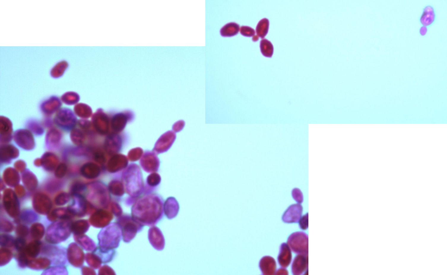
*note budding cells
Saccharomyces
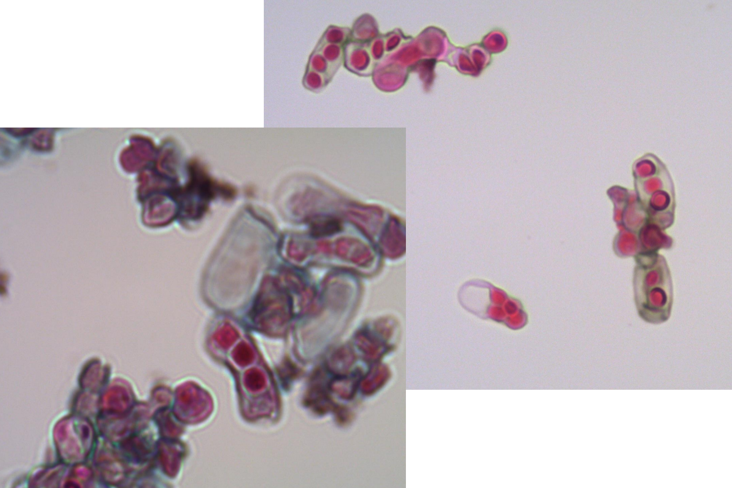
Schizosaccharomyces
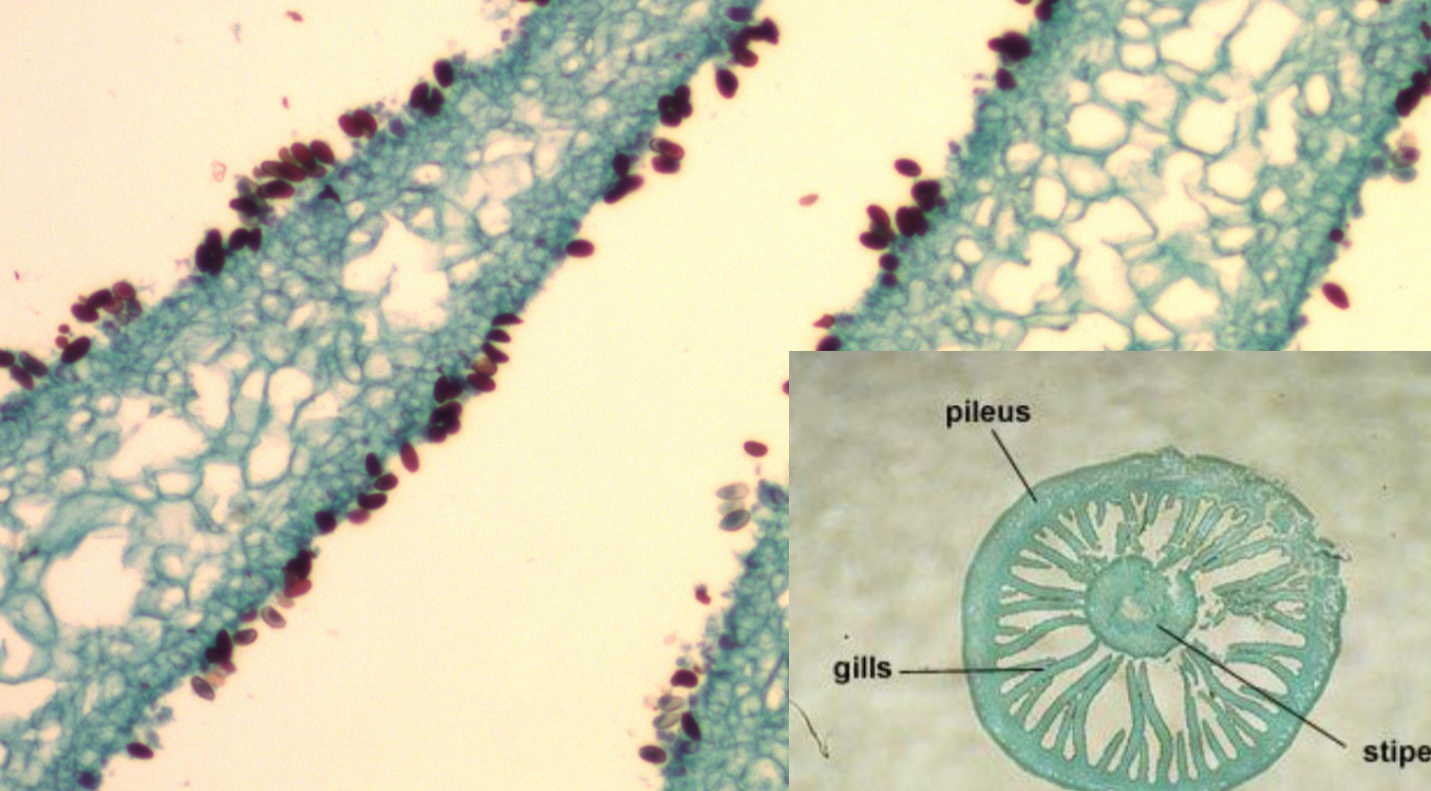
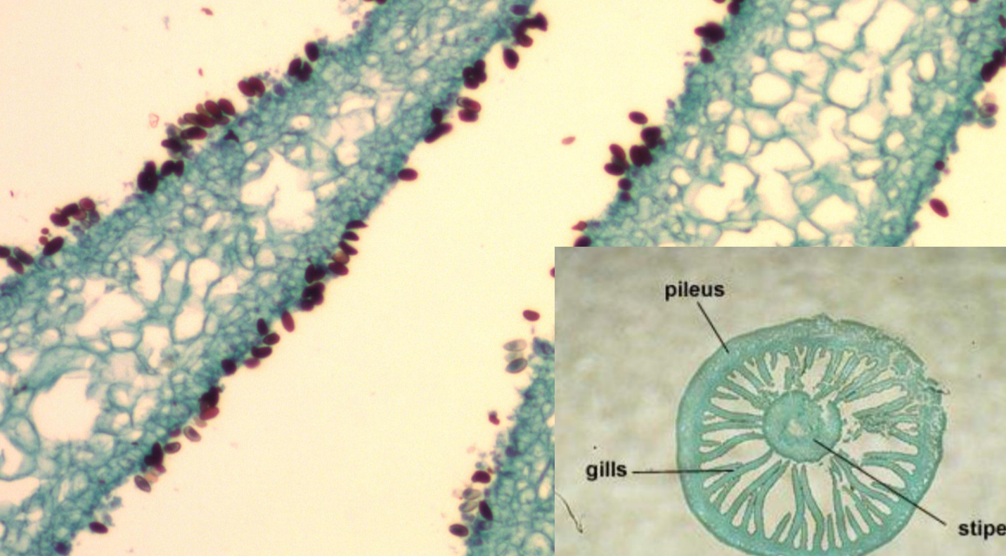
*note gills
Coprinus
red dots
basidiospores
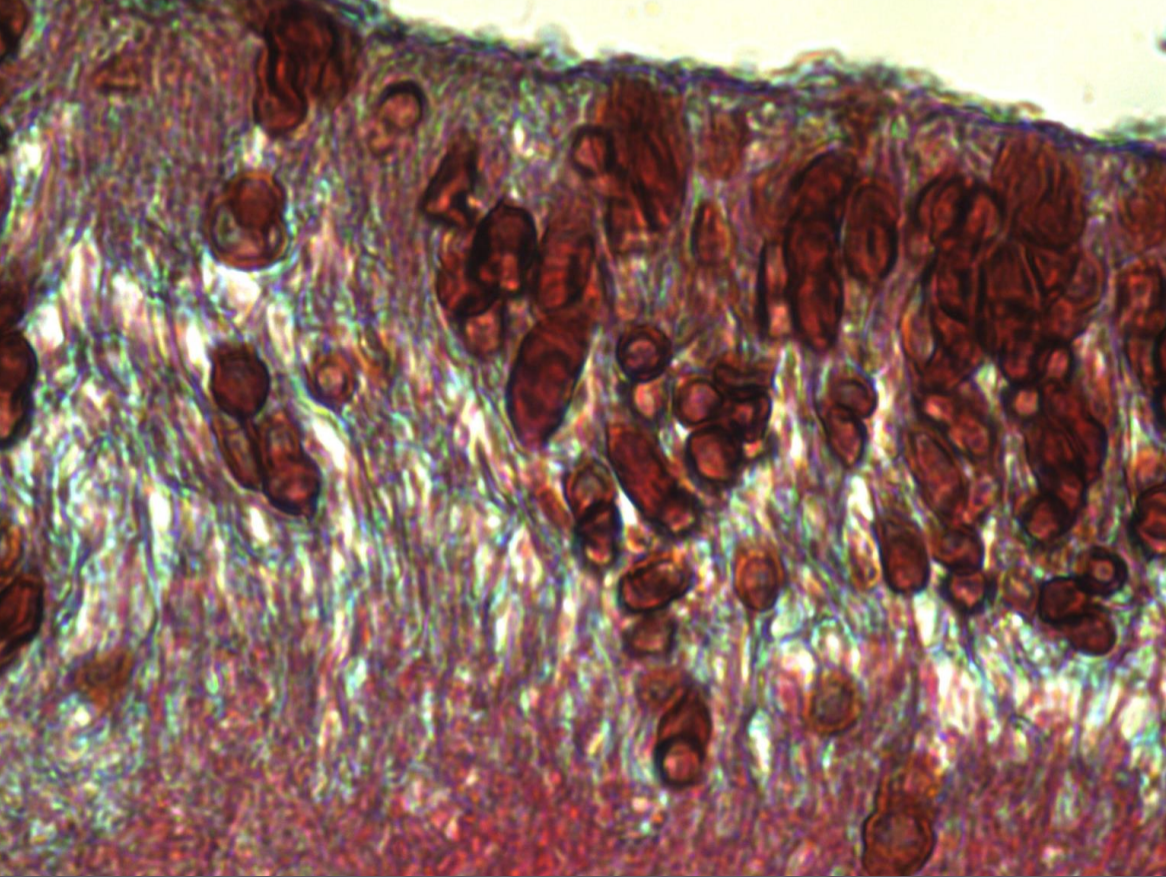
Physicia lichen
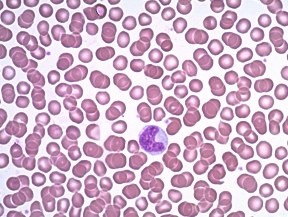
small pink
erythrocytes
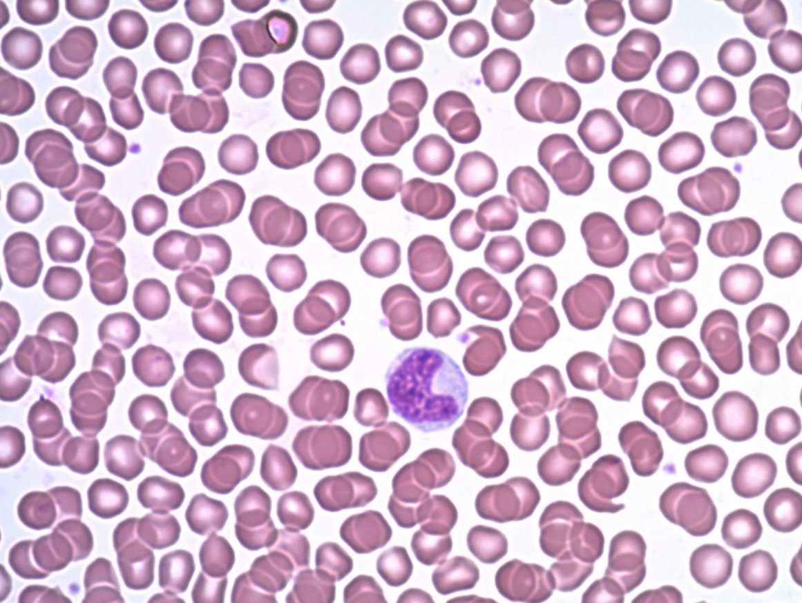
purple
leukocyte
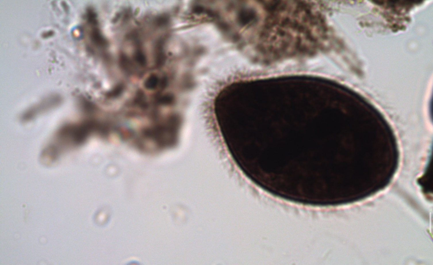
*note cilia
Balantidium coli
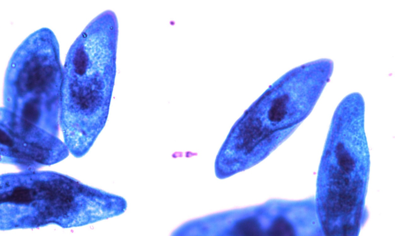
*note oral groove, nucleus, cilia (if visible)
Paramecium
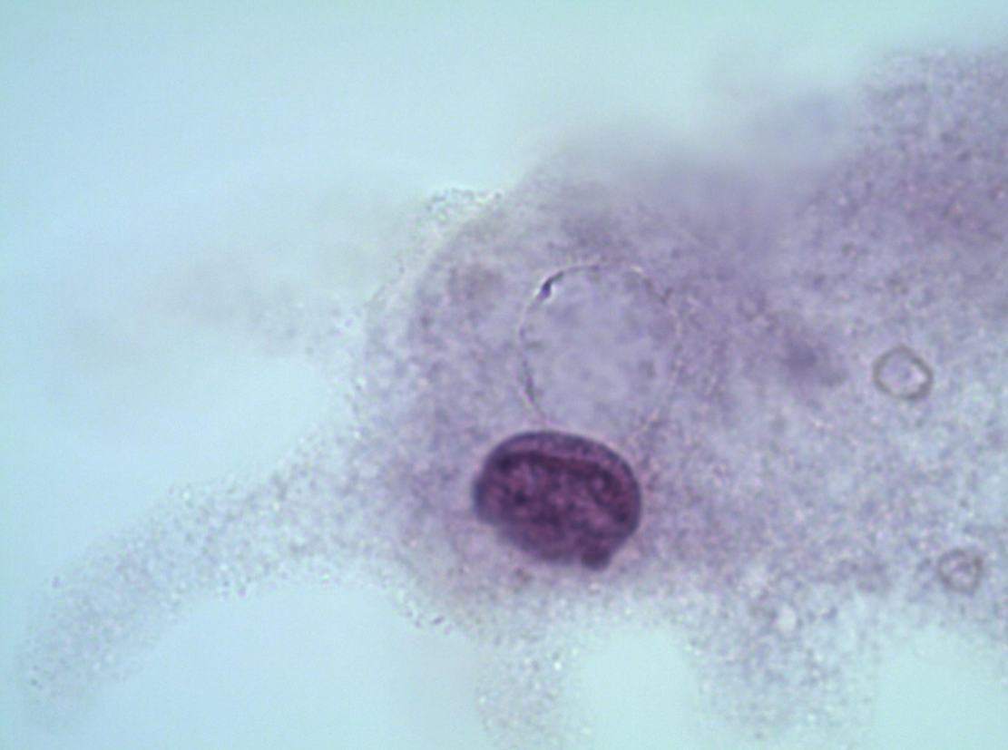
*note nucleus, pseudopodia
Amoeba
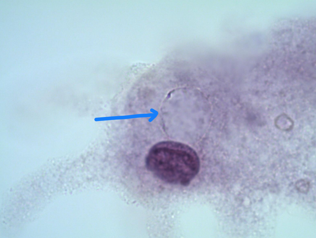
blue arrow
contractile vacuole
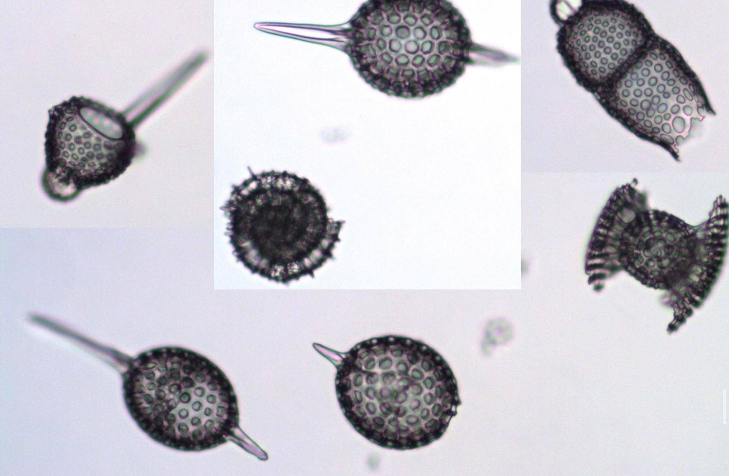
Radiolarians
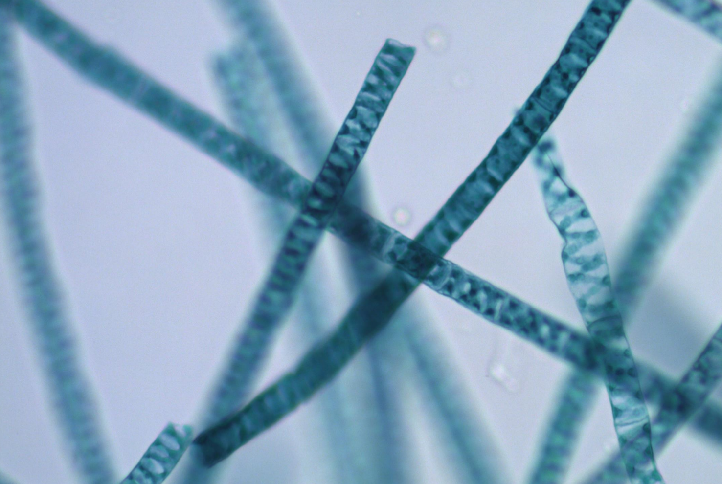
Spirogyra
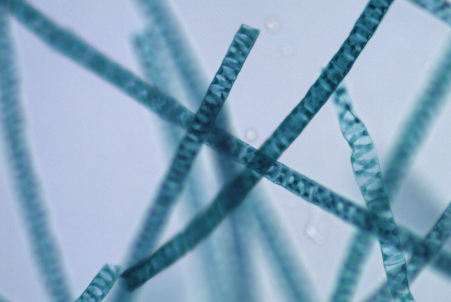
spirals inside
spiral chloroplast
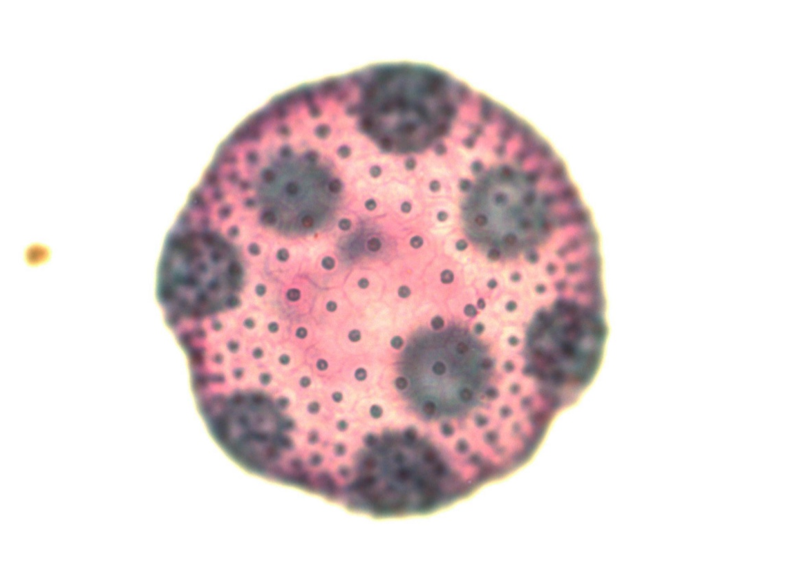
Volvox
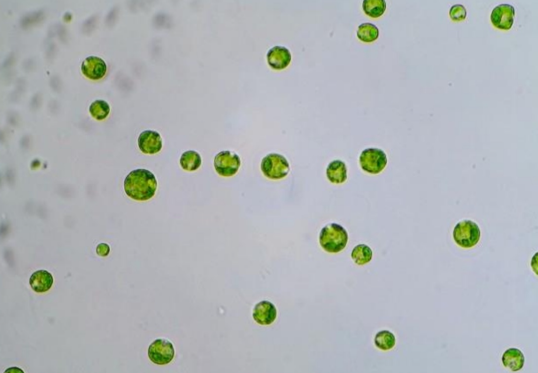
Chlamydomonas
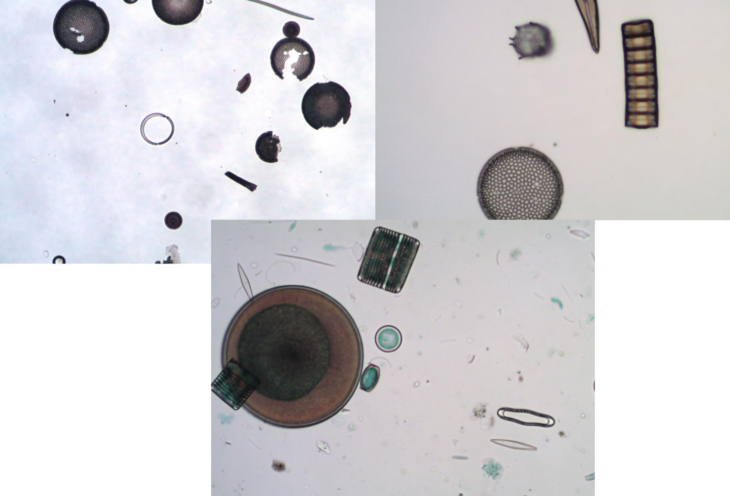
Diatoms
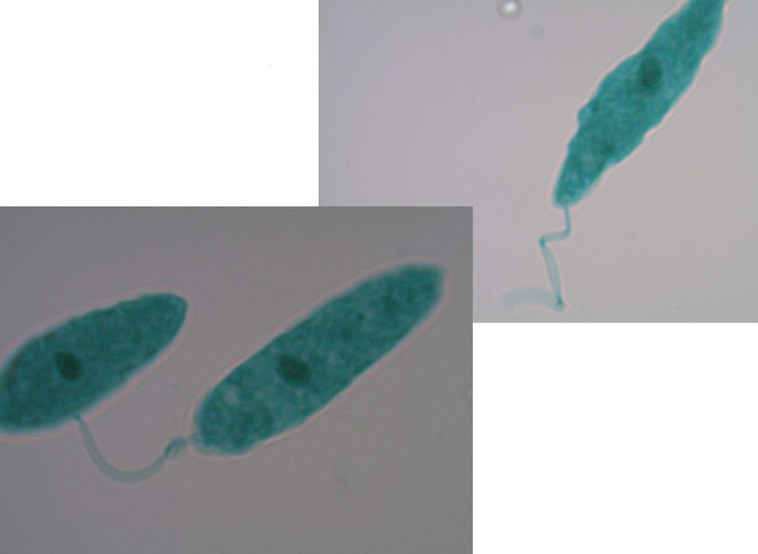
*note flagella, nucleus
euglena
normal flora
organisms that naturally reside on our skin; remain even after washing/disinfecting hands
degerming
mechanical action of removing microbes
aseptic technique
a way of working that reduces the chance of contamination by microbes
bunsen burner
used to heat-sterilize metal objects; hot air rises, creating convection current, creating sterile field around burner
incinerator
used to heat-sterilize metals while preventing aerosols
cofluency
growth that merges together to cover a surface; prevented through quad streaks (streak plates)
spectrophotometer
determines the absorbance of a bacterial culture; sends specific wavelength of light through a sample and a detector will read the amount of light that came through
serial dilutions (know how to calculate)
obtained by making a dilution, mixing then removing some of that material and diluting it in a new volume (repeated as needed)
direct methods of counting bacteria
microscopic counts, dilution plating and most probable number determinations
indirect methods of counting bacteria
biochemical readings and optical densities
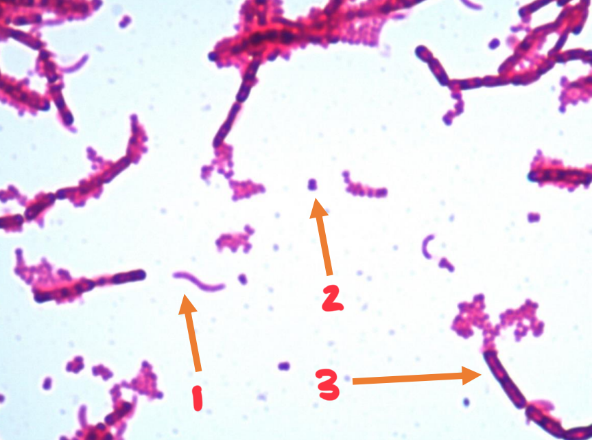
1
spirillum
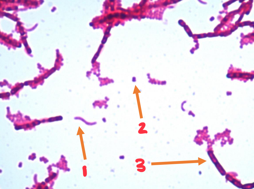
2
coccus
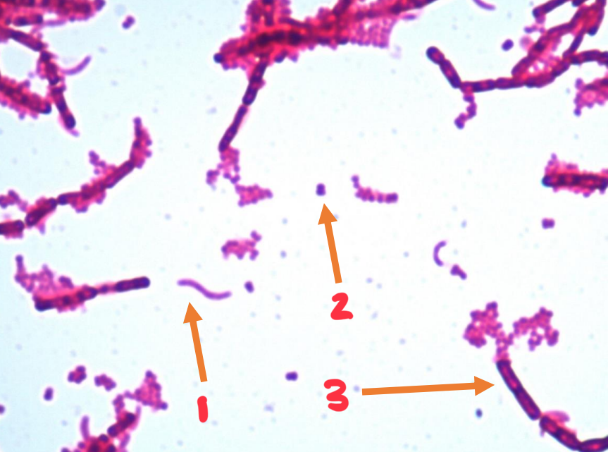
3
bacillus
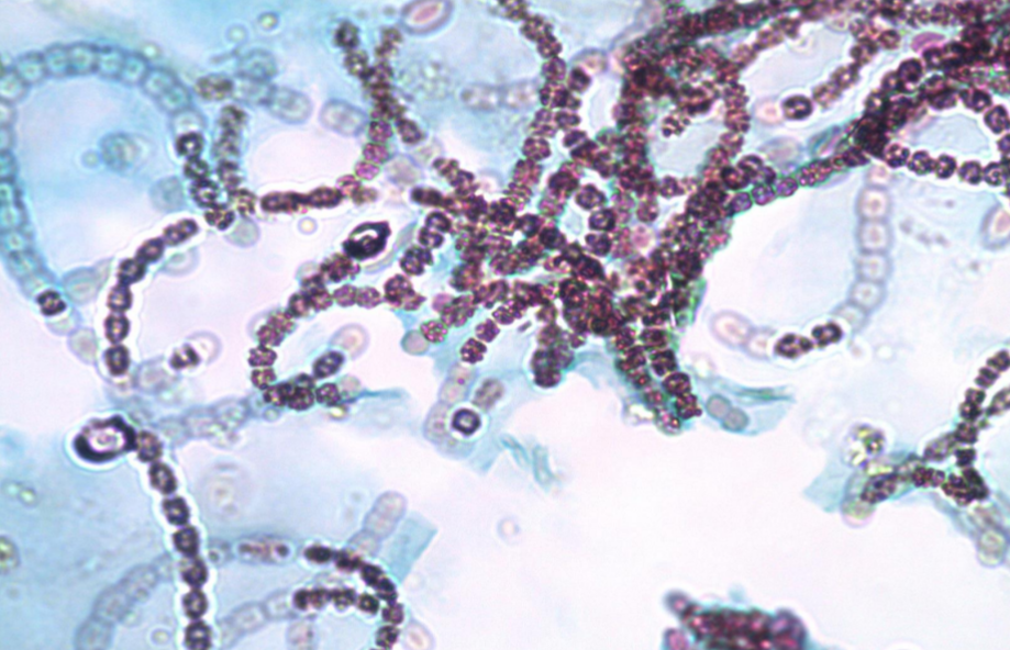
Nostoc
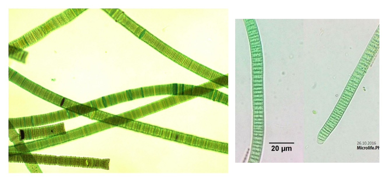
Oscillatoria
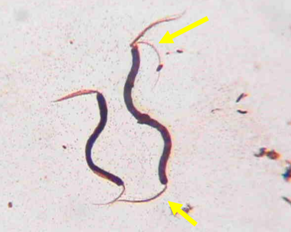
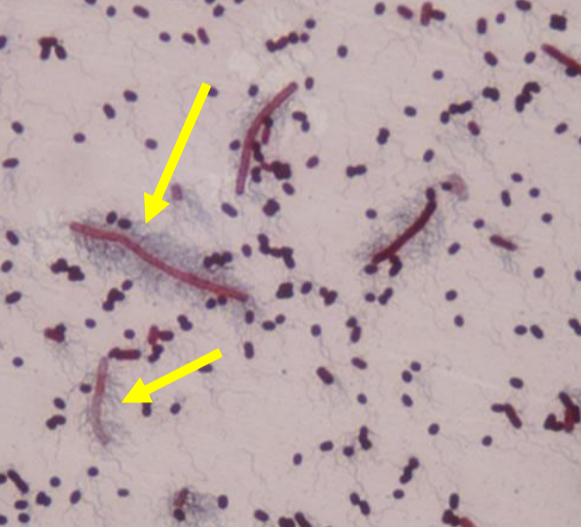
type of flagella
amphitrichous flagella
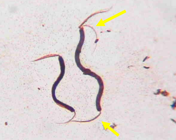
Spirillum volutans
type of flagella
peritrichous flagella
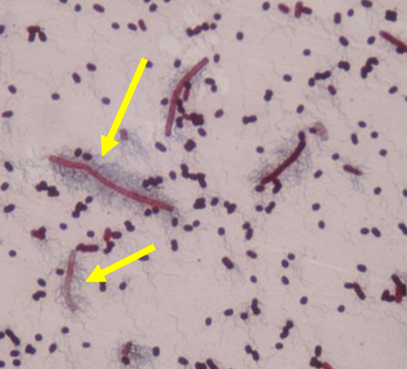
Proteus vulgaris
cluster of flagella on one side of bacteria
lophotrichous flagella
one flagella on one end of bacteria
monotrichous flagella
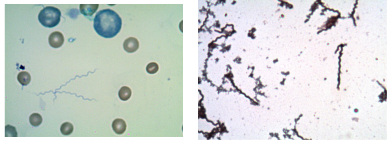
Spirochetes
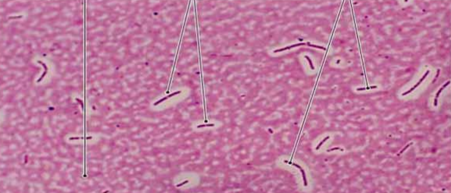
Klebsiella pneumoniae
chromophore
the colored molecule responsible for the visual of the stain
acidic dye
give off H+ and the chromophore is negatively charged; repelled by cellular structures so the background in stained
basic dye
gives off OH- and the chromophore is positively charged; attracted to cellular structures and stains the cell
smear
process of preparing a slide for a stain involving basic dyes; precedes simple, gram, endospore, and acid fast staining
heat fix
process of applying heat to a slide in order to kill the organisms and cause them to adhere well to the glass slide
bibulous paper
the absorbent paper used to blot excess water from slide before viewing
what dye did we use for the simple stain?
methylene blue
what is a simple stain?
a staining procedure that uses one (basic) dye
what is a negative stain?
stains the background with a negatively charged chromophore (acidic dye); negatively charged pigment is repelled from similarly charged bacterial cell
what dye did we use for negative stain?
nigrosin (india ink also works)
what is a capsule stain?
combination of a negative stain followed by a basic dye; there is no heat fix (to avoid shrinking organism)
differential stains
use more than one dye and are capable of revealing structure or characteristics of the stained cells
what is a gram stain?
offers insight into the type of cell envelope of your specimen; differentiates gram positive and gram negative bacteria
gram positive bacteria
have a thick cell wall (~20-80nm) located external to plasma membrane; wall is make of peptidoglycan
gram negative bacteria
have thinner cell wall (~10nm); have an additional outer membrane layer external to cell wall; primarily made of lipopolysaccharides (LPS) and membrane proteins
what is the primary stain in gram stains?
crystal violet; step 1, positively charged (basic) dye, interacts with gram positive and negative bacteria
what is the mordant in gram stains?
gram’s iodine; reacts with crystal violet to form iodine-crystal violet complex (I-CV complex) which stains peptidoglycan layer (harder to remove from gram positive)