Pelvic girdle and upper leg anatomy
1/100
There's no tags or description
Looks like no tags are added yet.
Name | Mastery | Learn | Test | Matching | Spaced | Call with Kai |
|---|
No analytics yet
Send a link to your students to track their progress
101 Terms
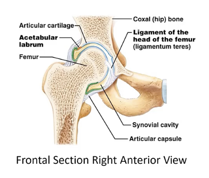
Acetabulofemoral joint
The joint between the acetabulum of the pelvis and the head of the femur
Facet of superior articular process of the sacrum
The smooth surface on the superior articular process of the sacrum that articulates with the inferior articular process of the last lumbar vertebra.
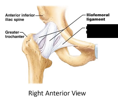
Iliofemoral ligament
A ligament extending from the ilium of the pelvis to the femur
Ligament of the head of femur
The ligament that connects the head of the femur to the acetabulum of the pelvis.
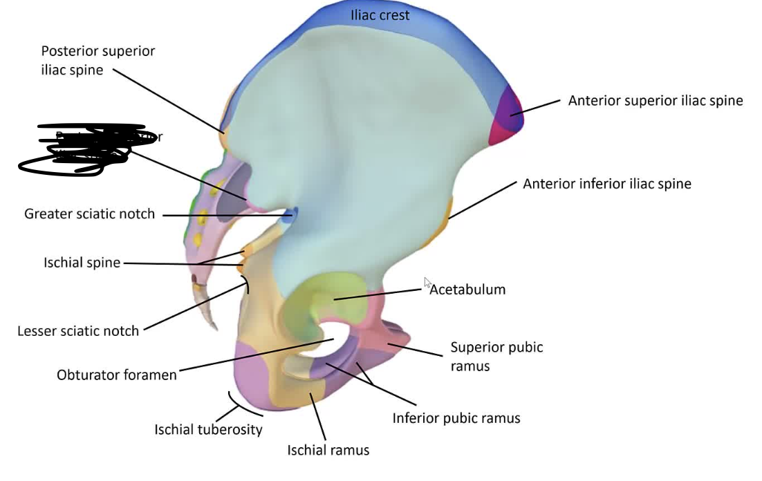
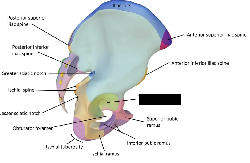
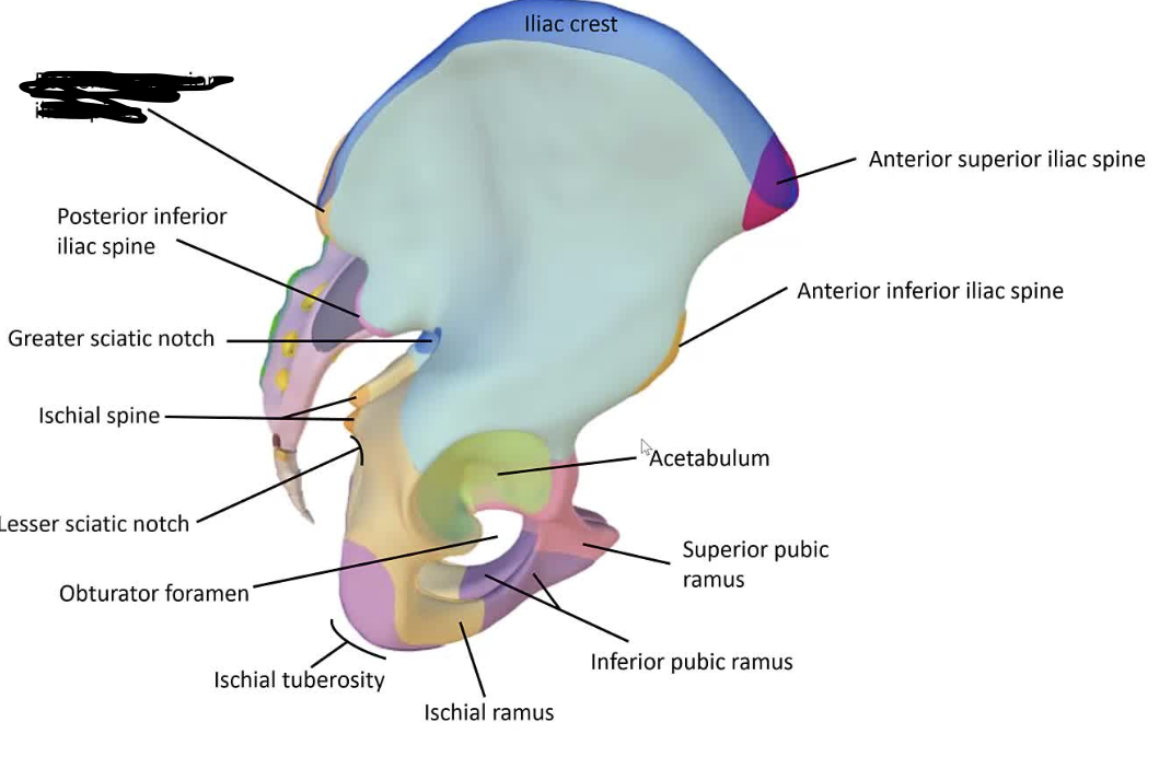
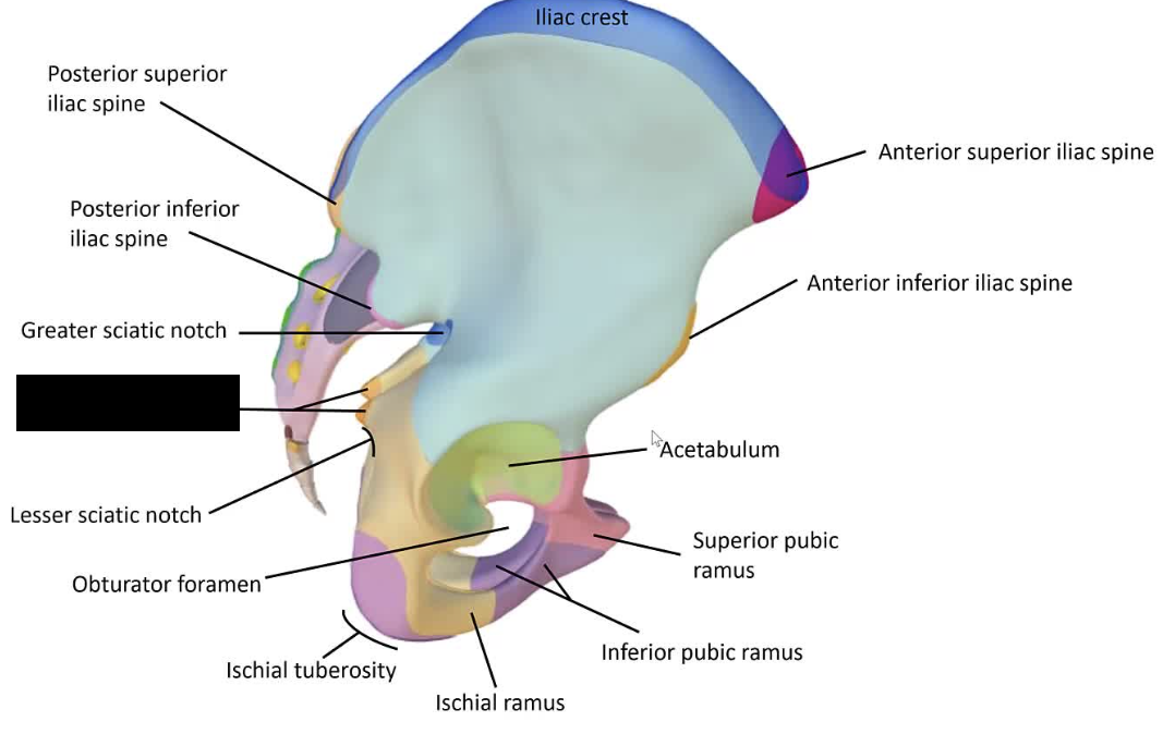
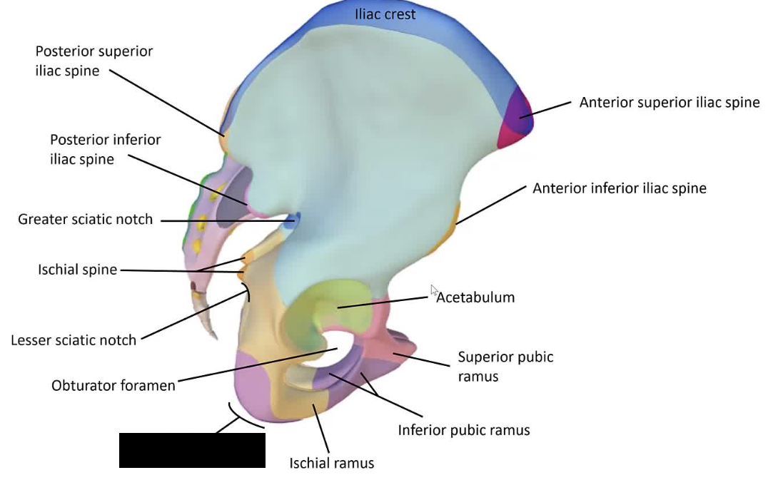
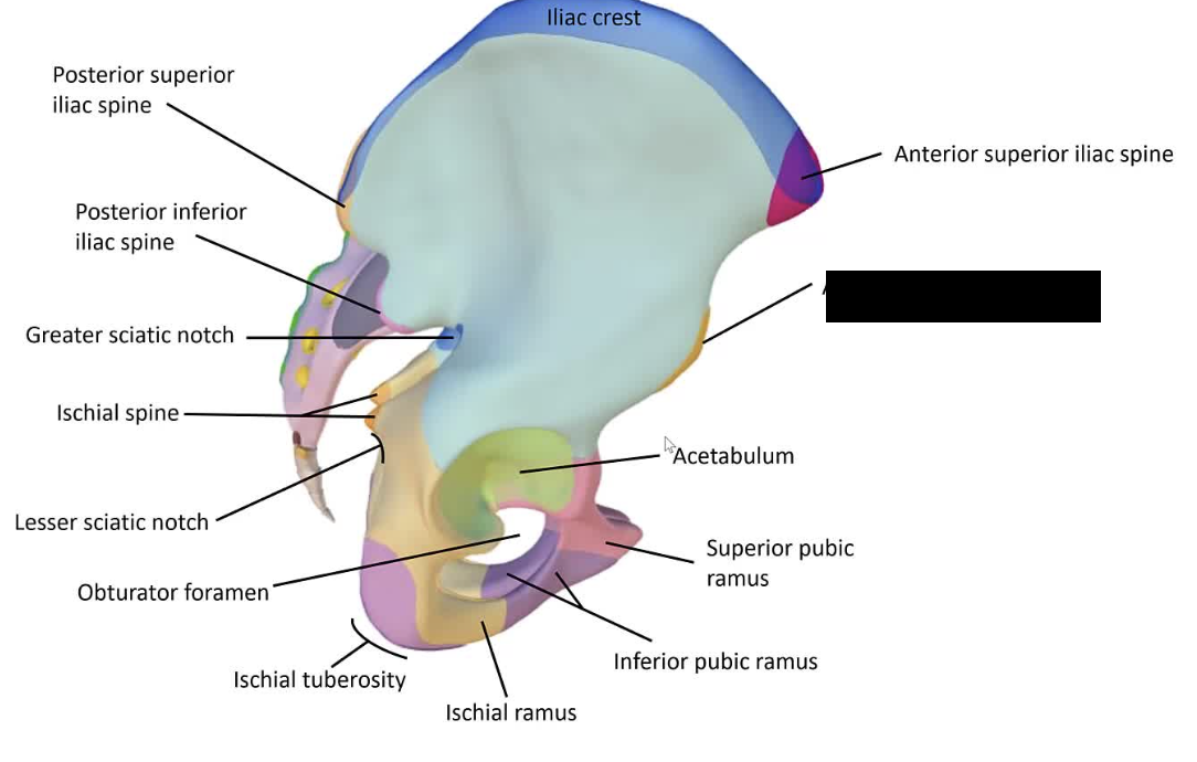
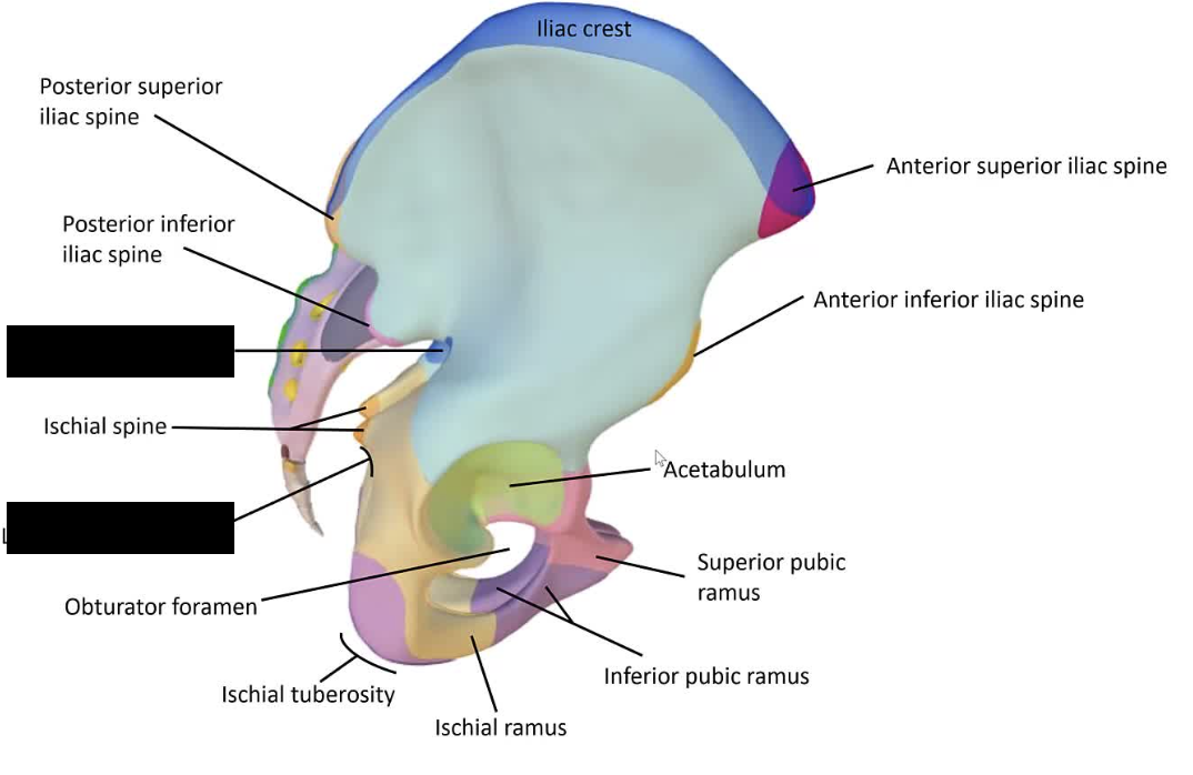
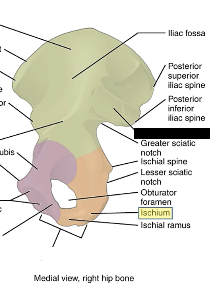
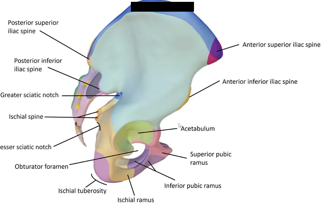
Posterior inferior iliac spine
The lower back projection of the iliac bone, situated below the posterior superior iliac spine.
Sacroiliac joint
The joint between the sacrum and the ilium of the pelvis.
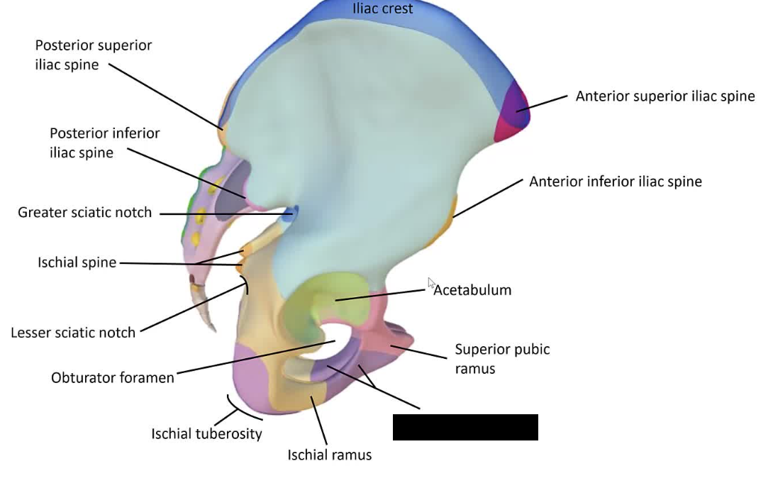
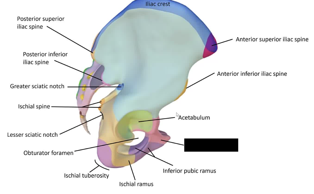
Acetabulum
The cup-shaped cavity on the lateral aspect of the pelvis that receives the head of the femur.
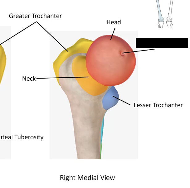
Femur
The thigh bone, extending from the hip to the knee.
Ilium
The broad, upper portion of the hip bone.
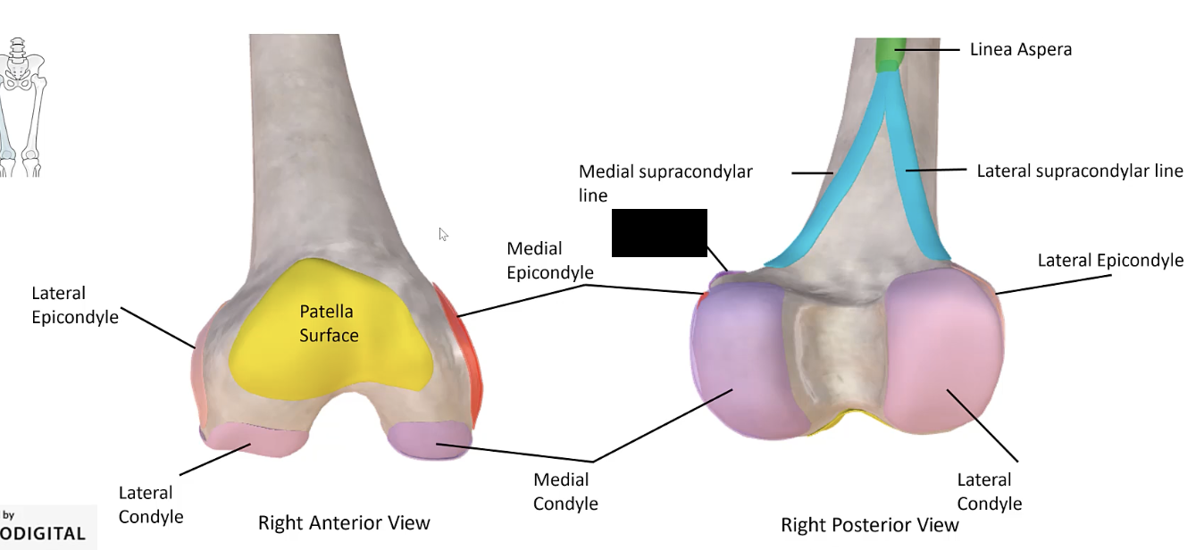
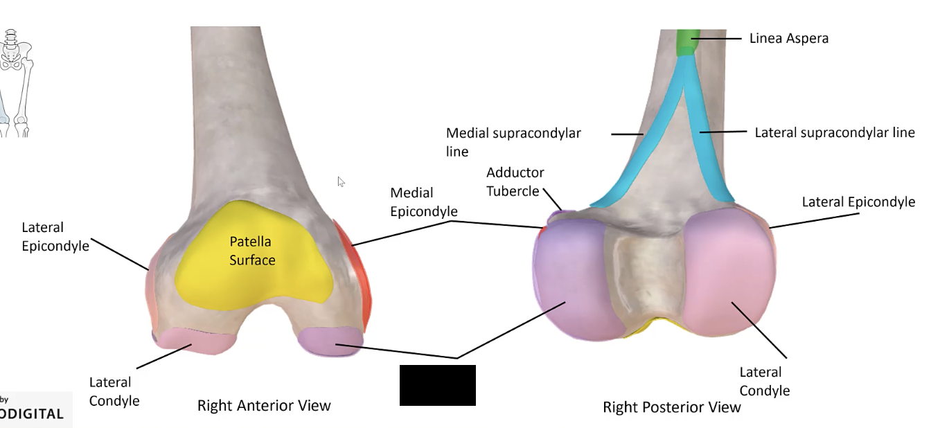
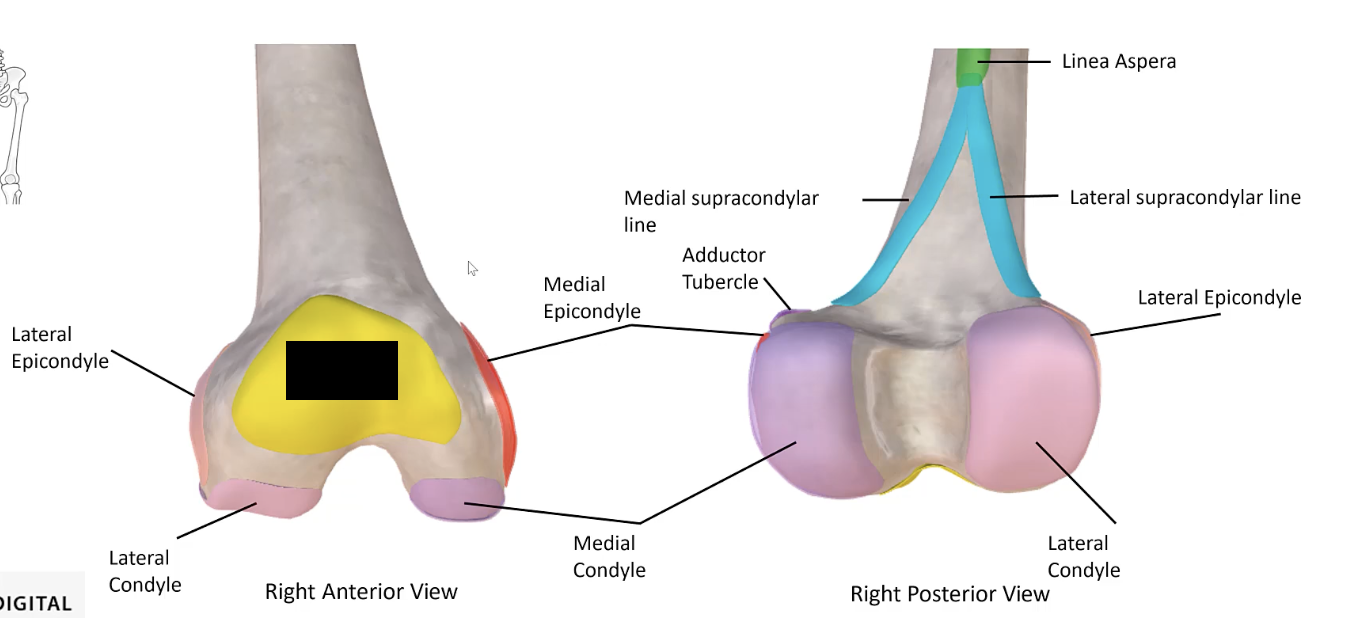
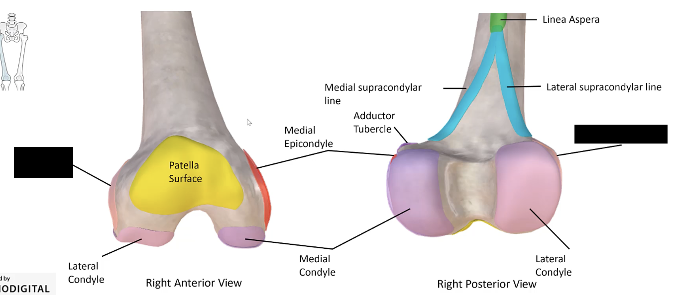
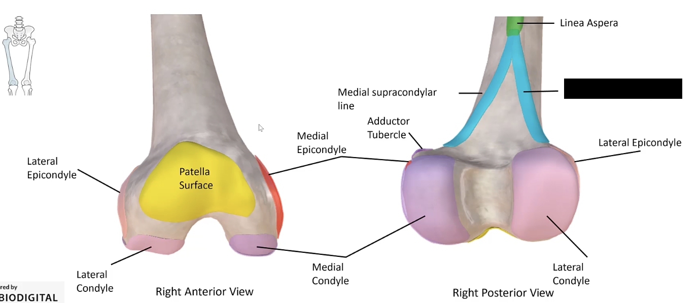
Linea aspera
The ridge along the posterior surface of the femur.
Posterior superior iliac spine
The upper back projection of the iliac bone.
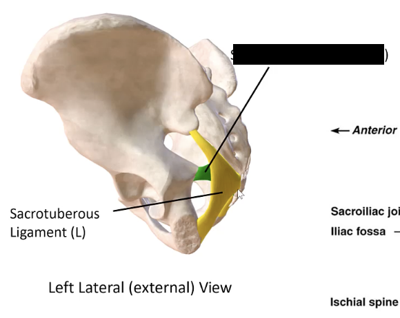
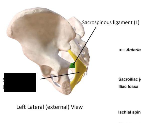
Sacrospinous ligament
The ligament that extends from the sacrum to the ischial spine.
Adductor tubercle
The small bump on the medial epicondyle of the femur important for muscle attachment.
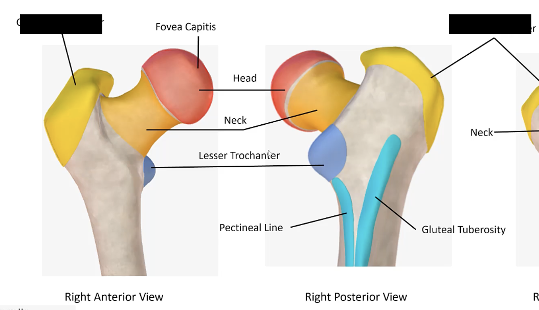
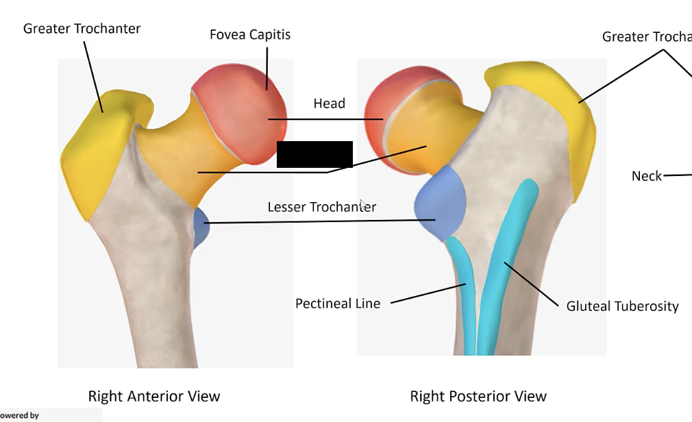
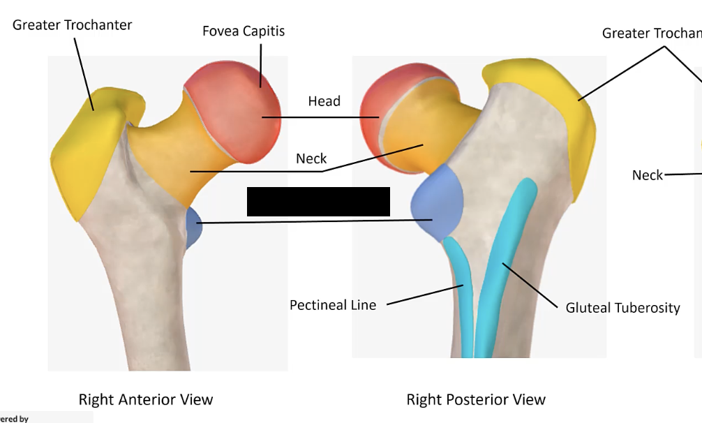
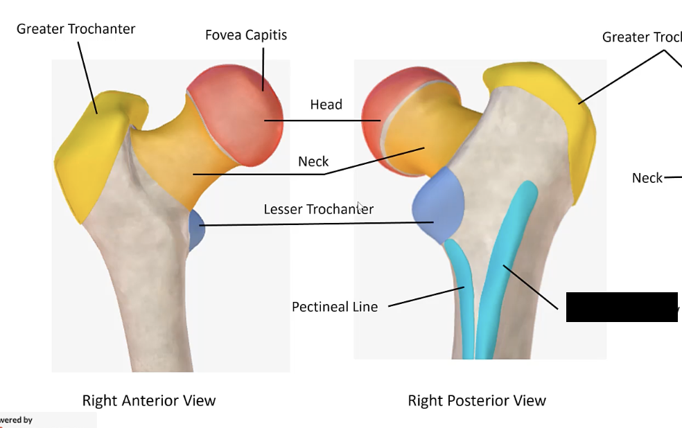
Fovea capitis
The small pit on the head of the femur. Point of attachment for ligament to acetabulum.
Ischial spine
The pointed projection from the posterior border of the ischium.
Medial condyle of femur
The rounded prominence at the medial side of the distal end of the femur.
Pubic arch
The arch formed by the inferior rami of the pubic bones.
Sacrotuberous ligament
The ligament that extends from the sacrum to the ischial tuberosity.
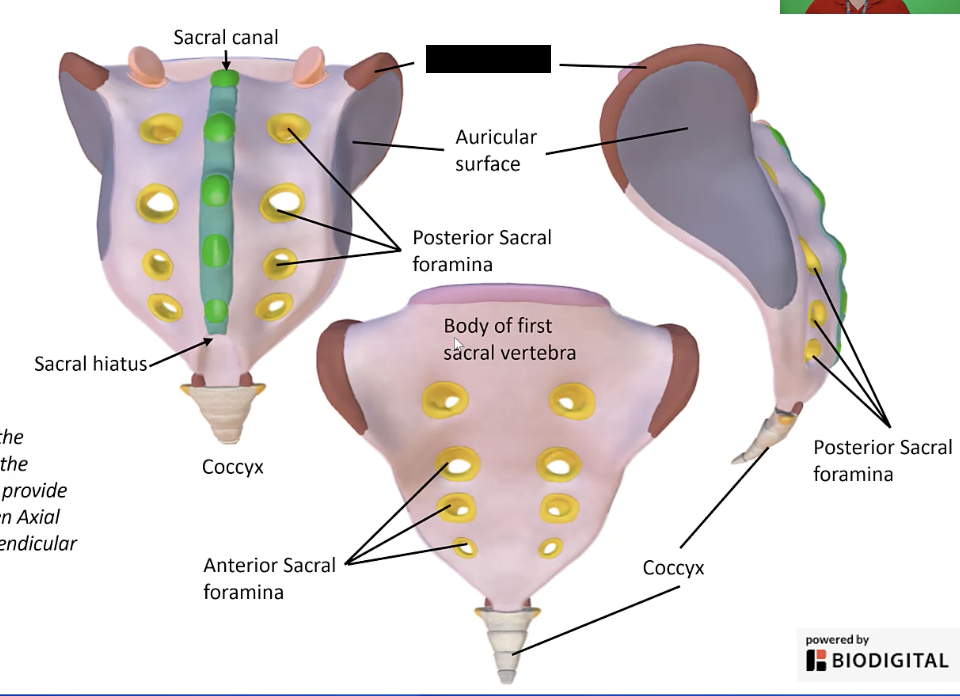
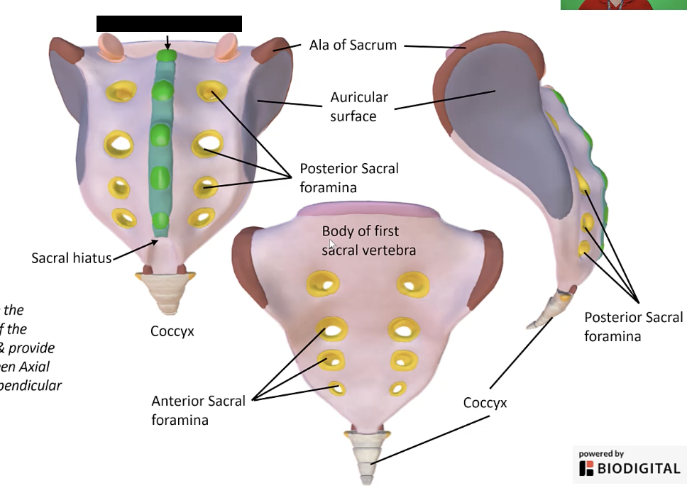
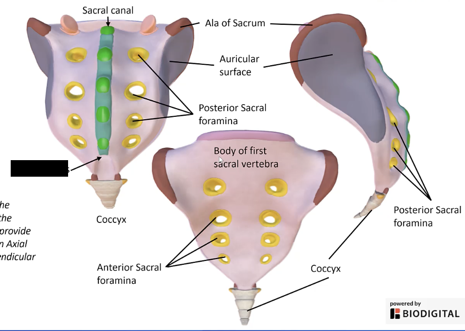
Ala of sacrum
The wing-like lateral extensions on either side of the sacrum where the sacrum articulates with the os coxae.
Greater trochanter
The large, prominent projection on the lateral aspect of the femur near its proximal end.
Ischial tuberosity
The roughened area on the inferior aspect of the ischium that forms muscle attachments
Neck of femur
The constricted part of the femur just below the head.
Pubic symphysis
The cartilaginous joint uniting the left and right pubic bones.
Sacrum
The triangular bone at the base of the spine, formed by the fusion of sacral vertebrae.
Anterior inferior iliac spine
The lower front projection of the iliac bone.
Head of femur
The rounded proximal end of the femur that fits into the acetabulum.
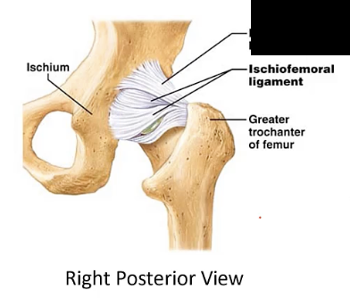
Ischiofemoral ligament
The ligament extending from the ischium to the femur.

Os coxae
The hip bone, formed by the fusion of the ilium, ischium, and pubis.
Pubis
The anterior part of the pelvic bone.
Sciatic foramen (greater and lesser)
The openings formed by the sacrotuberous and sacrospinous ligaments and the sciatic notch.
Anterior superior iliac spine
The upper front projection of the iliac bone.
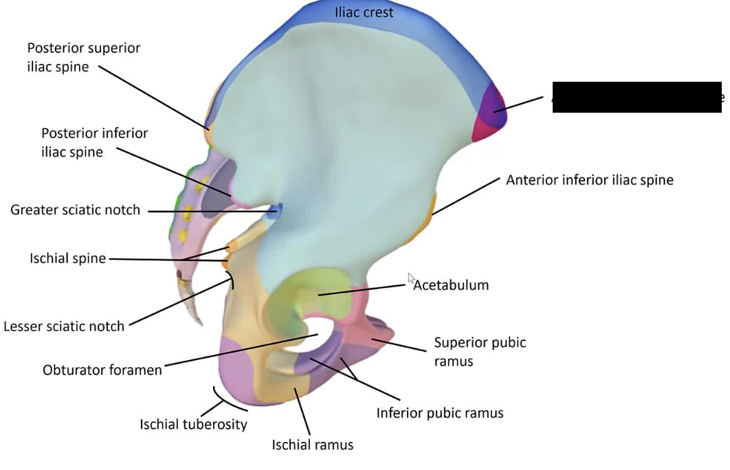
Ischium
The lower, posterior part of the hip bone.
Patella
The kneecap.
Sacral canal
The continuation of the vertebral canal in the sacrum.
Sciatic notch (greater and lesser)
The indentations on the ilium and ischium, converted to foramina by ligaments.
Auricular surface
The ear-shaped surface on the ilium that articulates with the sacrum.
Iliac crest
The upper curved edge of the ilium.
Lateral condyle of femur
The rounded prominence at the lateral side of the distal end of the femur.
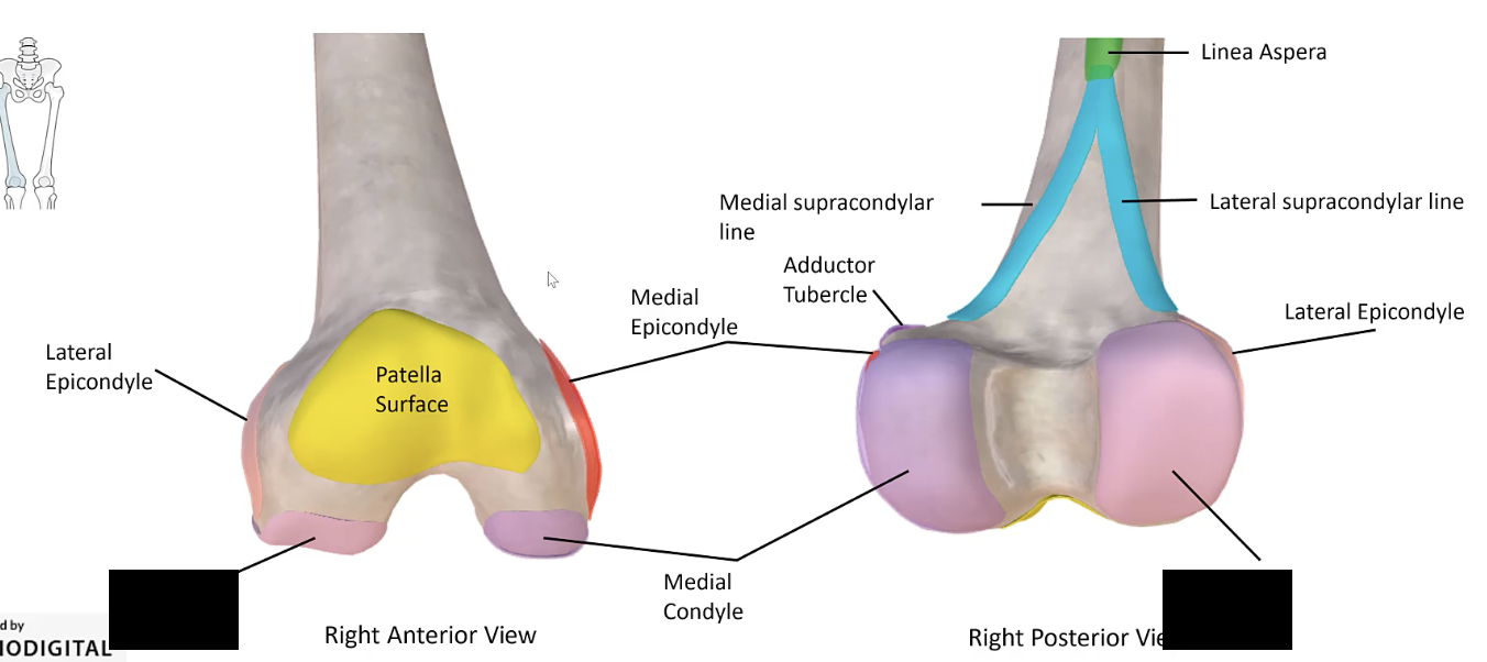
Patellar surface
The smooth area on the anterior distal femur that articulates with the patella.
Sacral foramina
The openings in the sacrum for the passage of nerves
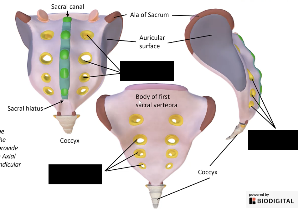
Coccyx
The small, triangular bone at the base of the vertebral column
Iliac fossa
The concave surface on the internal aspect of the ilium.
Lesser trochanter
The smaller projection on the medial aspect of the femur near its proximal end.
Pelvic girdle
The bony ring formed by the hip bones, sacrum, and coccyx.
Sacral hiatus
The opening at the inferior end of the sacral canal
Adductor brevis
A muscle located in the medial compartment of the thigh, originating from the pubis and inserting into the linea aspera of the femur.
Gluteus medius
A muscle located on the lateral aspect of the hip, originating from the ilium and inserting into the greater trochanter of the femur. Superficial to the gluteus minimus.

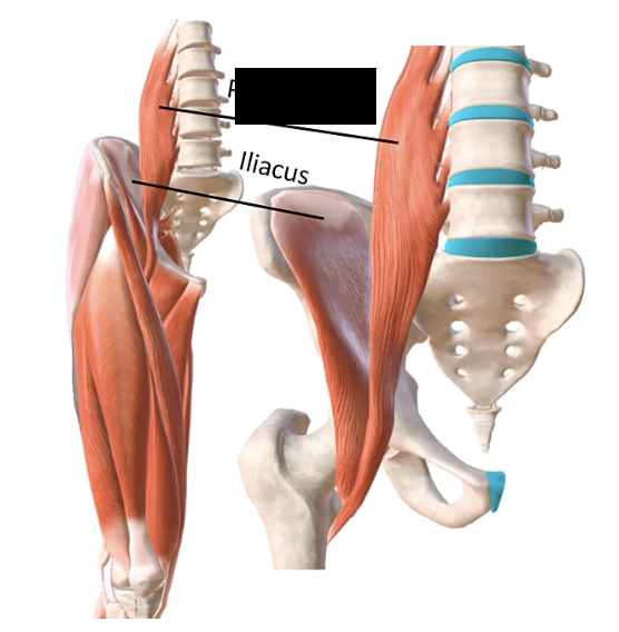
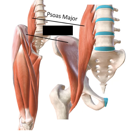
Iliopsoas
A muscle formed by the combination of the iliacus and psoas major muscles, extending from the lumbar spine and ilium to the lesser trochanter of the femur.
Pectineus
A muscle in the anterior part of the upper and medial aspect of the thigh, originating from the superior ramus of the pubis and inserting into the pectineal line of the femur.
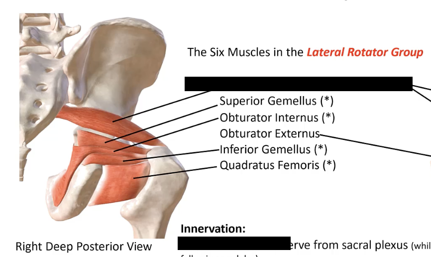
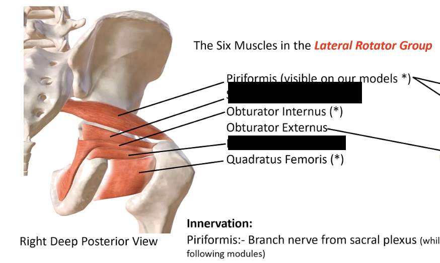
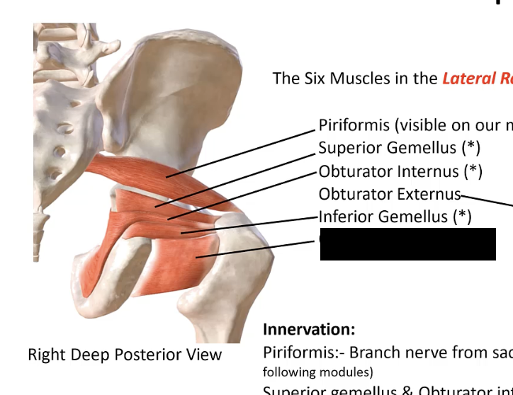
Superior gemellus
A small muscle in the gluteal region, originating from the ischial spine and inserting into the greater trochanter of the femur. Located between the piriformis and obturator internus.

Adductor longus
A muscle in the medial compartment of the thigh, originating from the pubis and inserting into the middle third of the linea aspera of the femur.
Gluteus minimus
A muscle located beneath the gluteus medius, originating from the ilium and inserting into the greater trochanter of the femur.
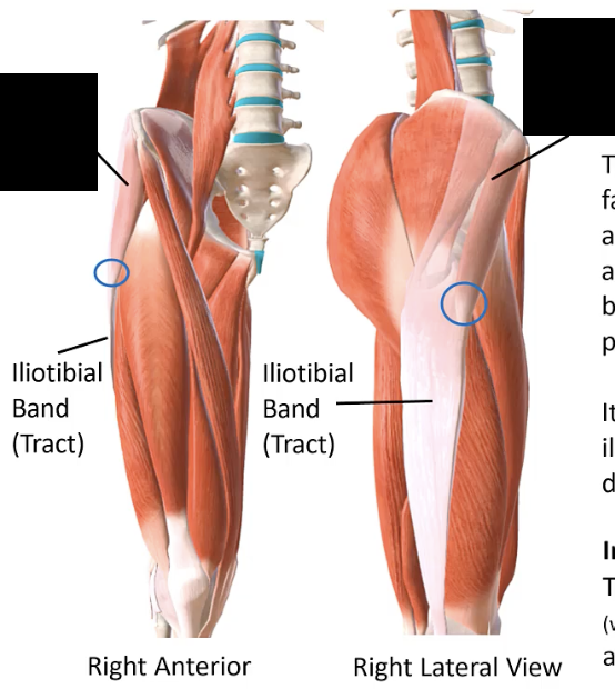
Iliotibial tract (band)
A thick band of fascia extending from the iliac crest to the lateral condyle of the tibia.
Piriformis
A deep muscle located in the gluteal region, originating from the sacrum and inserting into the greater trochanter of the femur.
Tensor fasciae latae
A muscle on the lateral aspect of the thigh, originating from the anterior superior iliac spine and inserting into the iliotibial tract.
Adductor magnus
A large muscle in the medial compartment of the thigh, originating from the pubis and ischium and inserting into the linea aspera and adductor tubercle of the femur.
Gracilis
A muscle in the medial compartment of the thigh, originating from the pubis and inserting into the medial surface of the tibia.
Inferior gemellus
A small muscle in the gluteal region, originating from the ischial tuberosity and inserting into the greater trochanter of the femur. Located between the Quadratus femoris and obturator internus.
Psoas major
A long muscle originating from the lumbar vertebrae and inserting into the lesser trochanter of the femur.
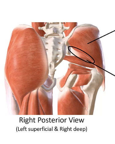
Gluteus maximus
The largest muscle in the gluteal region, originating from the ilium, sacrum, and coccyx, and inserting into the gluteal tuberosity of the femur and iliotibial tract.
Iliacus
A muscle located in the iliac fossa, originating from the iliac crest and inserting into the lesser trochanter of the femur.
Obturator internus
A muscle originating from the obturator membrane and surrounding bones, passing through the lesser sciatic foramen, and inserting into the greater trochanter of the femur.
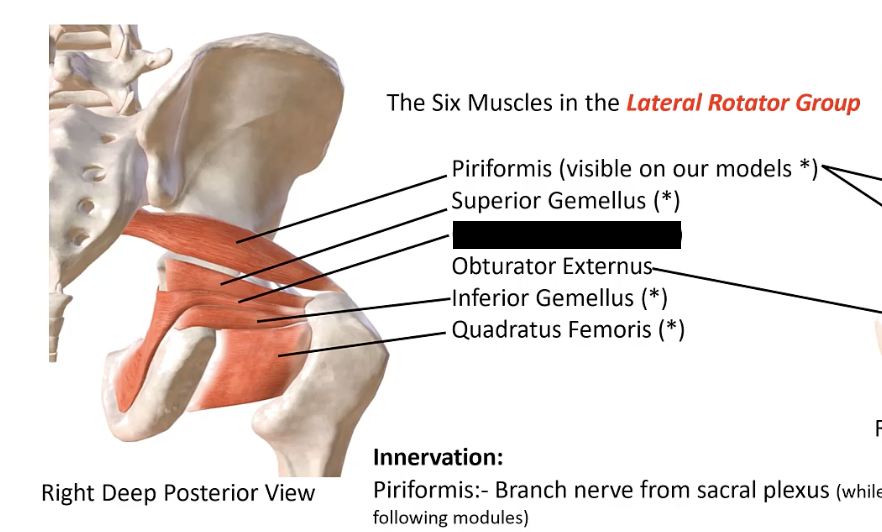
Quadratus femoris
A muscle in the gluteal region, originating from the ischial tuberosity and inserting into the intertrochanteric crest of the femur.
Biceps femoris - long head
A muscle in the posterior compartment of the thigh, originating from the ischial tuberosity and inserting into the head of the fibula.
Biceps femoris - short head
A muscle in the posterior compartment of the thigh, originating from the linea aspera of the femur and inserting into the head of the fibula.
Semitendinosus
A muscle in the posterior compartment of the thigh, originating from the ischial tuberosity and inserting into the medial surface of the proximal tibia.
Rectus femoris
A muscle in the anterior compartment of the thigh, originating from the anterior inferior iliac spine and inserting into the patella via the quadriceps tendon.
Vastus intermedius
A muscle located deep to the rectus femoris in the anterior compartment of the thigh, originating from the anterior and lateral surfaces of the femoral shaft and inserting into the patella via the quadriceps tendon.
Sartorius
A muscle in the anterior compartment of the thigh, originating from the anterior superior iliac spine and inserting into the medial surface of the proximal tibia.
Vastus lateralis
A muscle in the anterior compartment of the thigh, originating from the greater trochanter and linea aspera of the femur and inserting into the patella via the quadriceps tendon.
Semimembranosus
A muscle in the posterior compartment of the thigh, originating from the ischial tuberosity and inserting into the medial condyle of the tibia.
Vastus medialis
A muscle in the anterior compartment of the thigh, originating from the intertrochanteric line and linea aspera of the femur and inserting into the patella via the quadriceps tendon.
Anterior terminal branch
A branch of a nerve that typically refers to the continuation or division towards the anterior part of the body or limb.
Inferior gluteal nerve
A nerve originating from the sacral plexus (L5-S2) that innervates the gluteus maximus muscle.
Lumbosacral plexus
A network of nerve fibers that include the lumbar plexus (L1-L4) and sacral plexus (L4-S4), providing motor and sensory innervation to the lower limb.
Sacral plexus
A network of nerves emerging from the lower lumbar and sacral spinal nerves (L4-S4), innervating the pelvis, buttocks, genitals, thighs, calves, and feet.
Common fibular nerve
A nerve originating from the sciatic nerve, running along the lateral aspect of the knee and bifurcating into the superficial and deep fibular nerves.
Lateral terminal branches
Branches of a nerve that typically refer to the continuation or division towards the lateral part of the body or limb.
Medial terminal branch
A branch of a nerve that typically refers to the continuation or division towards the medial part of the body or limb.
Superficial fibular nerve
A nerve originating from the common fibular nerve, running down the lateral compartment of the leg, and providing sensory innervation to the dorsum of the foot
Deep fibular nerve
A nerve originating from the common fibular nerve, running down the anterior compartment of the leg, and providing motor innervation to the anterior muscles of the leg and sensory innervation to the web space between the first and second toes.
Lower leg terminal branches
The final branches of nerves that innervate the lower leg.
Obturator nerve
A nerve originating from the lumbar plexus (L2-L4), passing through the obturator foramen to innervate the medial compartment of the thigh.
Superior gluteal nerve
A nerve originating from the sacral plexus (L4-S1) that innervates the gluteus medius, gluteus minimus, and tensor fasciae latae muscles.
Femoral nerve
A nerve originating from the lumbar plexus (L2-L4) that innervates the anterior compartment of the thigh.
Lumbar plexus
A network of nerve fibers arising from the lumbar spinal nerves (L1-L4), providing motor and sensory innervation to the lower abdomen, pelvis, and anterior and medial thigh.
Posterior terminal branches
Branches of a nerve that typically refer to the continuation or division towards the posterior part of the body or limb.
Tibial nerve
A nerve originating from the sciatic nerve, running down the posterior compartment of the leg, and providing motor and sensory innervation to the posterior leg and sole of the foot.
Obturator foramen
The opening in the hip bone that allows for nerves and blood vessels to pass through.
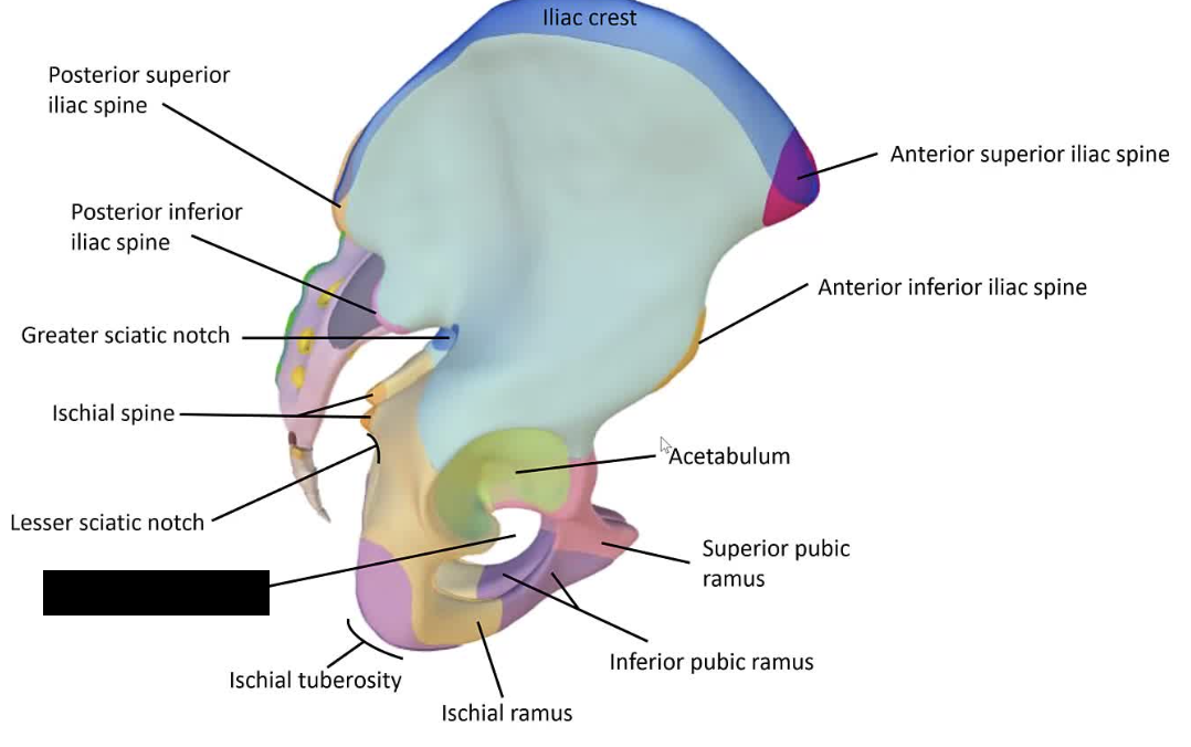
Ischial ramus
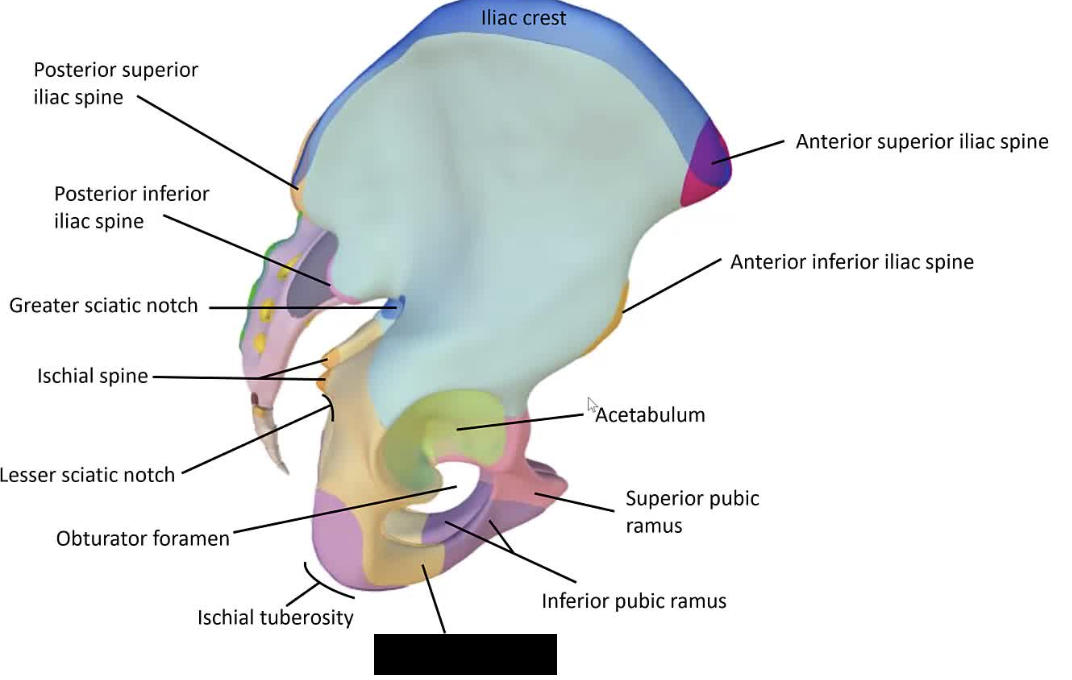
Inferior pubic ramus
Superior pubic ramus
Pectineal line of femur
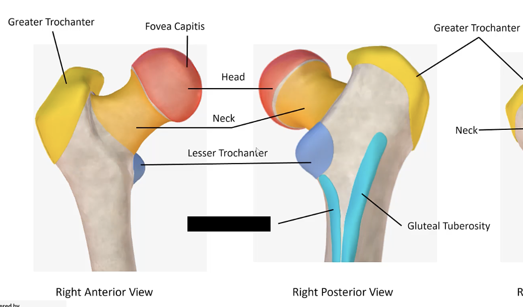
Gluteal tuberosity
Lateral epicondyle of femur
Medial epicondyle of femur
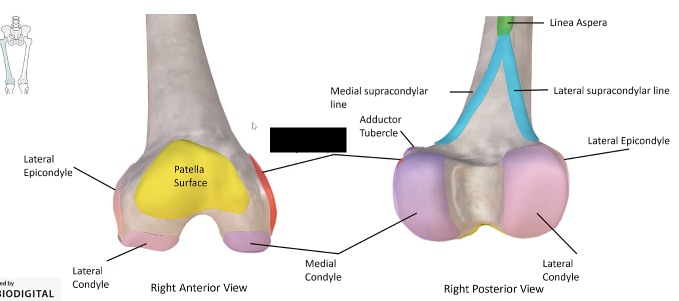
Medial supracondylar line
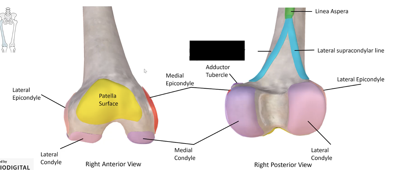
Lateral supracondylar line
Pubofemoral ligament
Tensor fascia latae
Muscle from anterolateral region of iliac crest and anterior superior iliac spine. Between gluteus medius (P) and sartorious (A). Attaches to and continuous with iliotibial tract.