Week 2 - Special Senses - Balance

0.0(0)
Studied by 0 people
call kaiCall Kai
learnLearn
examPractice Test
spaced repetitionSpaced Repetition
heart puzzleMatch
flashcardsFlashcards
GameKnowt Play
Card Sorting

1/14

encourage image

There's no tags or description

Looks like no tags are added yet.

Last updated 8:36 AM on 4/15/26
Name
Mastery
Learn
Test
Matching
Spaced
Call with Kai

No analytics yet

Send a link to your students to track their progress

15 Terms

1
New cards

Special Senses Breakdown

All special senses have corresponding ORGANS:

  • Organspecial sensestimulineural sensation

  • eyevisionlightcolour

  • Ear & cochleahearingsound wavespitch

  • Vestibular apparatusbalancehead movementmotion

  • Noes & olfactory epitheliumsmellairborne chemicalssmell

  • Tongue & taste budsgustationtastantsflavour

2
New cards

Balance: Vestibular system Definition

The sense of equilibrium, perception of balance and spatial orientation.

  • Head position and movement

  • Detecting if body is in motion

3
New cards

Balance: Factors effecting Balance

  • Base of Support: Area around the outside edges of the body in contact with the ground.

  • Centre of gravity: Average location of a body’s centre of mass.

  • Joint movement

  • Posture

Humans are unstable creatures (little base of support, high centre of gravity, lots of joints)

4
New cards

Balance: Vestibular apparatus/labyrinth Anatomy

Vestibular apparatus/labyrinth: Located in inner ear, sits next to the cochlea

  • Composed of vestibule (also called otolith organs) & 3 semicircular canals

  • Activates mechanoreceptors (hair cells)

  • Relies on other senses, such as sight (can cause motion sickness)

  • Made of 3 loops, called ‘semicircular canals’, each containing fluid

    • Provides proprioception of the head in 3 dimensions

<p><em><u>Vestibular apparatus/labyrinth</u></em>: Located in <strong>inner ear, </strong>sits <strong>next to the cochlea</strong></p><ul><li><p>Composed of <strong>vestibule (</strong>also called <strong>otolith organs)</strong> &amp; <strong>3 semicircular canals</strong></p></li><li><p>Activates <mark data-color="purple" style="background-color: purple; color: inherit;">mechanoreceptors (hair cells)</mark></p></li><li><p>Relies on other senses, such as <strong>sight</strong> (can cause motion sickness)</p></li><li><p>Made of 3 loops, called&nbsp;‘semicircular canals’, each containing fluid</p><ul><li><p>Provides proprioception of the head in 3 dimensions</p></li></ul></li></ul><p></p>
5
New cards

Balance: Vestibule / Otolith organs

Otolith organs are 2 membranous sacs - Utricle & saccule

Utricle: Sensitive to horizontal movements (e.g driving a car)

Saccule: Sensitive to vertical movements (e.g standing in an elevator)

  • Different functions due to different anatomical positions

Both surrounded by '“macula tissue” - hair cells (mechanoreceptors) surrounded by support cells

<p>Otolith organs are 2 <strong>membranous sacs</strong> - <mark data-color="yellow" style="background-color: yellow; color: inherit;">Utricle</mark> &amp; <mark data-color="yellow" style="background-color: yellow; color: inherit;">saccule</mark></p><p><mark data-color="yellow" style="background-color: yellow; color: inherit;">Utricle:</mark> Sensitive to <strong>horizontal movements </strong>(e.g driving a car)</p><p><mark data-color="yellow" style="background-color: yellow; color: inherit;">Saccule:</mark> Sensitive to <strong>vertical movements </strong>(e.g standing in an elevator)</p><ul><li><p>Different functions due to different anatomical positions</p></li></ul><p></p><p>Both surrounded by '“<strong><mark data-color="yellow" style="background-color: yellow; color: inherit;">macula</mark></strong><mark data-color="yellow" style="background-color: yellow; color: inherit;"> tissue</mark>” - <strong><mark data-color="purple" style="background-color: purple; color: inherit;">hair cells (mechanoreceptors)</mark></strong> surrounded by support cells</p><p></p>
6
New cards

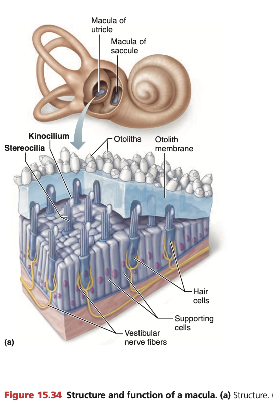
Balance: Otolith organs: Macula

Hair cells are embedded within the macula.

Stereocilia of the hair cells project into a gelatinous otolithic membrane laying on top.

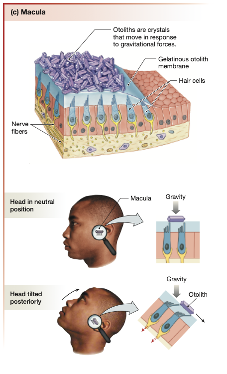
Resting above the viscous otolithic membrane are a layer of calcium carbonate crystals called “otoliths”.

  • Crystal otoliths make the otolithic membrane top-heavy, hence the gel moves in response to head movements in line with gravity

<p><mark data-color="yellow" style="background-color: yellow; color: inherit;">Hair cells</mark> are embedded within the <strong><mark data-color="yellow" style="background-color: yellow; color: inherit;">macula</mark>.</strong></p><p><strong><mark data-color="yellow" style="background-color: yellow; color: inherit;">Stereocilia</mark></strong> of the hair cells project into a <strong><mark data-color="green" style="background-color: green; color: inherit;">gelatinous otolithic membrane</mark></strong> laying on top<strong>.</strong></p><p>Resting above the viscous otolithic membrane are a layer of <strong><mark data-color="green" style="background-color: green; color: inherit;">calcium carbonate crystals</mark></strong> called “<mark data-color="green" style="background-color: green; color: inherit;">otoliths</mark>”.</p><ul><li><p>Crystal otoliths make the otolithic membrane <strong><em>top-heavy</em></strong>, hence the gel moves in response to <strong>head movements</strong> in line with <strong><u><mark data-color="red" style="background-color: red; color: inherit;">gravity</mark></u></strong></p></li></ul><p></p>
7
New cards

Balance: 3 Semicircular canals

3 membranous tubes.

Oriented at 90 degrees of one another. Oriented in the 3 different planes.

  • Filled with endolymph

  • At the base of each canal is a swelling called an ampulla

  • Inside each ampulla, is a gelatinous capsule called the cupula

Fluid movement in the:

Superor/Anterior semicircular canal: Nodding head - sagittal plane

Horizontal/Lateral semicircular canal: Shaking head - transverse plane

Posterior semicircular canal: Tilting head sideways - coronal plane

<p><mark data-color="yellow" style="background-color: yellow; color: inherit;">3 </mark><strong><mark data-color="yellow" style="background-color: yellow; color: inherit;">membranous tubes</mark></strong><mark data-color="yellow" style="background-color: yellow; color: inherit;">.</mark></p><p>Oriented at 90 degrees of one another. Oriented in the 3 different planes.</p><ul><li><p>Filled with <strong><mark data-color="yellow" style="background-color: yellow; color: inherit;">endolymph</mark></strong></p></li><li><p>At the base of each canal is a swelling called an <strong><mark data-color="yellow" style="background-color: yellow; color: inherit;">ampulla</mark></strong></p></li><li><p>Inside each ampulla, is a gelatinous capsule called the <strong><mark data-color="green" style="background-color: green; color: inherit;">cupula</mark></strong></p></li></ul><p></p><p><strong><em><u>Fluid movement</u></em></strong> in the:</p><p>Superor/Anterior semicircular canal: Nodding head - sagittal plane</p><p>Horizontal/Lateral semicircular canal: Shaking head - transverse plane</p><p>Posterior semicircular canal: Tilting head sideways - coronal plane</p><p></p>
8
New cards

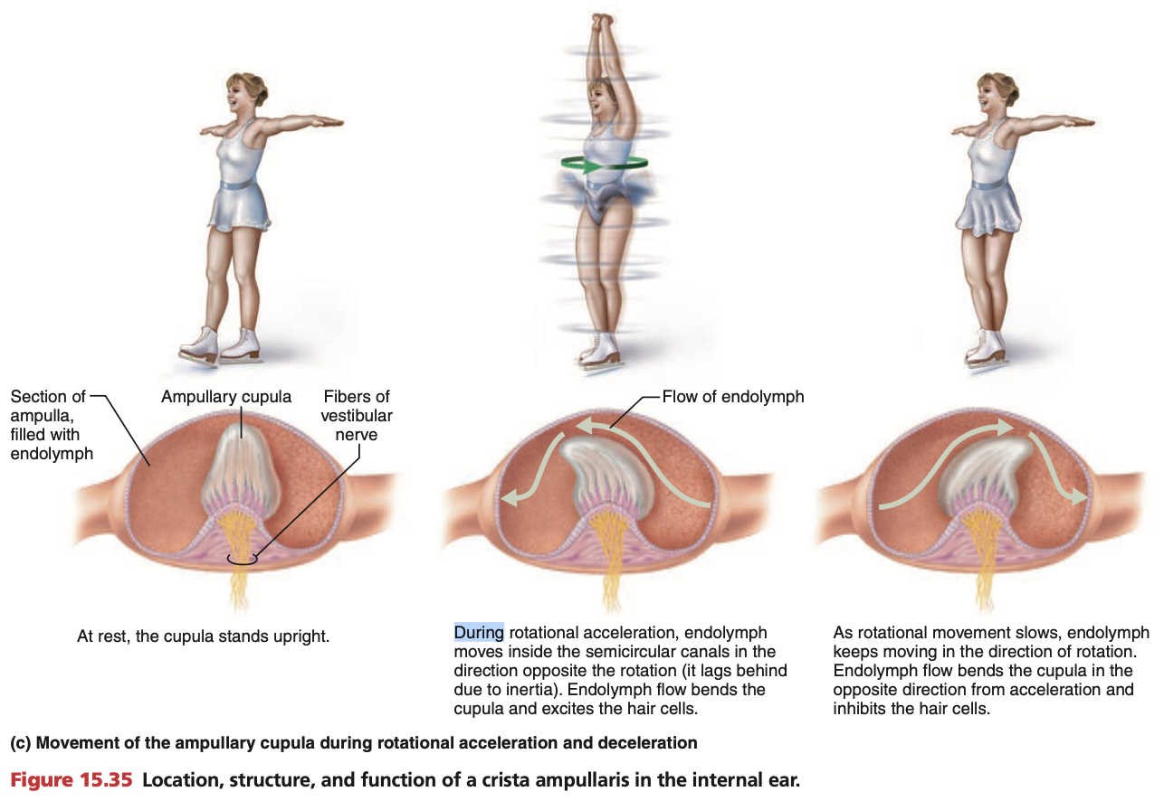
Balance: 3 Semicircular canals: Ampulla

  • Inside each ampulla, is a gelatinous capsule called the cupula

  • Hair cells (mechanoreceptors) extend into the cupula

  1. Head turns in a direction (e.g turns right) ←🙂‍↔

  2. Endolymph in the semicircular canals lags behind due to inertia. Semicircular canal continues moving regardless. (e.g horizontal semicircular canal)

  3. Endolymph is frozen for a few seconds, swishing into the cupula located within the ampula, causing the cupula to sway in the opposite direction momentarily.

    1. (e.g endolymph 💧 → ← semicircular canal & ampulla👂

    2. endolymph 💧 → ampulla & cupula momentarily 🪼 →)

  4. Movement of cupula causes stereocilia on the hair cells bends towards kinocilium, activating the hair cells

  5. Hair cells depolarise, firing action potentials

  6. As rotation slows down, endolymph finally moves in direction of rotation (e.g moves left)

  7. Cupula sways in direction of movement (e.g swishes left)

  8. Movement of cupula causes stereocilia on the hair cells bends away from kinocilium, activating the hair cells

  9. Hair cells hyperpolarise, deactivating and inhibiting hair cells.

<ul><li><p>Inside each <mark data-color="yellow" style="background-color: yellow; color: inherit;">ampulla</mark>, is a gelatinous capsule called the <strong><mark data-color="green" style="background-color: green; color: inherit;">cupula</mark></strong></p></li><li><p><mark data-color="purple" style="background-color: purple; color: inherit;">Hair cells (mechanoreceptors)</mark> extend into the <mark data-color="green" style="background-color: green; color: inherit;">cupula</mark></p></li></ul><p></p><ol><li><p>Head turns in a direction (e.g turns right) ←<span data-name="head_shaking_horizontally" data-type="emoji">🙂‍↔</span></p></li><li><p><strong><mark data-color="yellow" style="background-color: yellow; color: inherit;">Endolymph</mark></strong> in the semicircular canals lags behind due to <strong><u><mark data-color="red" style="background-color: red; color: inherit;">inertia</mark></u></strong>. Semicircular canal continues moving regardless. (e.g horizontal semicircular canal)</p></li><li><p>Endolymph is frozen for a few seconds, swishing into the cupula located within the ampula, causing the <strong><em><u>cupula to sway in the opposite direction</u></em></strong> momentarily.</p><ol><li><p>(e.g endolymph <span data-name="droplet" data-type="emoji">💧</span> → ← semicircular canal &amp; ampulla<span data-name="ear" data-type="emoji">👂</span></p></li><li><p>endolymph <span data-name="droplet" data-type="emoji">💧</span> → ampulla &amp; cupula momentarily <span data-name="jellyfish" data-type="emoji">🪼</span> →)</p></li></ol></li><li><p>Movement of cupula causes stereocilia on the hair cells bends <strong><em><u>towards</u></em></strong> kinocilium, activating the <strong><em><u><mark data-color="purple" style="background-color: purple; color: inherit;">hair cells</mark></u></em></strong></p></li><li><p>Hair cells <strong><em><u>depolarise</u></em></strong>, firing <strong><em><u><mark data-color="purple" style="background-color: purple; color: inherit;">action potentials</mark></u></em></strong></p></li><li><p>As rotation slows down, <strong><em><u>endolymph finally moves</u></em></strong> in direction of rotation (e.g moves left)</p></li><li><p><strong><em><u>Cupula sways in direction of movement</u></em></strong> (e.g swishes left)</p></li><li><p>Movement of cupula causes stereocilia on the hair cells bends <strong><em><u>away from</u></em></strong> kinocilium, activating the hair cells</p></li><li><p>Hair cells <strong><em><u>hyperpolarise</u></em></strong>, deactivating and <strong><em><u>inhibiting hair cells</u></em></strong>.</p></li></ol><p></p>
9
New cards

Balance: Endolymph & Dizziness

When the head suddenly stops after rotating for a while, the endolymph keeps moving through the canals, continuing to hyperpolarise hair cells and causing dizziness.

10
New cards

Balance: Push-pull system

As there are two vestibular labyrinths, one in each ear, when the head rotates in any direction, the hair cells on one side are stimulated, while the other is inhibited.

  • The brain compares info from both canals to understand how the head is moving

  • For turning the head right:

    • The right horizontal semicircular canal gets excited (more action potentials)

    • The left horizontal semicircular canal gets inhibited (less action potentials)

11
New cards

Balance: Mechanoreceptors & sensation

Mechanoreceptors within the vestibular apparatus sense:

  • Head position - utricle & saccule

    • Moving otolithic membranes in utricle & saccule bend stereocilia, causing some hair cells to depolarise as others hyperpolarise.

  • Head movement - semicircular canals

    • Comparing movement along all 3 planes of semicircular canals enables the brain to position the head in the 3D plane.

Movement of othilithic membranes and cupula in ampulla triggers mechanoreceptors (hair cells). At the base of each hair cell are fibres of the vestibular nerve.

Information travels down the vestibular branch of the Vestibulocochlear nerve (Cranial nerve 8).

12
New cards

Balance: Vestibular cortex - Temporal & parietal lobes

The vestibular cortex is located in several areas within the temporal and parietal lobes.

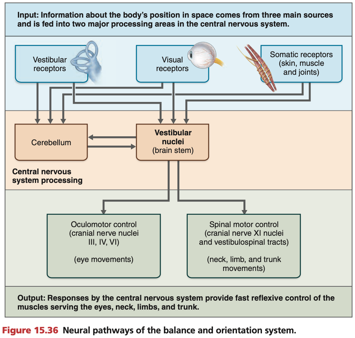
  • 3 aspects: vestibular, visual & somatosensory

Brain receives info from BOTH ears which is integrated at once.

13
New cards
<p><mark data-color="yellow" style="background-color: yellow; color: inherit;">Balance</mark>: <strong>Neural pathways</strong></p>

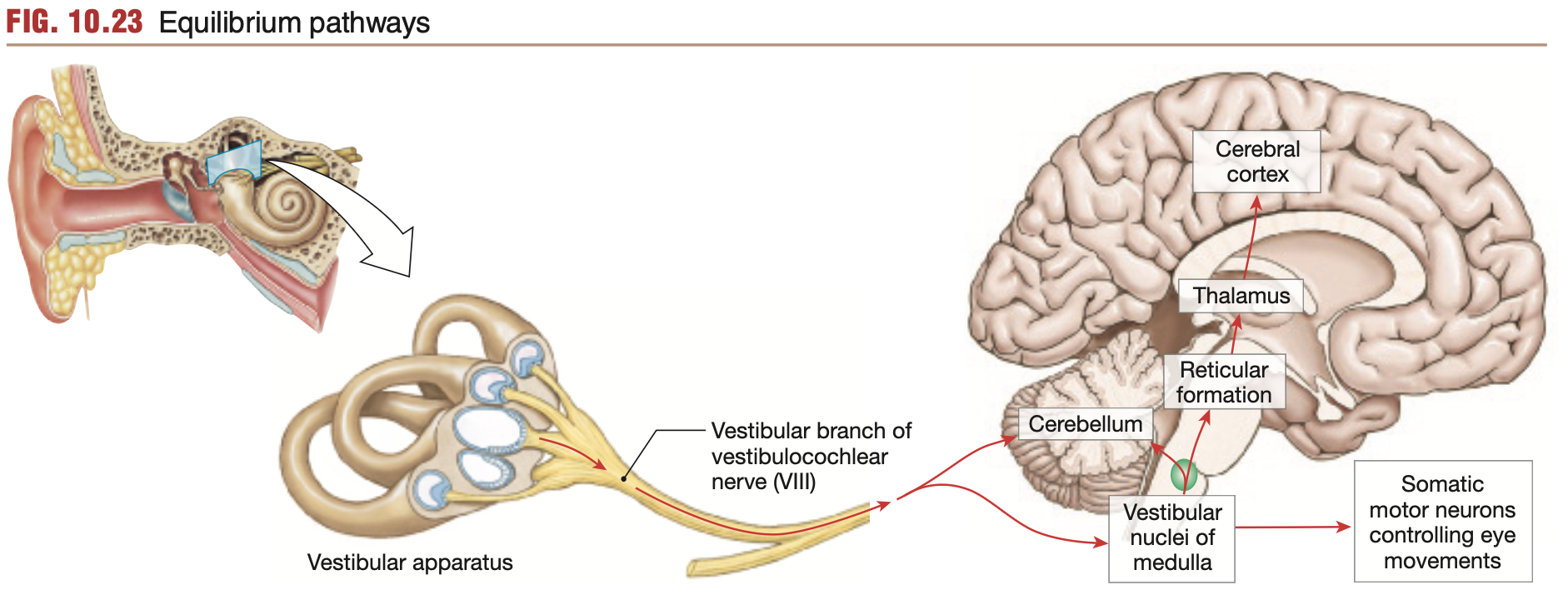
Balance: Neural pathways

Vestibular fibres (vestibulocochlear nerve (Cranial nerve 8)) congregate in the vestibular nuclei within the brainstem’s medulla

Fibres propagate in 4 directions:

  1. Cerebellum: To sense balance

  2. Thalamus: To send signals to sensory and motor areas of the face

  3. Extraocular motor neurons: To move the eyes along with the head

  4. Motor neurons of limbs and neck: To maintain body balance and head position

Reflexes:

  • Sometimes impulses travel directly to reflex centres in brain stem

<p><strong><u>Vestibular fibres</u></strong> (<strong><u><mark data-color="blue" style="background-color: blue; color: inherit;">vestibulo</mark></u></strong><mark data-color="blue" style="background-color: blue; color: inherit;">cochlear</mark><strong><mark data-color="blue" style="background-color: blue; color: inherit;"> nerve (Cranial nerve 8)</mark></strong>) congregate in the <strong><u>vestibular nuclei</u></strong> within the brainstem’s <strong><mark data-color="purple" style="background-color: purple; color: inherit;">medulla</mark></strong></p><p>Fibres propagate in 4 directions:</p><ol><li><p><strong><em><u><mark data-color="purple" style="background-color: purple; color: inherit;">Cerebellum</mark></u></em></strong><mark data-color="purple" style="background-color: purple; color: inherit;">:</mark> To sense <strong>balance</strong></p></li><li><p><mark data-color="purple" style="background-color: purple; color: inherit;">Thalamus:</mark> To send signals to sensory and motor areas of the <strong>face</strong></p></li><li><p><mark data-color="purple" style="background-color: purple; color: inherit;">Extraocular motor neurons:</mark> To move the eyes along with the head</p></li><li><p><mark data-color="purple" style="background-color: purple; color: inherit;">Motor neurons of limbs and neck:</mark> To maintain body balance and head position</p></li></ol><p></p><p><strong><em><u><mark data-color="green" style="background-color: green; color: inherit;">Reflexes:</mark></u></em></strong></p><ul><li><p>Sometimes impulses travel <strong>directly to reflex centres in brain stem</strong></p></li></ul><p></p>
14
New cards

Balance Disorders: Vestibular labyrinth malfunctions

Inner ear problems can result from otoliths detaching from otolith organs and entering semicircular canals.

  • Stroke is a common cause of disorders with the vestibular neural pathway.

15
New cards

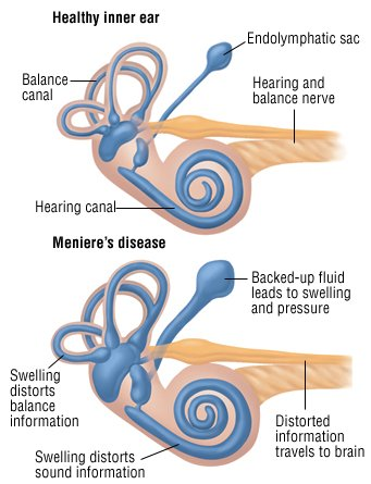
Balance Disorders: Meniere’s disease

  • Buildup of endolymph within the inner ear

  • Disrupts functioning of hair cells

  • Unclear cause

<ul><li><p>Buildup of <strong>endolymph</strong> within the <strong>inner ear</strong></p></li><li><p>Disrupts functioning of <strong>hair cells</strong></p></li><li><p>Unclear cause</p></li></ul><p></p>