Unit 5 - Intro & Autonomic NS
1/22
There's no tags or description
Looks like no tags are added yet.
Name | Mastery | Learn | Test | Matching | Spaced | Call with Kai |
|---|
No analytics yet
Send a link to your students to track their progress
23 Terms
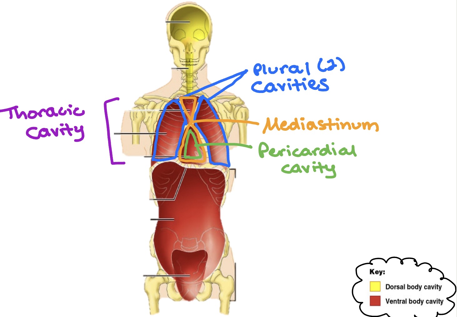
Body cavities - thoracic
Thoracic cavity:
Plural cavities (2) → each surrounds a lung
Mediastinum → surrounds heart & other structures
Pericardial cavity → central portion of thoracic within the mediastinum that houses ONLY the heart
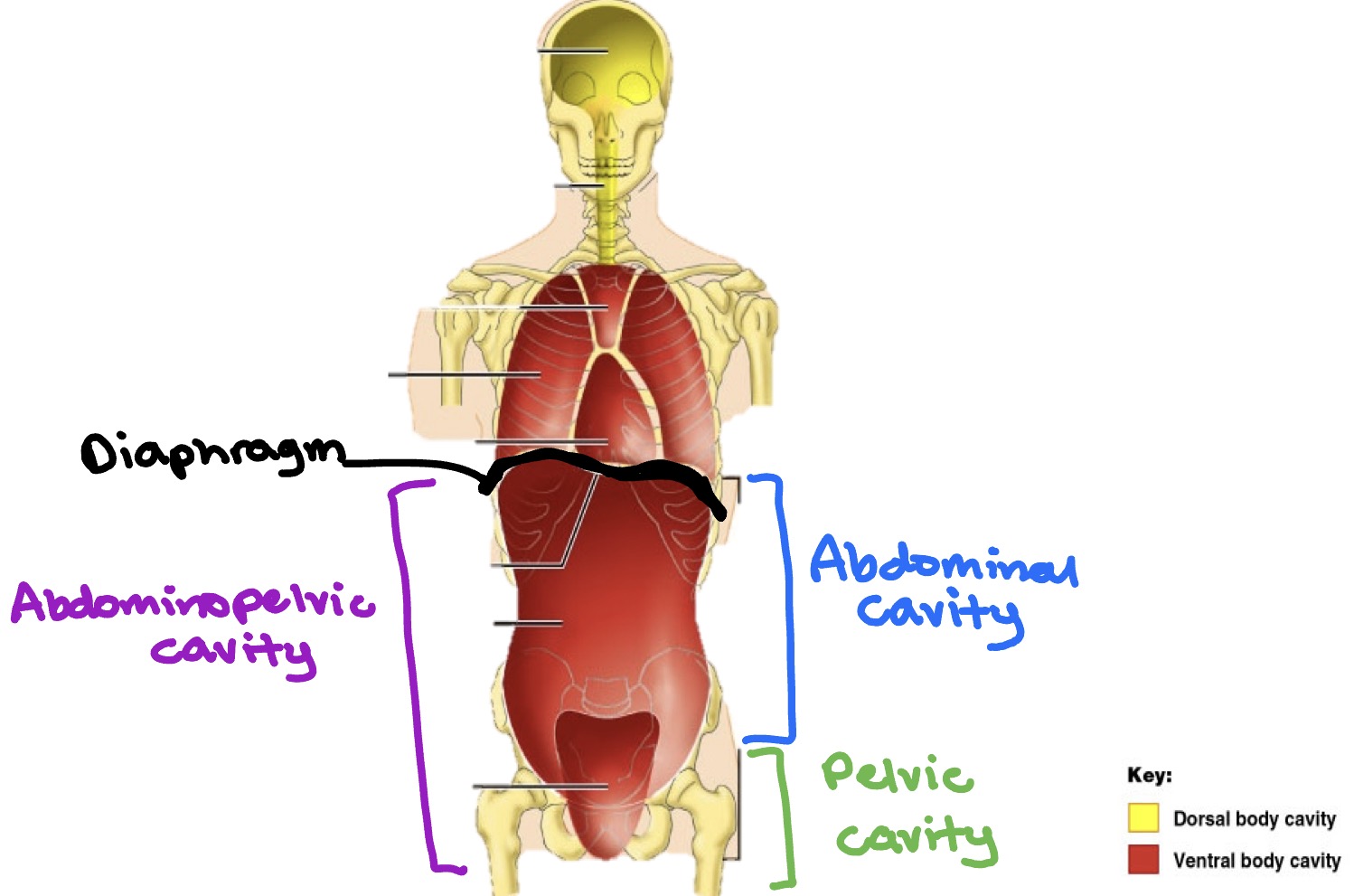
Body cavities - abdominopelvic
(Diaphragm splits thoracic and abdominopelvic cavities)
Abdominopelvic cavity
Abdominal cavity → stomach, spleen, liver, gallbladder, small intestine, most of large intestine
Pelvic cavity → portions of large intestine, urinary bladder, internal reproductive organs
Ventral body cavity - chart
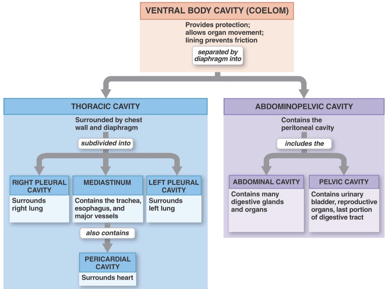
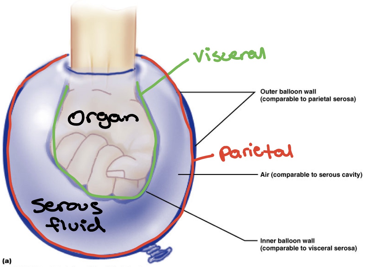
Membranes in Ventral Body Cavity
Viscera (organs) & walls of ventral body cavity are lined in a serous membrane
Parietal layer - lines cavity walls
Visceral layer - adheres to surface of organ
Serous fluid is b/w the 2 layers to reduce friction
Named Serous Membranes
Parietal/visceral pleura - pleural cavity & lung surface
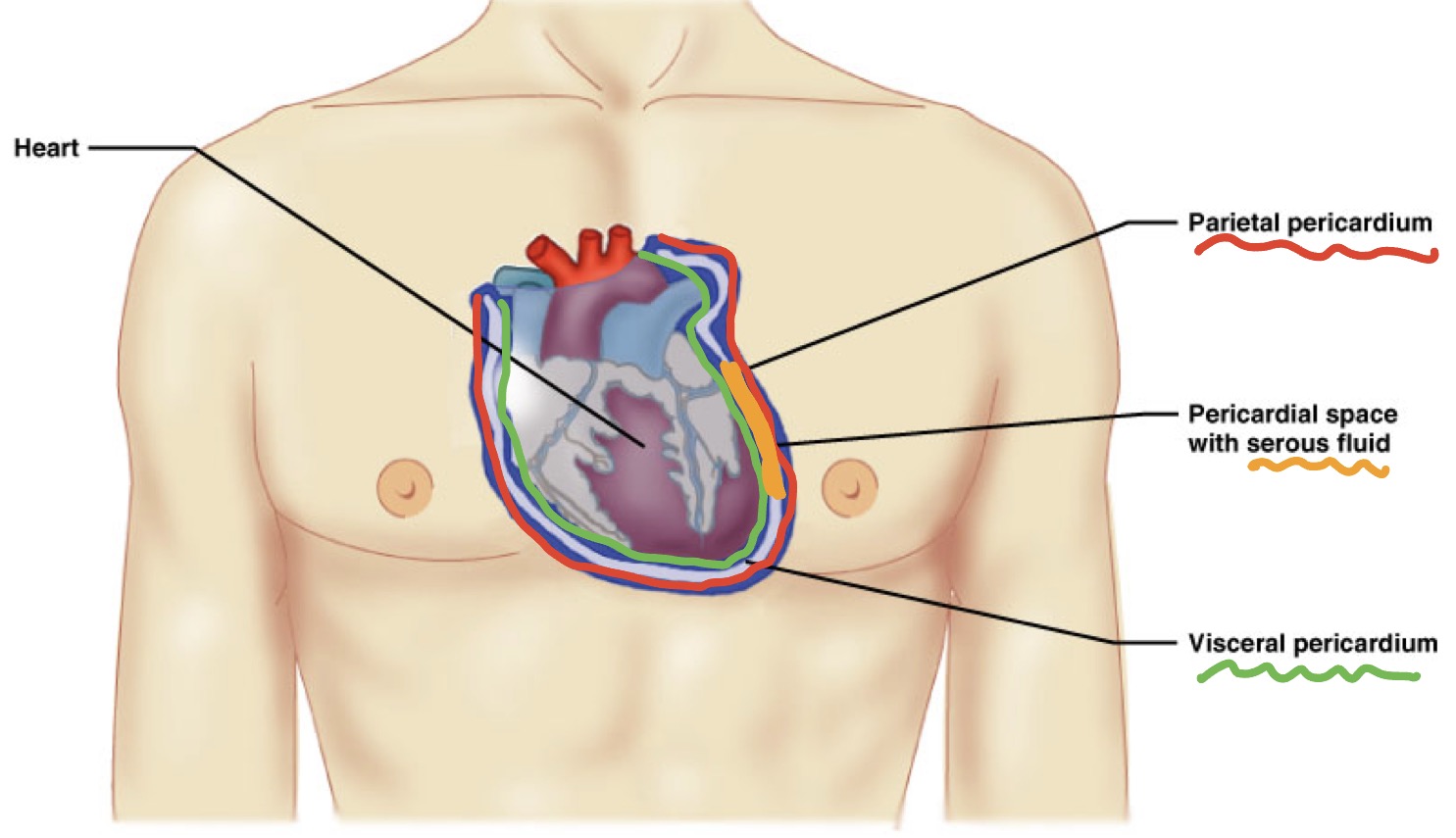
Parietal/visceral pericardium - pericardial cavity & heart surface
Parietal/visceral peritoneum - abdominopelvic cavity & viscera inside it
Membranes in Ventral Body Cavity - representation
Membranes in Ventral Body Cavity - heart visual
Organization of NS - chart
Autonomic
Motor: smooth muscle, cardiac muscle, glands
Sensory: from organs
Divides into…
Sympathetic - fight or flight
Parasympathetic - rest & digest
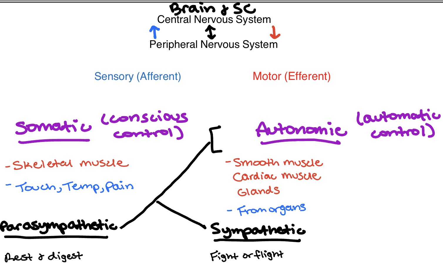
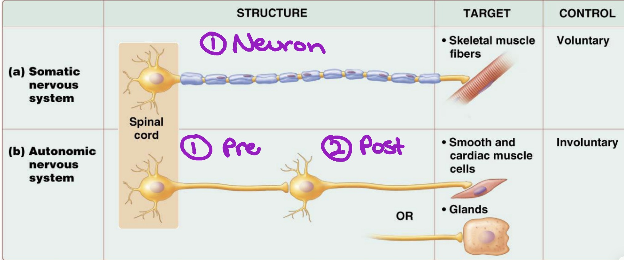
Somatic NS
Conscious control
Effector: skeletal muscle
Efferent pathway: one neuron system
Neurotransmitter & response of target organ: acetylcholine (excitatory)

Autonomic NS
Automatic control
Effector: cardiac & smooth muscle & glands
Efferent pathway: 2 neuron system (presynaptic & postsynaptic)
Neurotransmitter & response of target organ:
Presynaptic neuron: acetylcholine (excitatory)
Postsynaptic neuron: varies b/w sympathetic (epi & norepi) & parasympathetic (acetylcholine)
Either excitatory or inhibitory
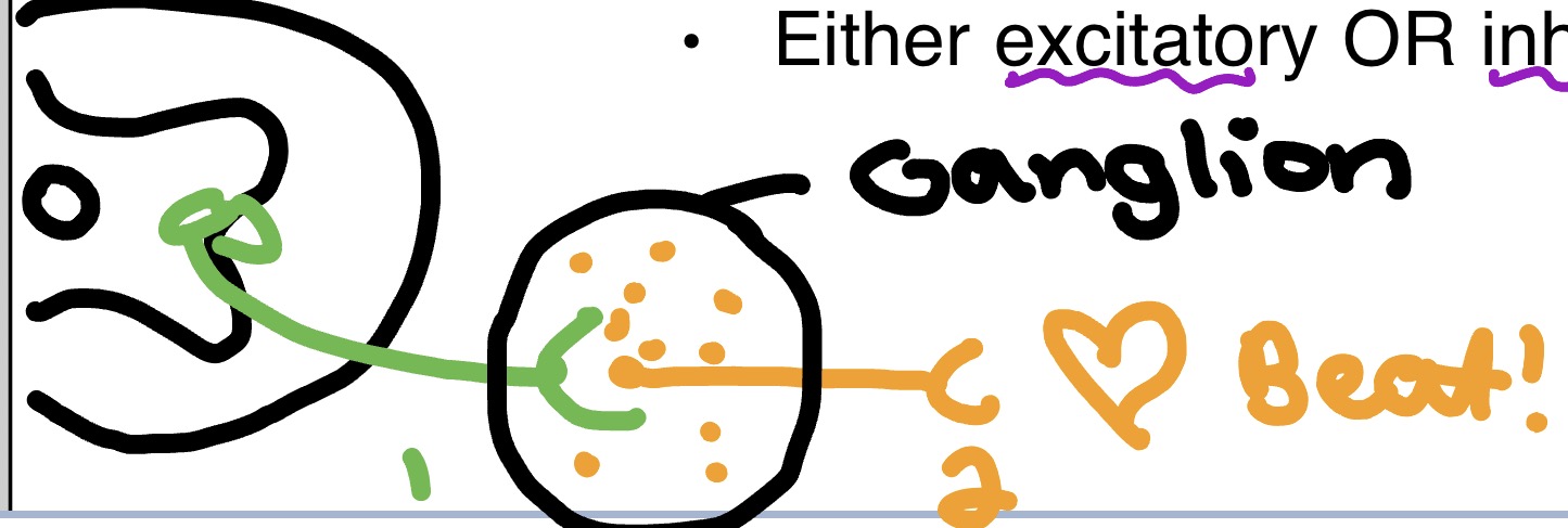
Somatic vs. Autonomic - neurons
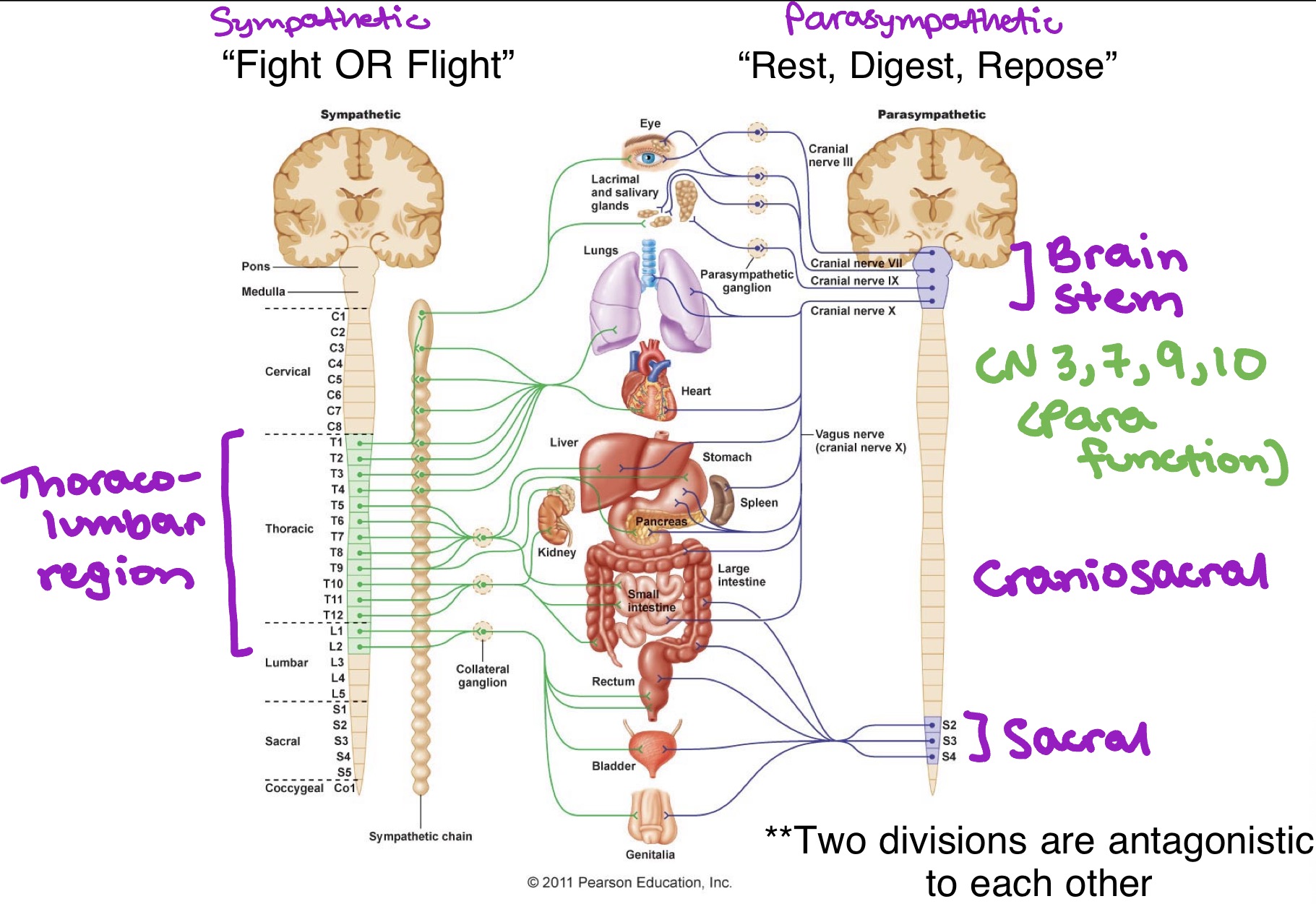
2 divisions of Autonomic NS
Sympathetic (fight of flight)
Thoracolumbar region (T1-L2)
Parasympathetic (reset & digest)
Brain stem & sacral (S2-S4) → craniosacral
CN 3, 7, 9, 10 = parasympathetic function
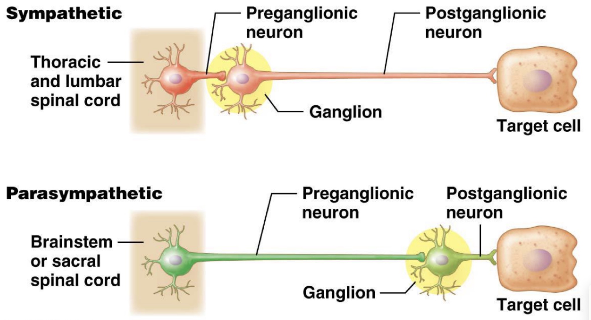
Sympathetic NS - neurons
Origin of presynaptic neuron cell body:
Thoracolumbar region of spinal cord
Fiber length:
Presynaptic → short
Postsynaptic → long
Location of ganglia:
Close to spinal cord
Parasympathetic NS - neurons
Origin of presynaptic neuron cell body:
Brain & sacral spinal cord (craniosacral division)
Fiber length:
Presynaptic → long
Postsynaptic → short
Location of ganglia:
In/near visceral effector organs
Sympathetic & parasympathetic neurons - compared
Parasympathetic NS - info
Only innervates internal organs
Generally inhibits body function
Only major body function not inhibited is digestion
Location of presynaptic neuron cell bodies: craniosacral
Nuclei for CN 3, 7, 9, 10
S2-S4 spinal cord levels
Synapse b/w pre & post synaptic ganglia:
At terminal ganglia
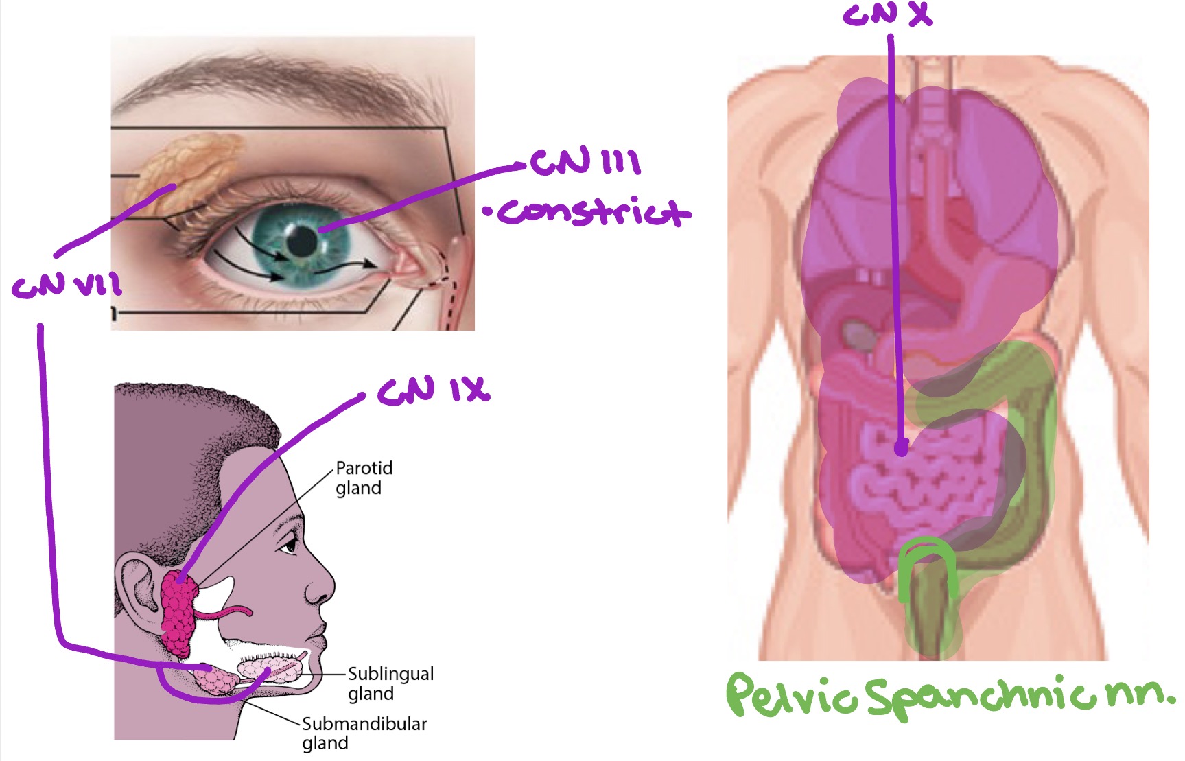
Parasympathetic NS - cranial outflow
Presynaptic fibers run via:
Oculomotor n. (CN 3) - smooth muscle in eye
Facial n. (CN 7) - lacrimal, submandibular, & sublingual glands
Glossopharyngeal n. (CN 9) - parotid gland
Vagus n. (CN 10) - organs in thorax & GI tract → 2/3 tranverse colon
Parasympathetic NS - sacral outflow
Presynaptic neurons originate from S2-S4 level of spinal cord
Fibers of presynaptic neurons travel through ventral root → spinal n. → ventral rami
Exit ventral rami as pelvic splanchnic nerves
Pelvic splanchnic nerves synapse in intramural ganglia (at/near effector)
Postsynaptic fibers innervates remainder of GI tract from distal 1/3 of transverse colon & pelvic viscera
Parasympathetic NS - summary
Sympathetic NS - info
More widespread than parasympathetic NS
Generally stimulates body functions
Only major body function not stimulated is digestion
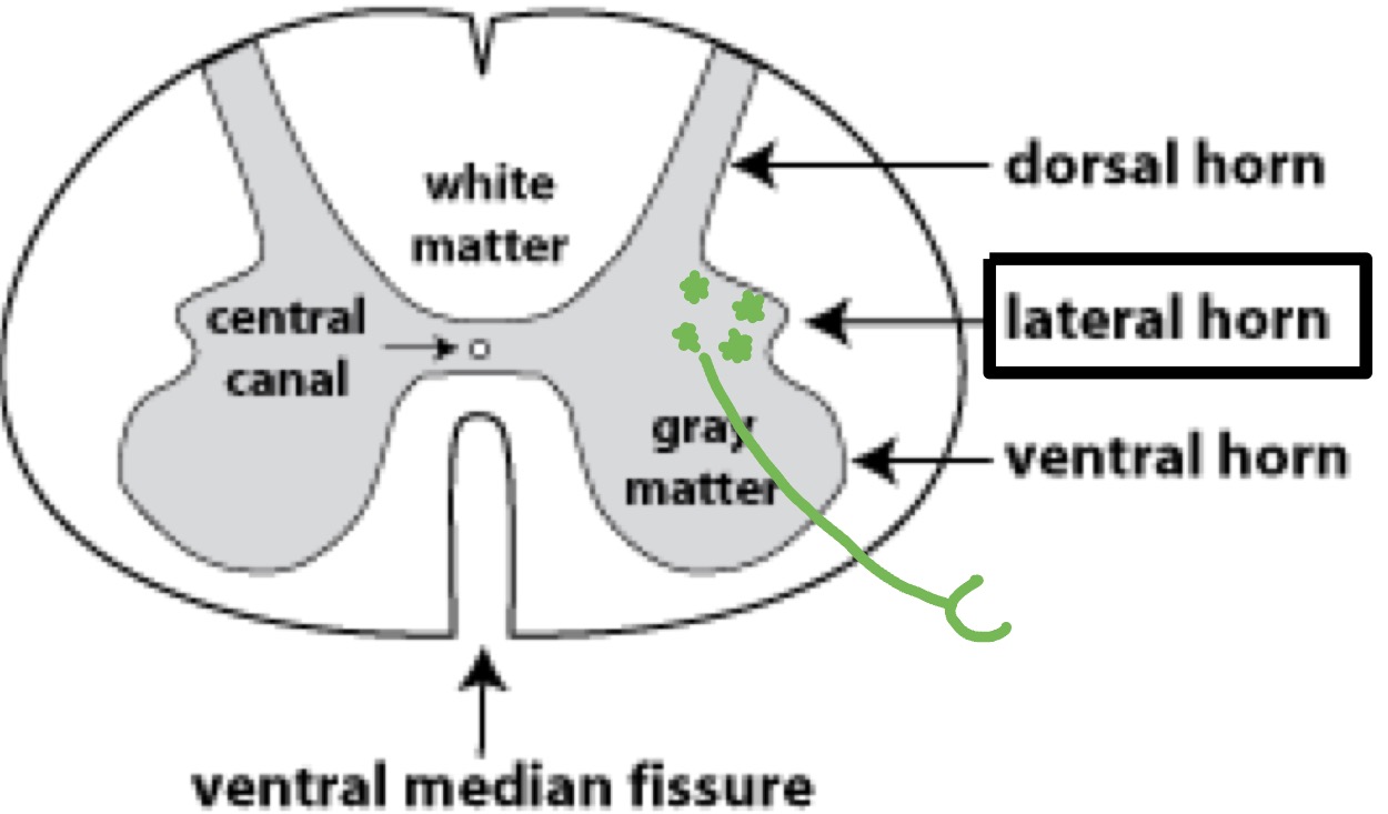
Location of presynaptic neuron cells bodies:
Thoracolumbar region of spinal cord (T1-L2) → lateral horn
Synapse b/w pre & post synaptic neurons:
Paravertebral ganglia: sympathetic chain
Prevertebral ganglia: on abdominal aorta
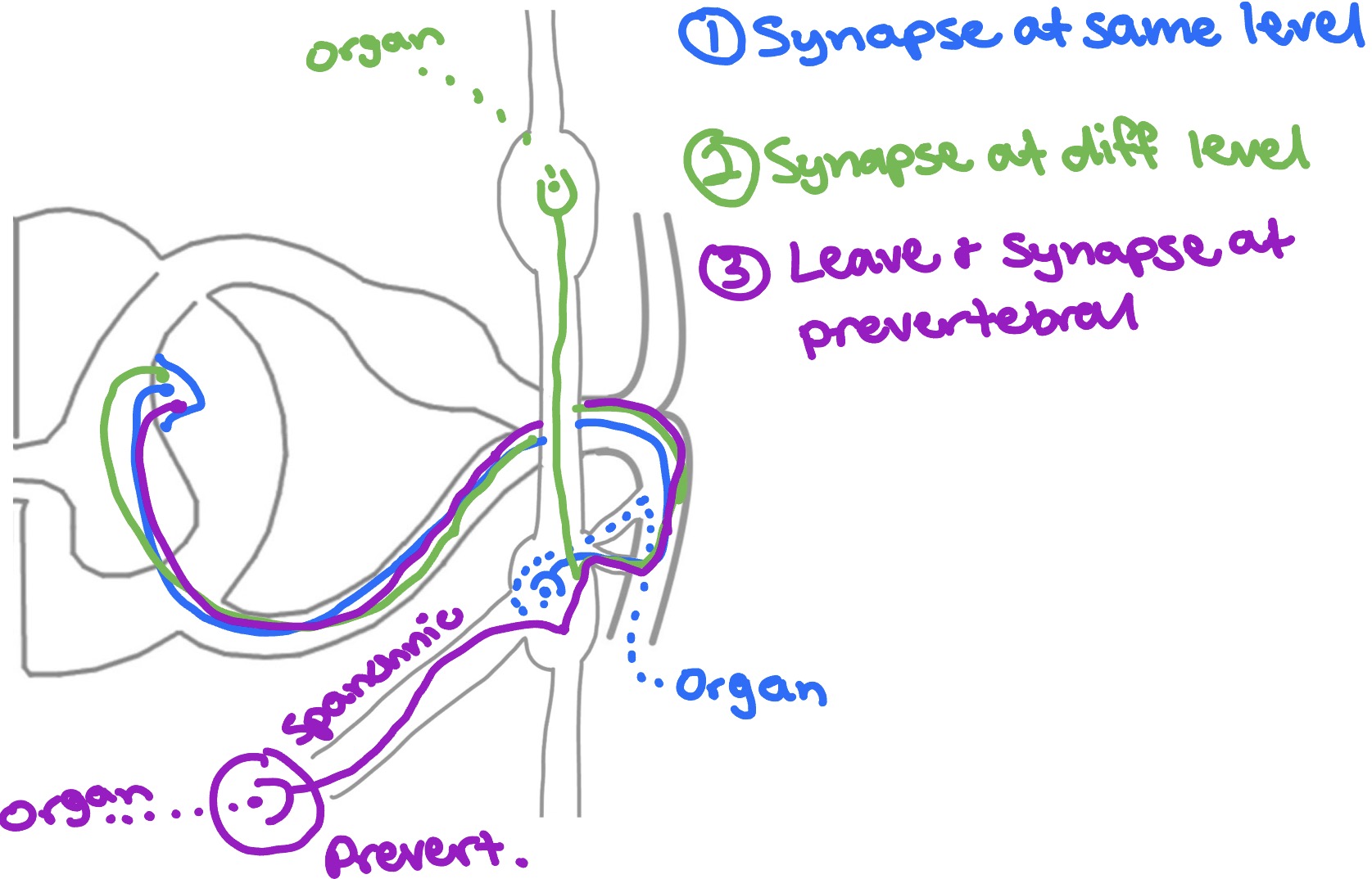
Sympathetic NS - outflow
3 options for pathways:
1. Synapse at same level
2. Synapse at different level
3. Leave & synapse at prevertebral (Spanchnic nerve)
Adrenal Medulla
Postsynaptic sympathetic neurons
Located in medulla of adrenal gland
Does not develop axons, but still produces epinephrin & norepinephrine which will have sympathetic effects on target structures

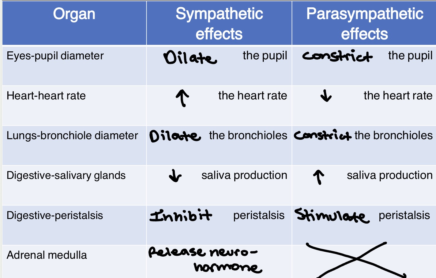
Effects of Autonomic Divisions - chart