lab 5 exam
1/44
There's no tags or description
Looks like no tags are added yet.
Name | Mastery | Learn | Test | Matching | Spaced | Call with Kai |
|---|
No analytics yet
Send a link to your students to track their progress
45 Terms
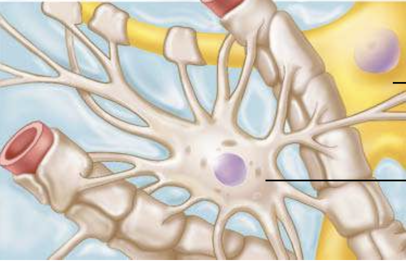
ID the cell type, location of cell type, function
Astrocytes (CNS) - supports neurons, anchor neurons to capillaries, and help form blood-brain barrier
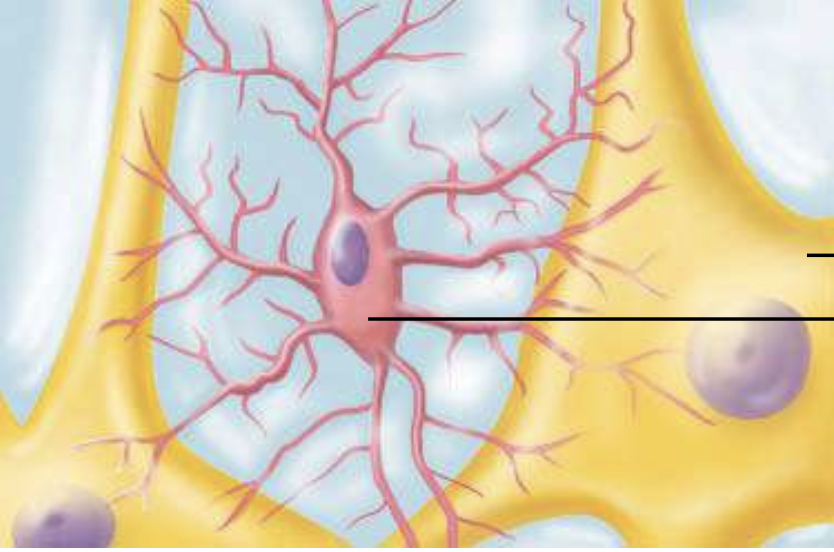
ID the cell type, location of cell type, function
Microglia (CNS) – immune defense, engulf bacteria/debris (phagocytosis)
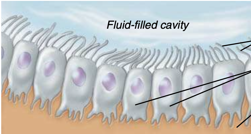
ID the cell type, location of cell type, function
Ependymal (CNS) – produce and circulate CSF (cerebrospinal fluid)
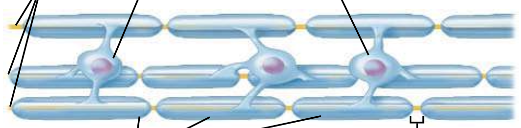
ID the cell type, location of cell type, function
Oligodendrocytes (CNS) – forms the myelin sheath around axons (insulation → faster signals)

ID the cell type(purple), location of cell type, function
Satellite (PNS ganglia) –support and protect neuron cell bodies
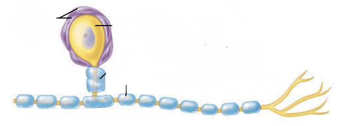
ID the cell type(blue), location of cell type, function
Schwann (PNS) – form myelin sheath in PSN
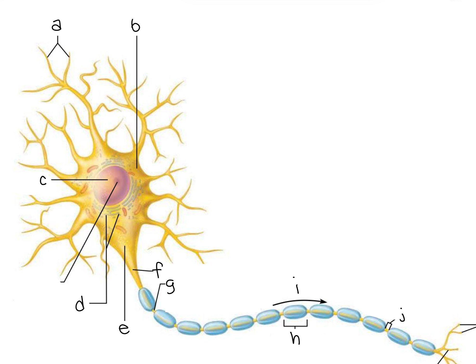
label
a. dendrites
b. cell body
c. nucleus
d. chromatophilic substance (rough endoplasmic reticulum, RER)
e. axon hillock
f. initial segment of axon = axon hillock
g. axon
h. schwann cell
i. impulse direction
j. myelin sheath gap = node of ranvier
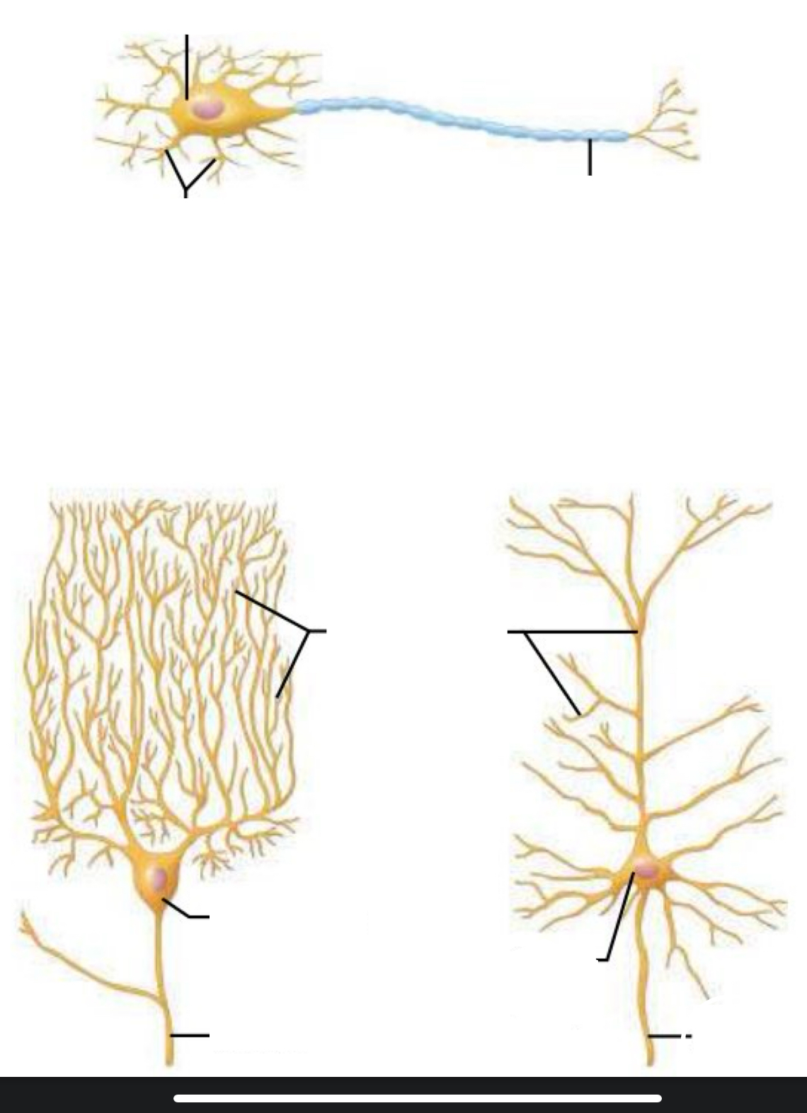
Identity neuron type and location
multipolar
location: purkinje cell of cerebellum (brain) + pyramidal cell (CNS)
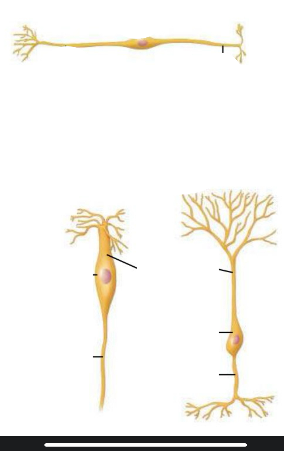
Identity neuron type and location
bipolar
location: olfactory cell (nose) + retinal cell (eye)
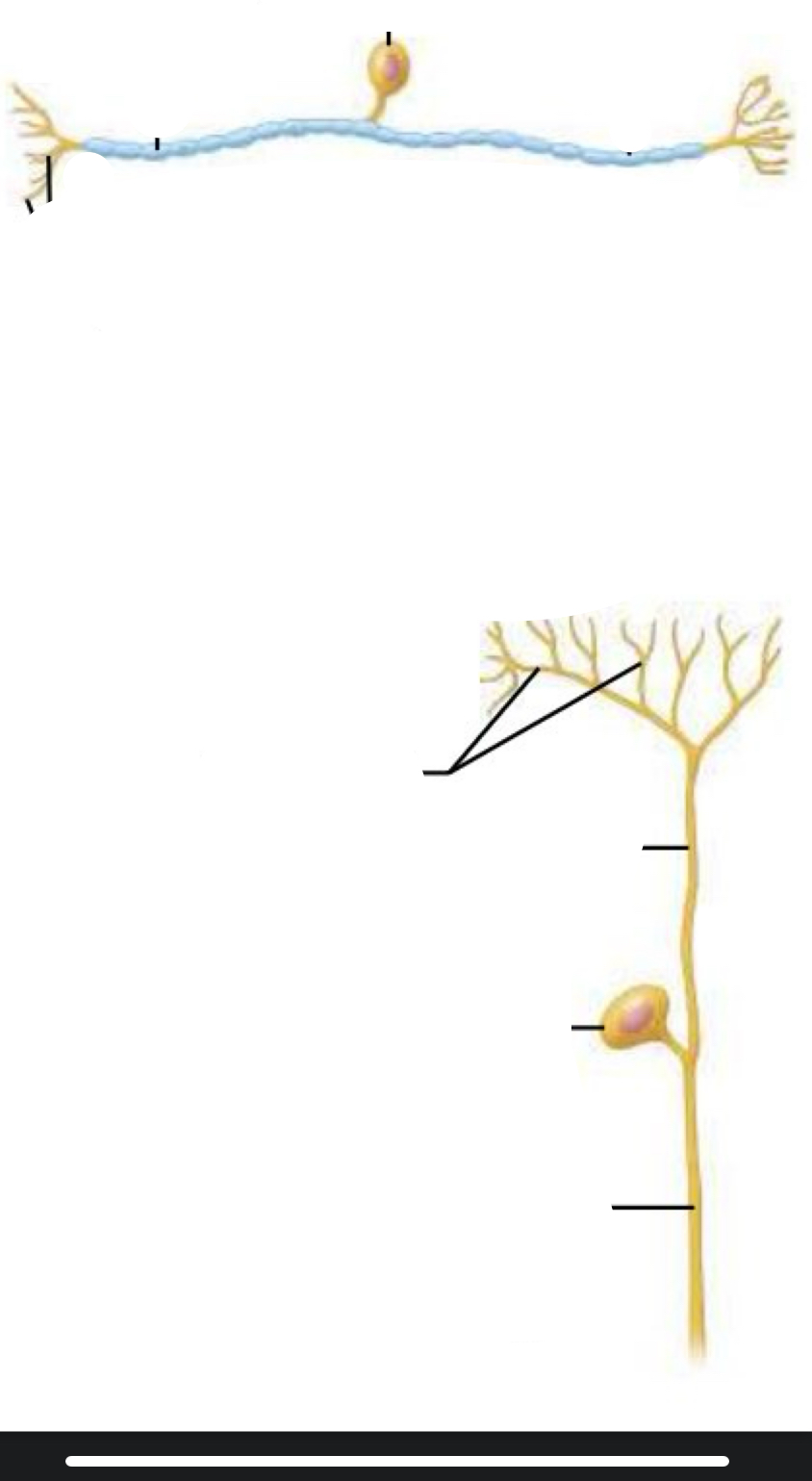
Identity neuron type and location
unipolar
location: dorsal root ganglion cell (PNS)
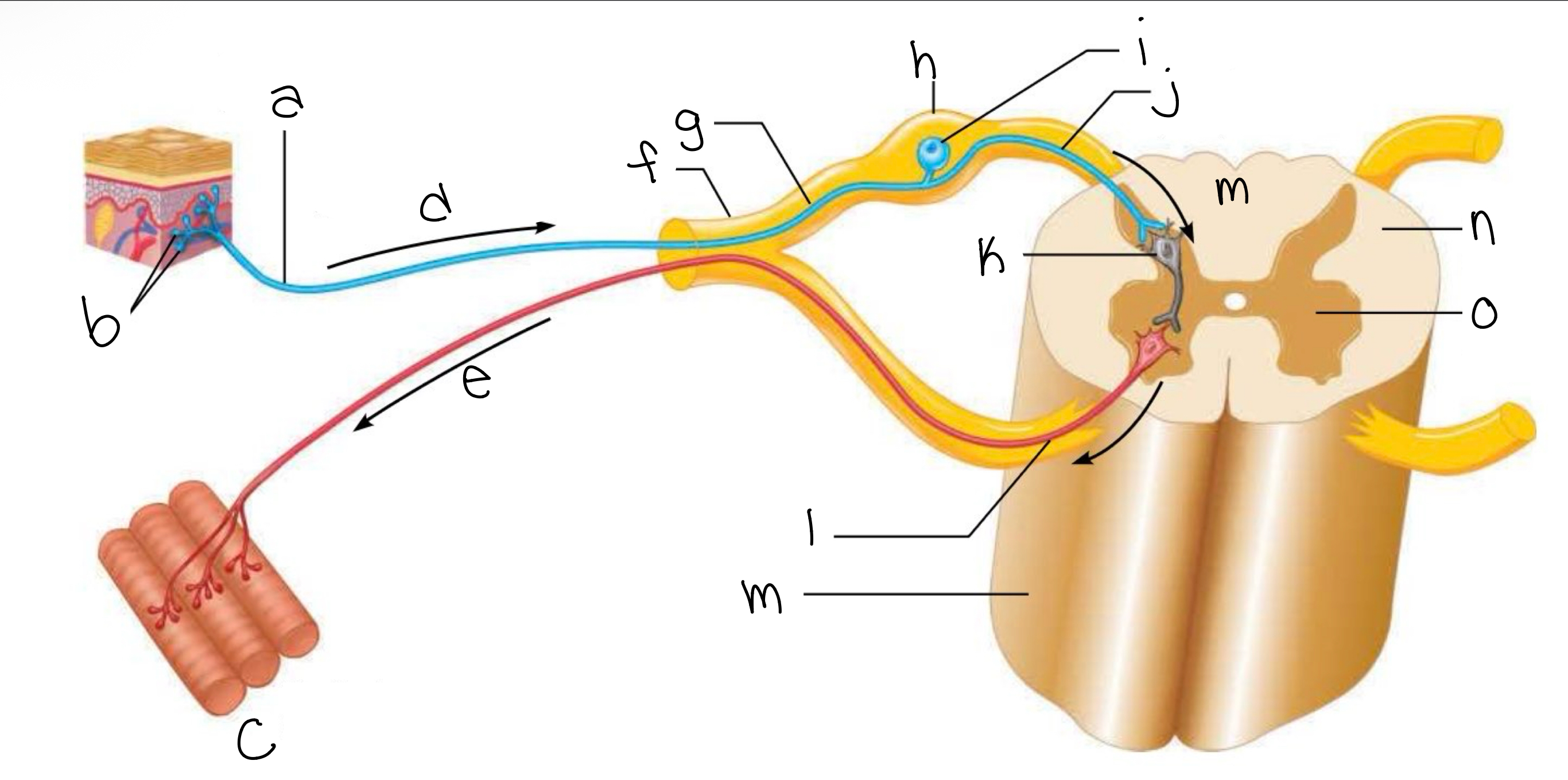
label picture
a. peripheral process (axon)
b. receptive endings
c. skeletal muscles
d. afferent transmission (sensory input)
e. efferent transmission (motor output)
f. spinal nerve
g. afferent sensory neuron
h. dorsal root ganglion
i. cell body
j. central process (axon)
k. between interneuron or association neuron
l. efferent motor neuron
m. spinal cord
n. white mater
o. gray mater
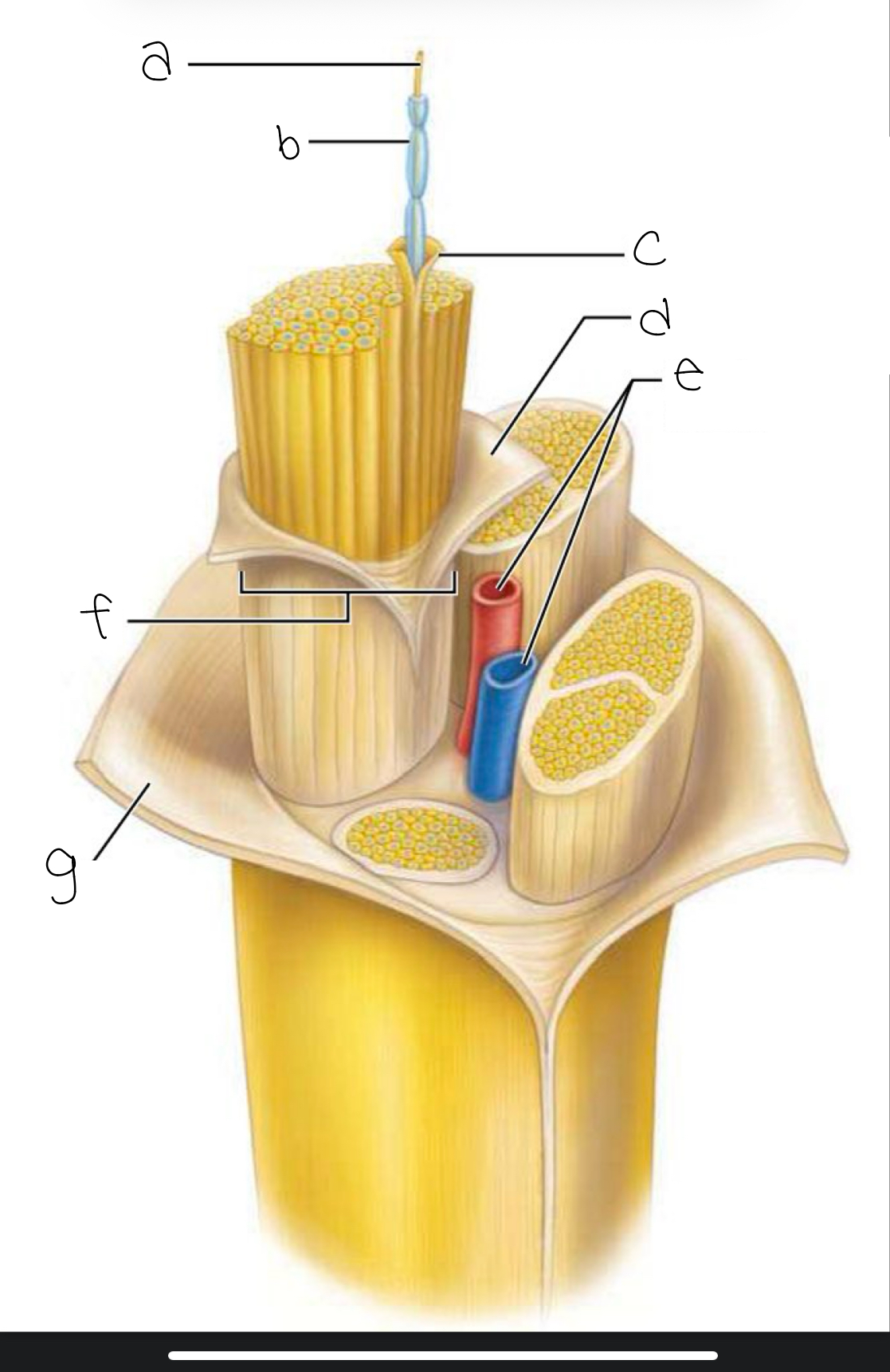
label picture
a. axon = nerve fiber
b. myelin sheath
c. endoneurium
d. perineurium
e. blood vessels
f. fascicle
g.epineurium
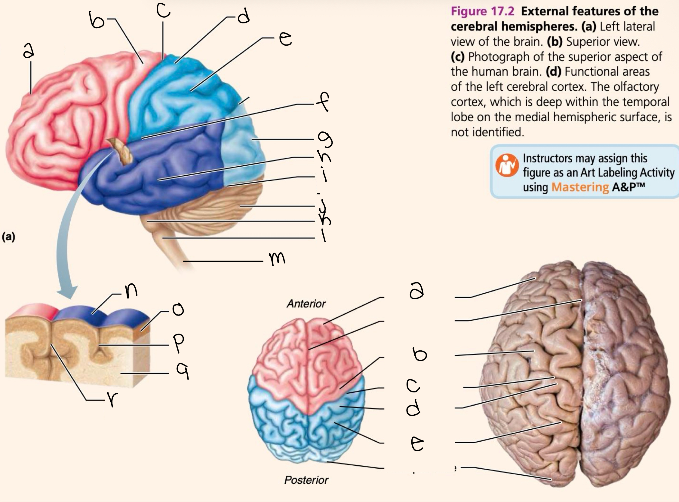
label picture
a. frontal lobe
b. precentral gyrus
c. central sulcus
d. postcentral gyrus
e.parietal lobe
f. lateral sulcus
g. occipital lobe
h. temporal lobe
i. transverse cerebral fissure
j. cerebellum
k. pons
l. medulla oblongata
m. spinal cord
n. gyrus
o. cortex (gray mater)
p. sulcus
q. white mater
r. fissure
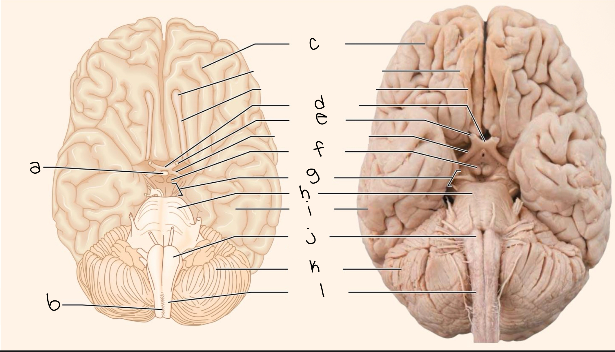
label picture
a. pituitary gland
b. decussation of pyramids
c. frontal lobe
d. optic chiasma
e. optic nerve
f. mammillary body
g. midbrain
h. pons
i. temporal bone
j. medulla oblongata
k. cerebellum
l. spinal cord
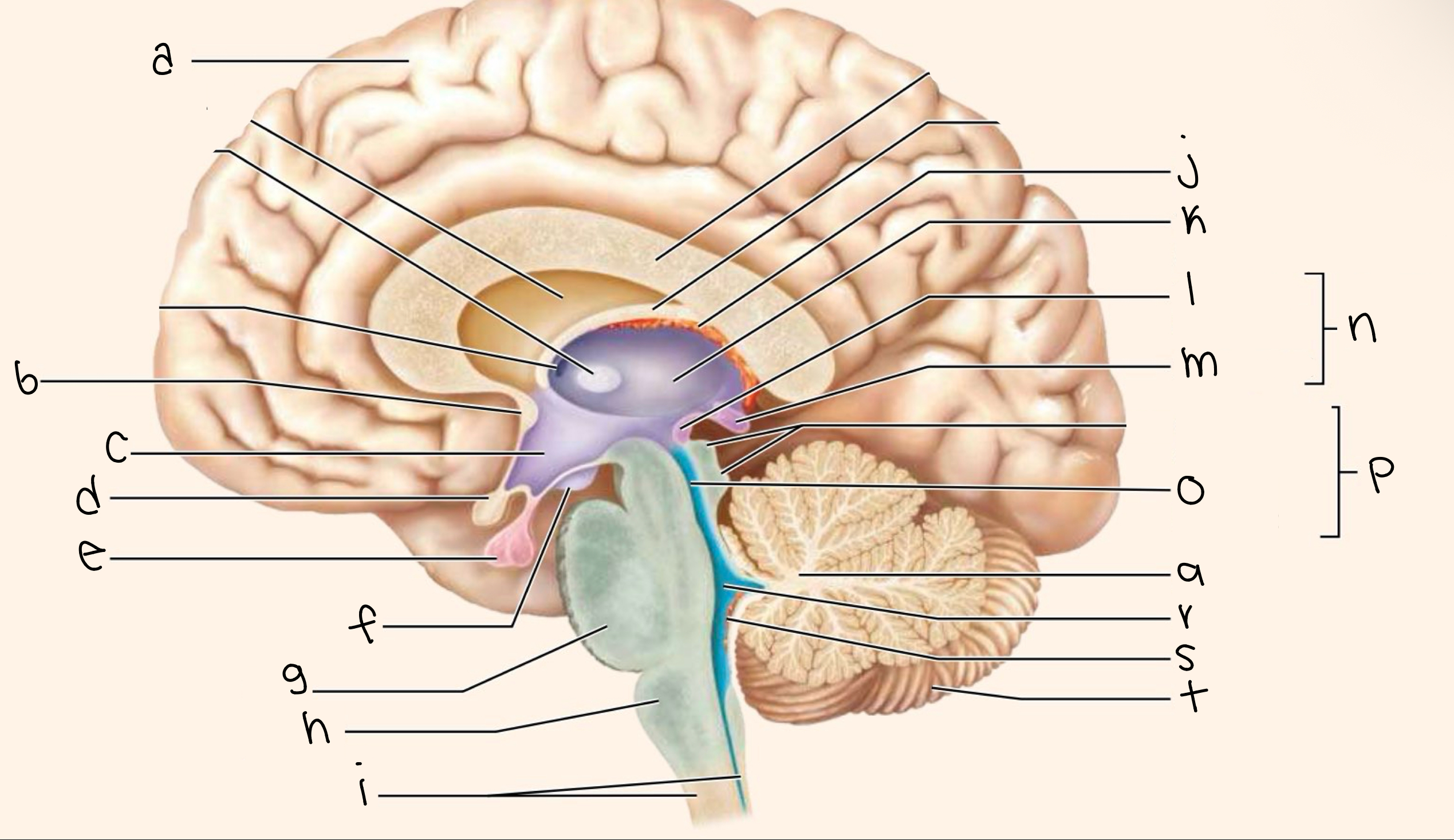
label picture
a. cerebral hemisphere
b. anterior commissure
c. hypothalamus
d. optic chiasma
e. pituitary gland
f. mammillary
g. pons
h.. medulla oblongata
i. spinal cord
j. choroid plexus of 3rd ventricle
k. thalamus
l. posterior commissure
m. pineal gland (melatonin)
n. epithalamus
o. cerebral aqueduct
p. midbrain
q. arbor vitae of cerebellum (tree of life)
r. fourth ventricle
s. choroid plexus
t. cerebellum
name the 6 pituitary gland hormones
gh (growth hormone)
fsh (follicle stimulating hormone)
lh (luteinizing hormone)
act h (adrenocorticotropic hormone)
ths (thyroid stimulating hormone)
prl (prolactin)
what hormone does the pituitary gland release to stimulate bone and tissue growth?
gh (growth hormone)
which pituitary hormone targets the reproductive organs and stimulates follicle development?
fsh (follicle stimulating hormone)
which pituitary hormone acts on reproductive organs to trigger ovulation or testosterone production?
lh (luteinizing hormone)
which pituitary hormone stimulates the adrenal glands?
act h (adrenocorticotropic hormone)
which pituitary hormone targets the thyroid gland?
ths (thyroid stimulating hormone)
which pituitary hormone acts on the mammary glands to produce milk?
prl (prolactin)
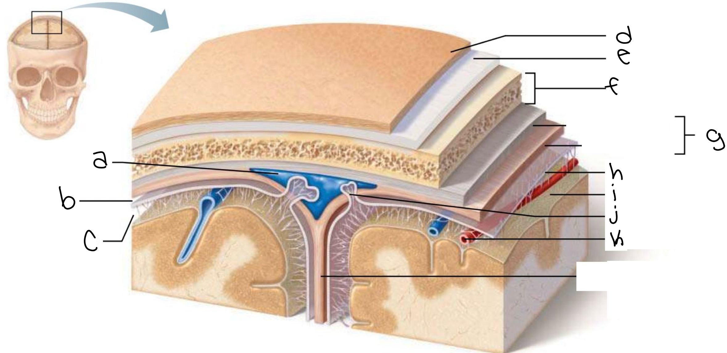
label picture
a. superior sagittal sinus
b. subdural space
c. subarachnoid space
d. skin of scalp
e. periosteum
f. bone of skull
g. dura mater
h. arachnoid mater
i. pia mater
j. arachnoid granulation
k. blood vessel
what are the 5 levels of brain protection
skin of scalp
periosteum + bone of skull
brain meninges (dura mater, arachnoid mater, pia mater)
CFS in brain ventricles
BBB (blood brain barrier)
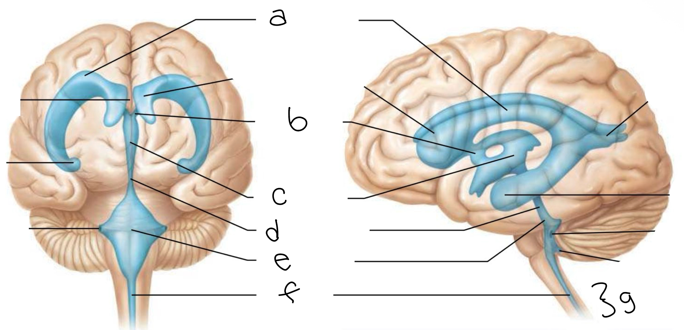
label picture
a. lateral ventricle
b. interventricular foramen
c. third ventricle
d. cerebral aqueduct
e. fourth ventrical
f. central canal
g. spinal cord
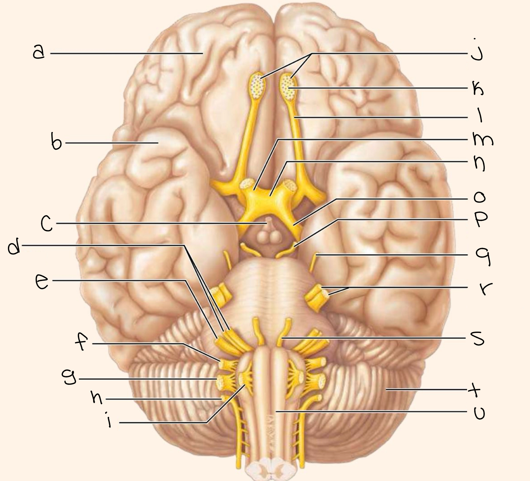
label picture
a. frontal lobe
b. temporal lobe
c. infundibulum
d. facial nerve (VII)
e. vestibulocochlear nerve (VIII)
f. glossopharyngeal nerve (IX)
g. vagus nerve (X)
h. accessory nerve (XI)
i. hypoglossal nerve (XII)
j. filaments of oifactory nerve (I)
k. olfactory bulb
l. olfactory tract
m. optic nerve (II)
n. optic chiasma
o. optic tract
p. oculomotor nerve (III)
q. trochlear nerve (IV)
r. trigeminal nerve (V)
s. abducens nerve (VI)
t. cerebellum
u. medulla oblongata
nerve type? function? location?
I. olfactory
sensory
smell
nose → olfactory bulb
nerve type? function? location?
II. optic
sensory
vision
retina → brain
nerve type? function? location?
III. oculomotor
motor
eye movement + pupil constriction
midbrain → eye muscle
nerve type? function? location?
IV. trochlear
motor
moves eye down
midbrain → superior oblique
nerve type? function? location?
V. trigeminal
mixed
face sensation + chewing
face (3 branches=ophthalmic, maxillary, and mandibular)
nerve type? function? location?
VI. abducens
motor
moves eye outward
pons → lateral rectus
nerve type? function? location?
VII. facial
mixed
facial expression + taste (front tongue) + saliva/tears
pons → face
nerve type? function? location?
VIII. vestibulocochlear
sensory
hearing + balance
inner ear → brain
nerve type? function? location?
IX. glossopharyngeal
mixed
taste (back tongue) + swallowing + saliva
medulla → throat
nerve type? function? location?
X. vagus
mixed
controls organs (heart, lungs, digestion)
medulla → thorax and abdomen
nerve type? function? location?
XI. accessory
motor
head turn + shoulder shrug
spinal cord → neck muscles
nerve type? function? location?
XII. hypoglossal
motor
tongue movement
medulla → tongue
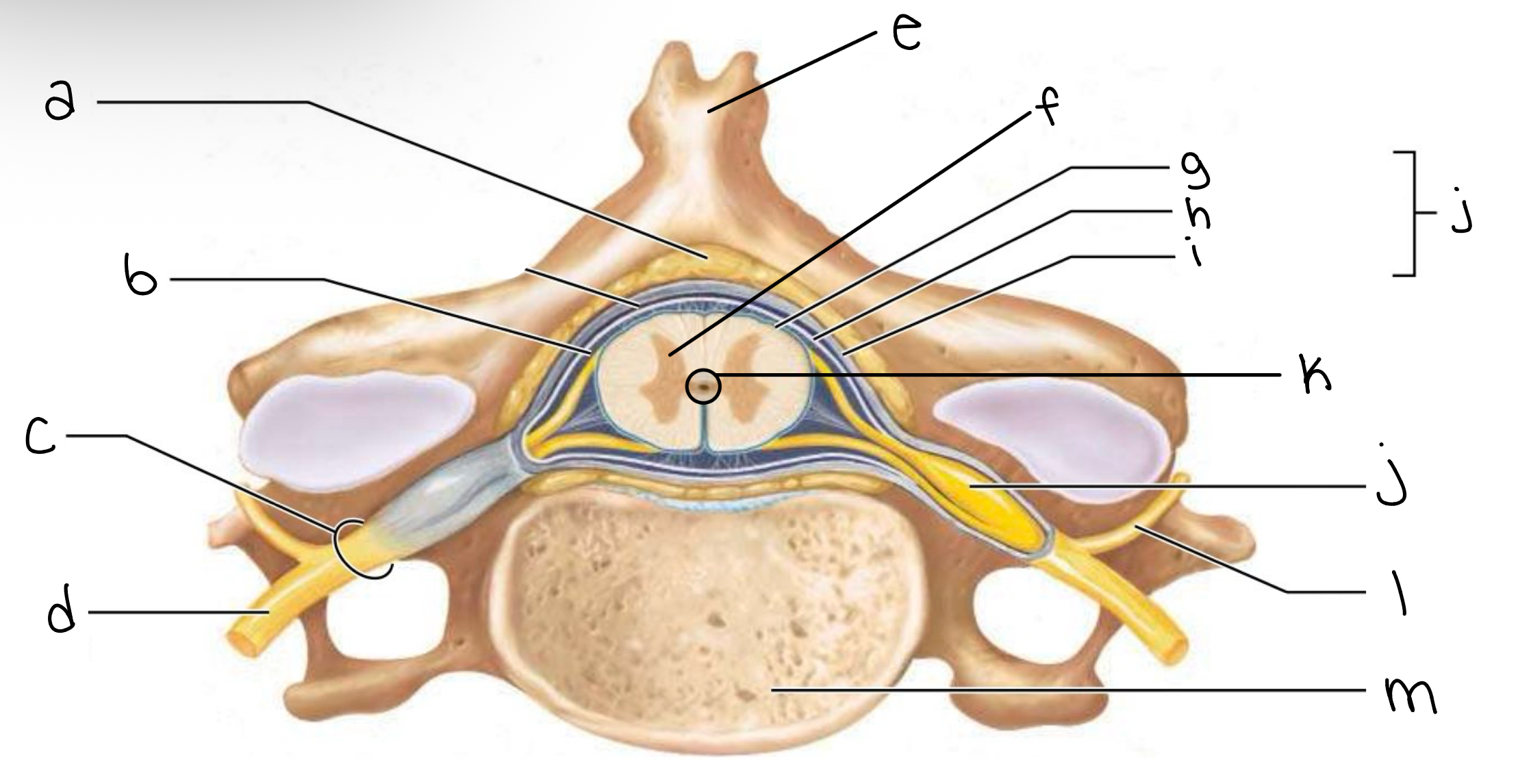
label picture
a. epidural space (contains fat)
b. subarachnoid space
c. spinal nerve
d. ventral ramus of spinal nerve
e. spinous process of cervical vertabrae
f. gray commissure
g. pia mater
h. arachnoid mater
i. dura mater
j. spinal meninges
k. central canal
l. dorsal ramus of spinal nerve
m. body of vertebra
define plexus
is a (complex) network of spinal serves which together serve a particular body region
list the 4 plexuses
cervical plexus (C1-C5)
brachial plexus (C5-T1)
lumbar plexus (L1-L4)
sacral plexus (L4-S4)
name the working area and one nerve of the cervical plexus
neck + shoulder
phrenic nerve
name the working area and one nerve of the brachial plexus
shoulders + arms + upper limbs
axillary nerve
name the working area and one nerve of the lumbar plexus
lower abdomen + anterior thigh
femoral nerve
name the working area and one nerve of the sacral plexus
posterior thigh + lower leg + foot
sciatic nerve