lutfi back and spinal chord 1
1/27
There's no tags or description
Looks like no tags are added yet.
Name | Mastery | Learn | Test | Matching | Spaced | Call with Kai |
|---|
No analytics yet
Send a link to your students to track their progress
28 Terms
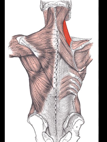
the nuchal ligament/ line is the origin of what muscle ? (along with spinusproces of t7-t12
trpezius musccle
OINF of trapezius muscle ?
O-ligamentum nuchae, superior nuchal line, spinous processes of C7- T12 •
I-lateral 1/3 of the clavicle, acromion, crest of the scapula
N-accessory spinal (CN XI)
F-elevates the shoulder, pulls the scap

OINF of latissimus dorsi?
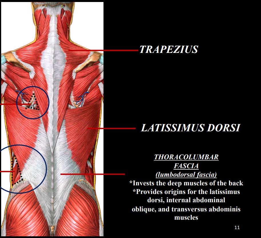
O-spinous processes of the lumbar vertebrae, lower 6 thoracic vertebrae, thoracolumbar fascia, iliac crest •
I-floor of the bicipital groove of humerus •
N-thoracodorsal n. •
F-adduction and extension of the UE
which two muscles form the axillary fold (armpit crease)
latissimus dorsi and teres major
borders of the TRIANGLE OF AUSCULTATION ?
Lateral border of trapezius = medial side
Superior border of latissimus dorsi = base
Medial border of scapula = lateral side
*Floor = rhomboid major and ICS 6
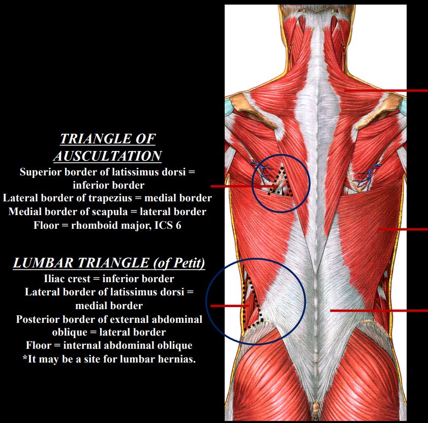
borders of the INFERIOR LUMBAR TRIANGLE (of Petit) ?
Iliac crest = inferior border •
Lateral border of latissimus dorsi = medial border •
Posterior border of external abdominal oblique = lateral border •
Floor = internal abdominal oblique

borders of the SUPERIOR LUMBAR TRIANGLE (Grynfeltt-Lesshaft) ?
Roof: external oblique •
Floor: transversalis fascia •
Superiorly: 12th rib •
Medially: quadratus lumborum
• Laterally: internal oblique
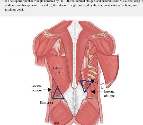
what triangle does the TAP (Transversus Abdominus Plane) anesthesia go through
the triangle of petit or inferiors lumbar triangle
how deep does the TAP anehtsia needle go in the triangle of petit ?
through the exterior oblique , then the interior oblique , then the needle stops before going through the transverse abdominus.
OINF of levator scapule ?
• O-posterior tubercles of transverse processes of C1-C4 •
I-superior angle of the scapula and upper medial border of the scapula •
N-C3-C4 •
F-elevation of the scapula
OINF of Rhomboid minor
O-spinous process of C7, ligamentum nuchae, T1 •
I-spine of the scapula •
N-dorsal scapular nerve •
F-retract and fix the scapula
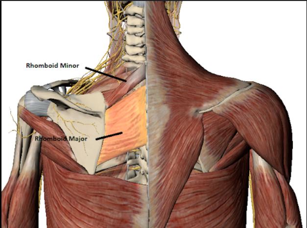
OINF of RHOMBOID MAJOR:?
O-spinous processes of T2-T5 •
I-medial border of scapula •
N-dorsal scapular nerve •
F-retract and fix the scapula

where are the serratus muscles ?
SERRATUS POSTERIOR SUPERIOR: • Deep to the rhomboids. C7, ribs 2- 5, ligamentum nuchae. Supplied by ventral rami of C8-T3 •
SERRATUS POSTERIOR INFERIOR: • Deep to the lastissimus dorsi. Attaches to the lower 4 ribs. Supplied ventral rami of T9-T11
what nerve are all the Intrinsic (Deep) Muscles of the Back supplied by?
the dorsal rami of the spinal nerves.
the odd illiocostalis muscle ?
lumborum = lateral
the three main intrinsic muscle groups of the back are ?
LATERAL COLUMN: • ILIOCOSTALIS MUSCLE: •
INTERMEDIATE COLUMN: • LONGISSIMUS MUSCLE: •
• MEDIAL COLUMN: • SPINALIS MUSCLE: •
the erector spinea or sacropinalis
Deep to the sacrospinalis muscle we find the?
TRANSVERSOSPINALIS MUSCLES:
the multifidus muscle of the back is most robust in what part of the spine?
lumbar
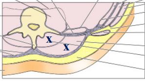
muscles denoted by the x’s deep then superficial?
deep= TRANSVERSOSPINALIS MUSCLE and then superfiscial to that is the SACROSPINALIS OR ERECTOR SPINAE MUSCLE.
boundaries of the sub occipital triangle ?
Rectus Capitis Posterior Major muscle = forms the medial border • (in blue)
superior oblique (Obliquus Capitis Superior) muscle = forms the lateral border • (in green)
inferior oblique (Obliquus Capitis Inferior) muscle = forms the inferior border (in red)
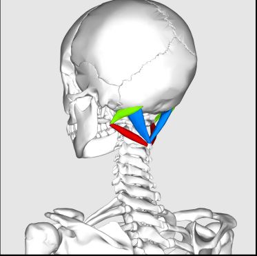
floor of the sub occipital triangle ?
The region has a floor formed by the posterior arch of the atlas and the posterior atlanto-occipital membrane
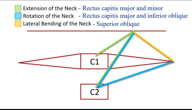
muscles responsible for extension of the neck?
- Rectus capitis major and minor

muscles responsible for rotation of the neck?
Rectus capitis major and inferior oblique

muscle responsible for lateral bending of the neck?
Superior oblique
insertion of the inferior oblique ?
transverse process of atlas
contents of suboccipital triangle?
the vertebral artery, suboccipital nerve (C1), and suboccipital venous plexus.
in the suboccipital triangle , a traumatic injury May occur from a car wreck (rupture of the cruciform ligament) or, from rheumatoid arthritis. this injury is called?
Atlantoaxial dislocation (subluxation)
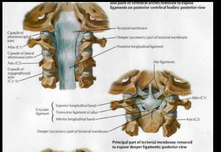
Components of the Occipitoaxial Ligament?
1-Cruciform Ligament a-Transverse lig b-Longitudinal lig 2-Apical Ligament 3-Alar Ligament 4-Tectorial Membrane