Human Anatomy Exam 2
1/169
There's no tags or description
Looks like no tags are added yet.
Name | Mastery | Learn | Test | Matching | Spaced | Call with Kai |
|---|
No analytics yet
Send a link to your students to track their progress
170 Terms
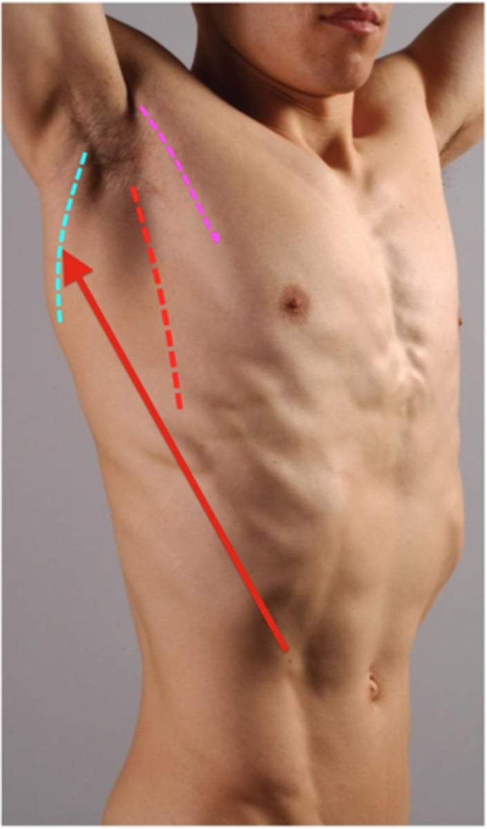
boundaries of the deltopectoral triangle
Clavicle, deltoid, pectoralis major
contents of the deltopectoral triangle
cephalic vein
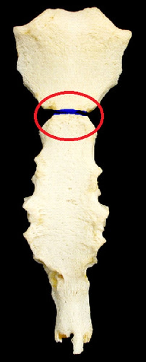
sternal angle
horizontal ridge between the manubrium and body of the sternum
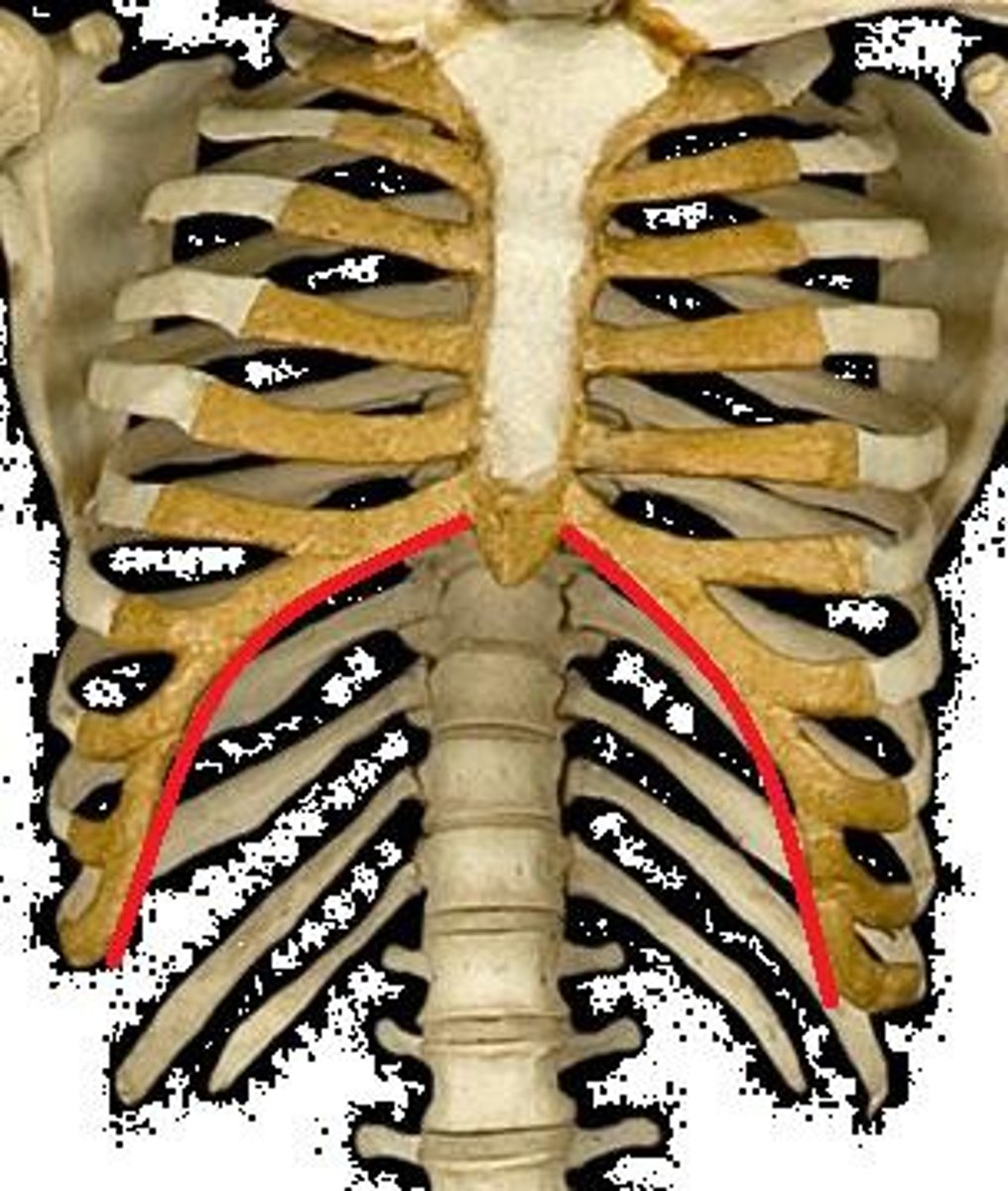
costal margin
lower border of rib margin formed by the medial edges of the 8th, 9th, and 10th ribs - creates the costal angle
vertebral level of iliac crest
L4
vertebral level of inferior angle of scapula
T8
boundaries of the triangle of auscultation
medial border of scapula (superolateral), lateral border of trapezius (superomedial), superior border of latissimus dorsi (inferior)
- thickening of fascia/less muscle here --> you can listen to breathing sounds of the thorax
anterior axillary fold
surface anatomy landmark formed by the pectoralis major

posterior axillary fold
surface anatomy landmark formed by the latissimus dorsi and teres major
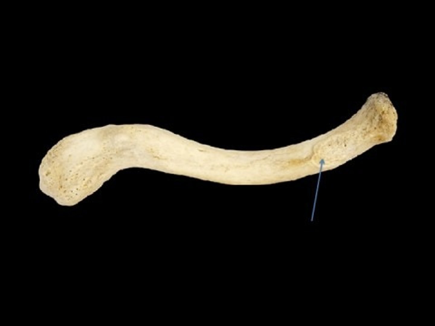
costal tuberosity
broad, roughened spot on the inferior surface of the medial aspect of the clavicle (attachment point for the costoclavicular ligament)
C3 dermatome
lateral neck, base of neck
C4 dermatome
Shoulder area, clavicular area, upper scapular area
C5 dermatome
lateral aspect of the arm
C6 dermatome
lateral forearm and thumb
C7 dermatome
middle 3 fingers and center of posterior aspect of forearm
C8 dermatome
little finger, medial side of the hand and forearm
T1 dermatome
medial aspect of forearm and inferior arm
T2 dermatome
medial aspect of superior arm and skin of axilla
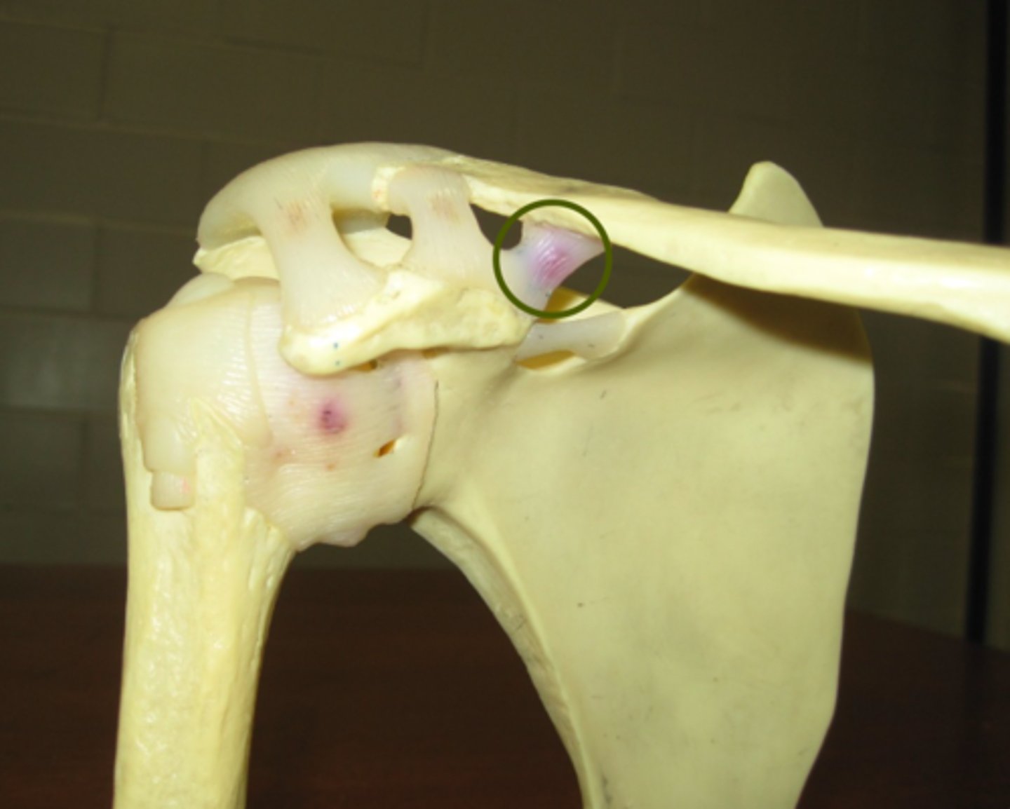
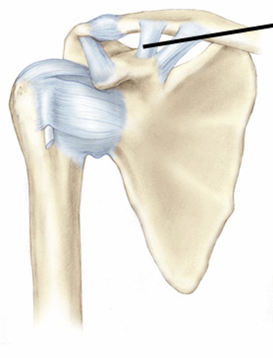
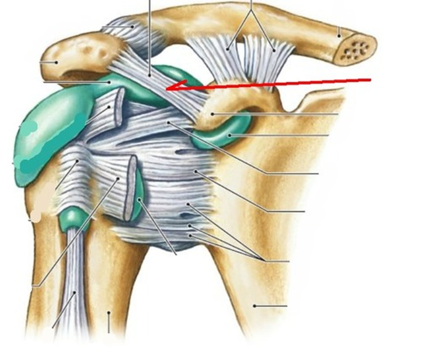
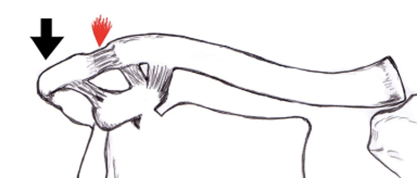
superior transverse scapular ligament
ligament that bridges the suprascapular notch
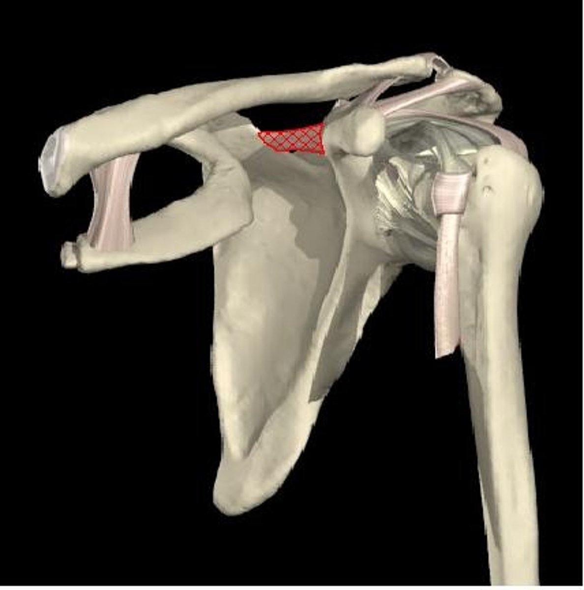
suprascapular artery
branch off subclavian a. --> runs above the superior transverse scapular ligament over the suprascapular notch - supplies blood to supraspinatus and infraspinatus mm. (army goes over the bridge...)
suprascapular nerve
runs below the superior transverse ligament in the suprascapular notch - innervates supraspinatus and infraspinatus mm. (...navy goes under the bridge)
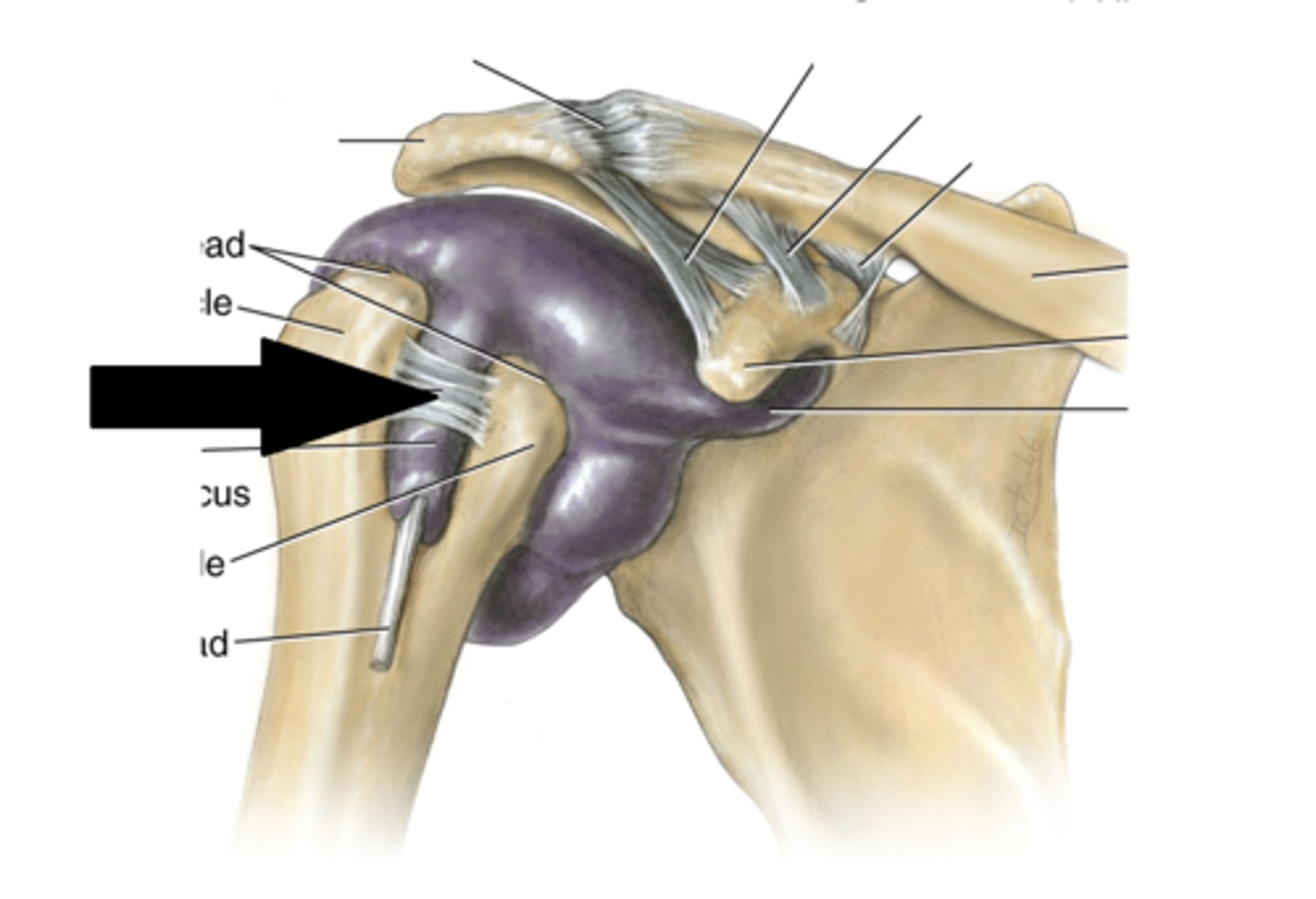
biceps LH tendon v. triceps LH tendon
the biceps LH tendon is nested within the glenohumeral joint, the triceps LH tendon stays separate - leads to more complicated injuries involving the bicep v. the tricep
functions of scapular movement
- keeps glenoid fossa and humeral head congruent
- aligns angle of pull for shoulder joint muscles
- increases ROM of GH joint
- reduces subluxation of humerus
Limb Girdle Muscular Dystrophy
heritable disorder with onset in early 20s, no effect on life expectancy - start w/ fatigue/shakiness when picking up light objects --> rounded slumped shoulder girdle --> muscles wither/weaken (progressive, can take years to decades) - orthopedic surgery to fix the scapula to the thoracic cage --> restores function of UE muscles (explains why proximal function is crucial for distal function)
shoulder girdle
scapula and clavicle
- gives structure and support
- structures originate on axial skeleton and insert on appendicular skeleton (but don't cross the joint ex. rhomboids, trapezius)
- scapula provides position and support for glenohumeral joint
- muscles control movement of the SCAPULA
shoulder joint
glenohumeral joint
- responsible for ROM
- structures originate on axial and appendicular skeleton and cross the joint to insert on the appendicular skeleton
- SITS muscles
- muscles control movement of the ARM
articulations of the sternoclavicular joint
- medial (sternal) end of the clavicle
- clavicular notch of the manubrium,
- the cartilage of the 1st rib
ligaments of the sternoclavicular joint
- anterior sternoclavicular ligament
- posterior sternoclavicular ligament
- interclavicular ligament
- costoclavicular ligament
costoclavicular ligament
ligament that connects costal cartilage of the 1st rib to clavicle (costal tuberosity)
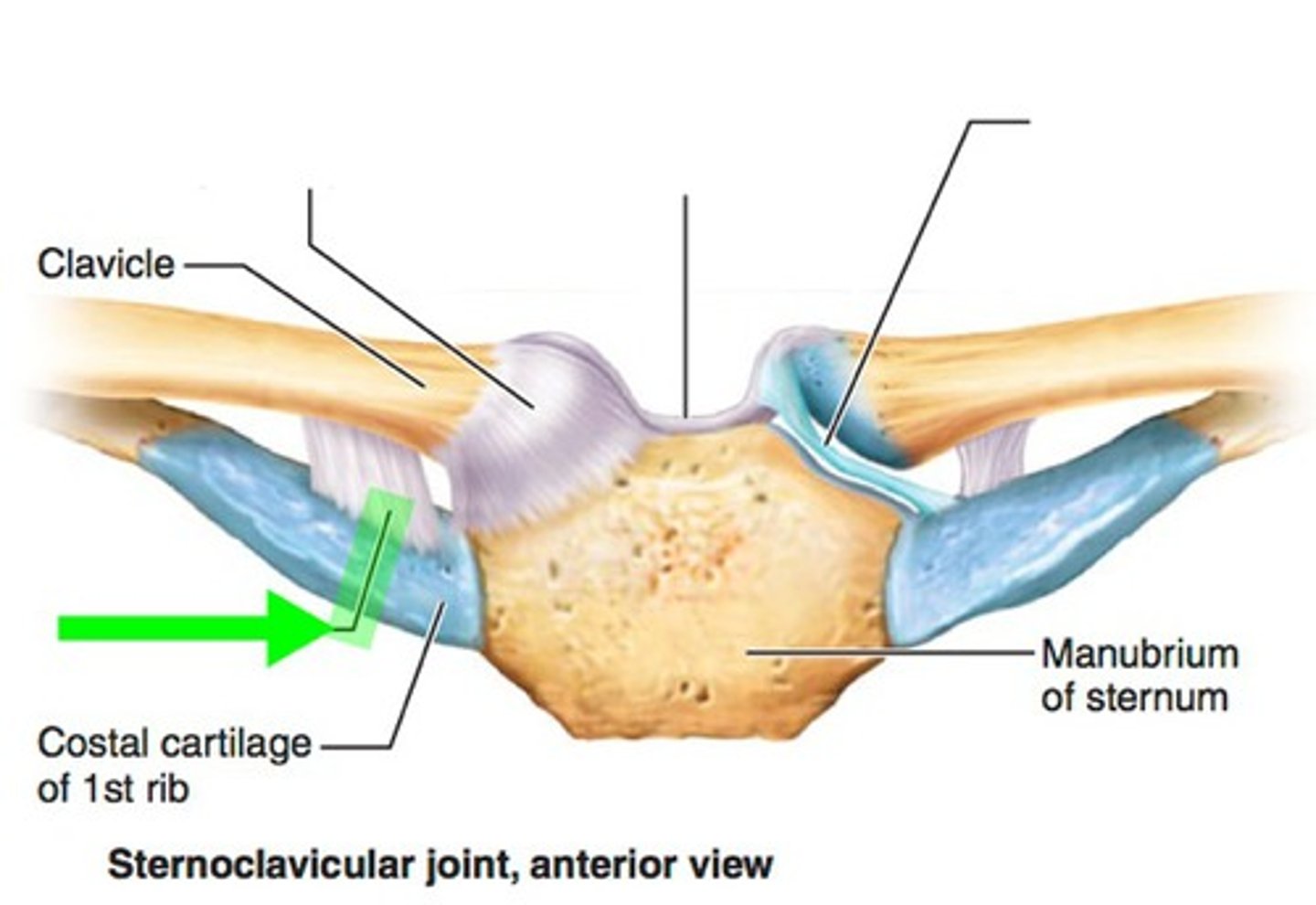
function of the sternoclavicular joint
link upper limb to axial skeleton (very stable joint) - integrity of this joint necessary for functional movement distally
motions of the sternoclavicular joint
protraction/retraction, elevation/depression
articulations of the acromioclavicular joint
acromion process of the scapula, lateral (acromial) end of the clavicle
ligaments of the acromioclavicular joint
coracoclavicular (conoid, trapezoid) ligament, acromioclavicular ligament
conoid ligament
medial part of coracoclavicular ligament - attaches to coracoid process of scapula and conoid tubercle of the clavicle
trapezoid ligament
lateral part of the coracoacromial ligament - attaches to coracoid process of scapula and trapezoid line of the clavicle
acromioclavicular ligament
connects the clavicle to the acromion process of the scapula - part of the ceiling of the supraspinous fossa
function of the acromioclavicular joint
- keep the glenoid fossa aligned with the humeral head
- suspends UE from trunk
actions of the acromioclavicular joint
rotation in anterior/posterior directions, spins, glides
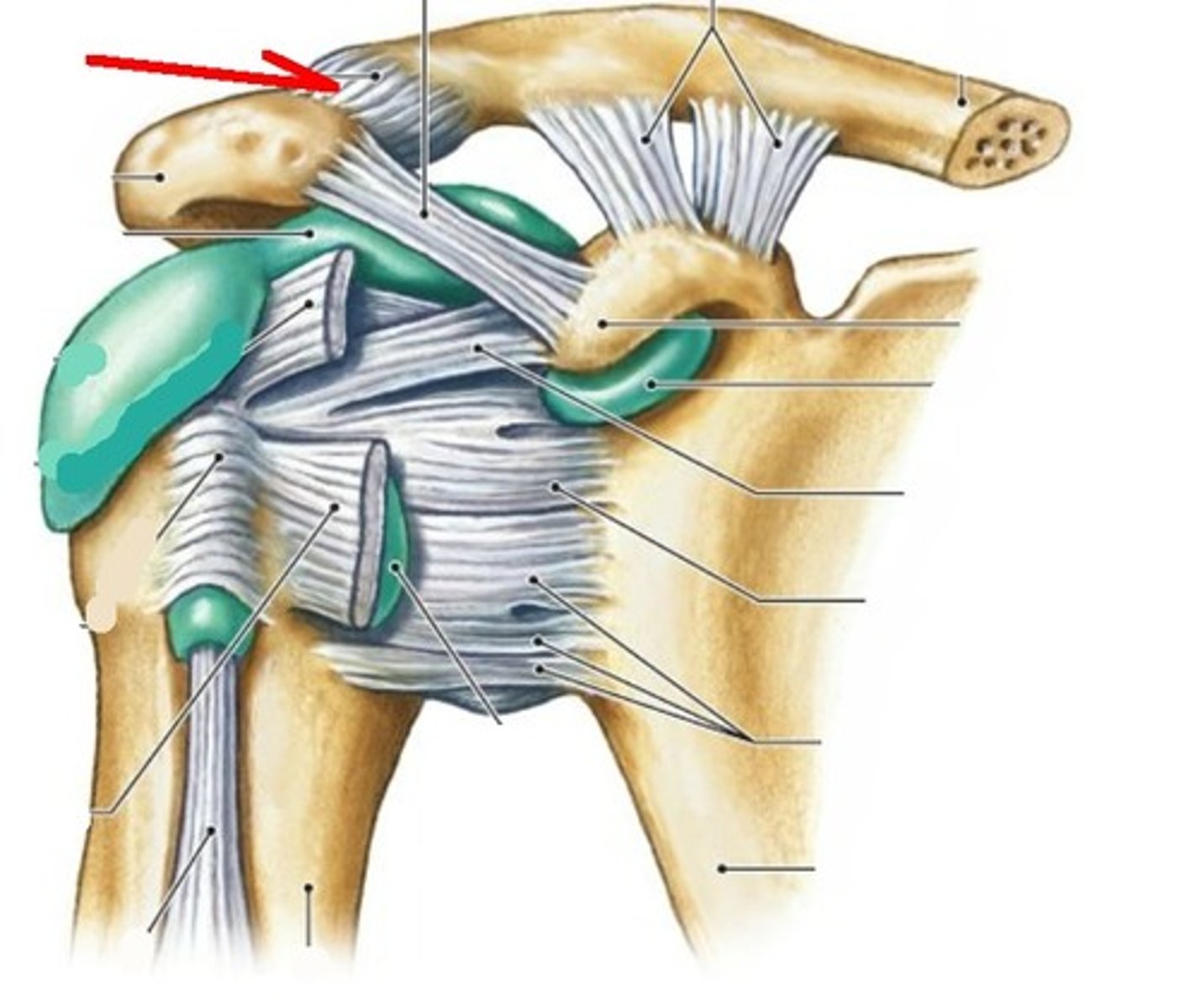
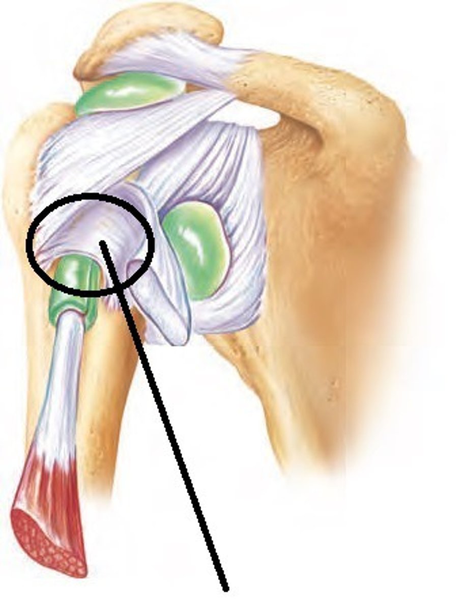
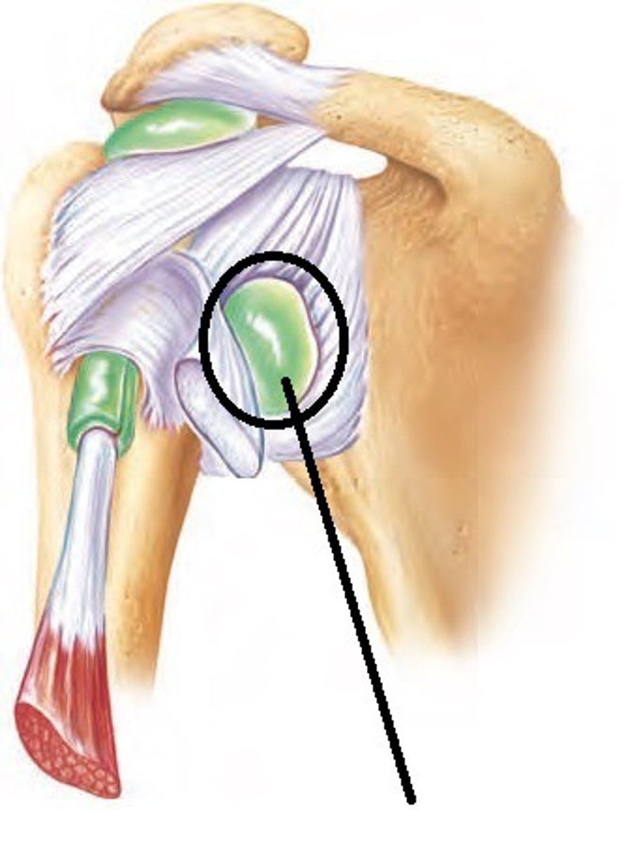
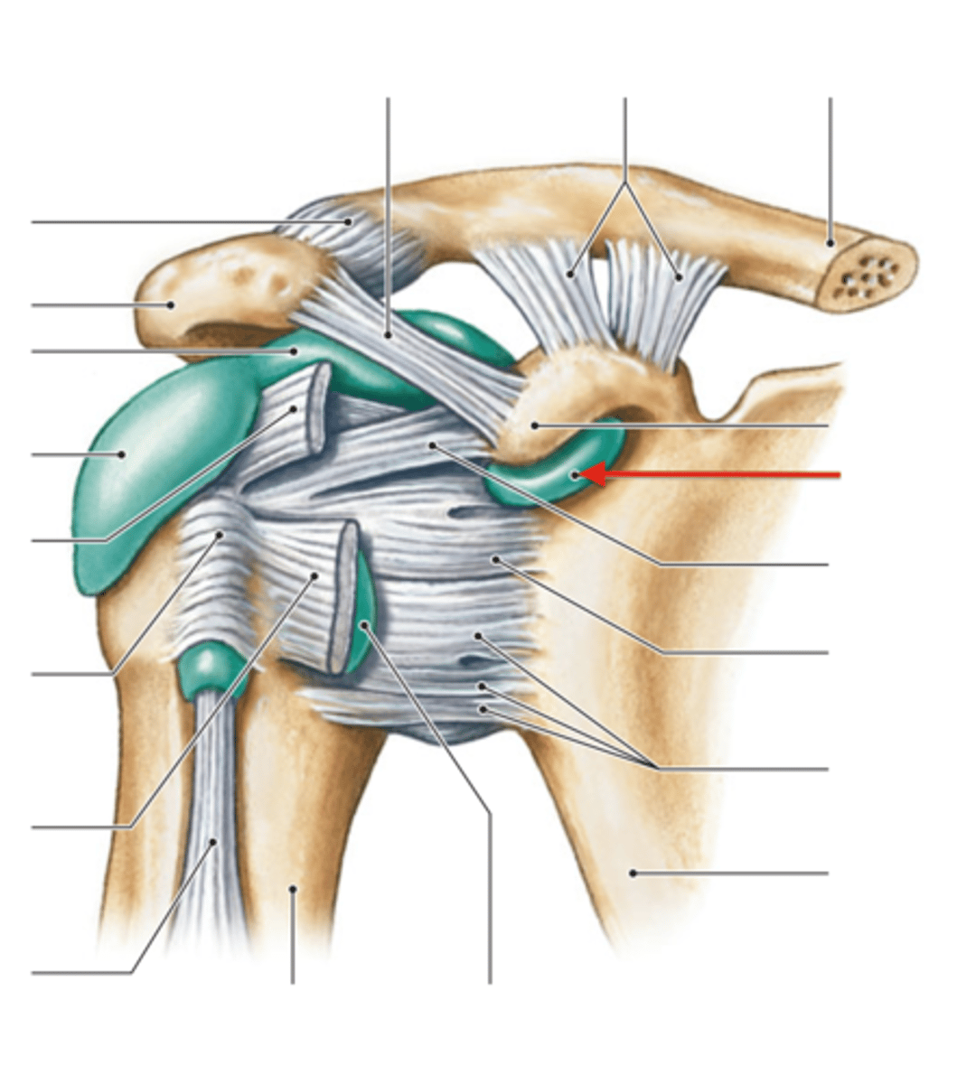
transverse humeral ligament
narrow ligament that extends between the greater and lesser tubercles of the humerus, biceps tendon runs through
coracoacromial ligament
ligament that connects the coracoid process to acromion - part of the ceiling for the supraspinous fossa
coracohumeral ligament
Connects head of humerus to the coracoid process

scapulothoracic joint
"quasi-joint" between anterior scapula and thoracic wall - no synovial joint/2 articular surfaces but there is sliding/gliding motion
transverse humeral joint
"quasi-joint" between the tubercles of the humerus and the bicipital groove - the transverse humeral ligament + synovial membrane cross the bicipital groove and biceps tendon passes through, no cartilage associated
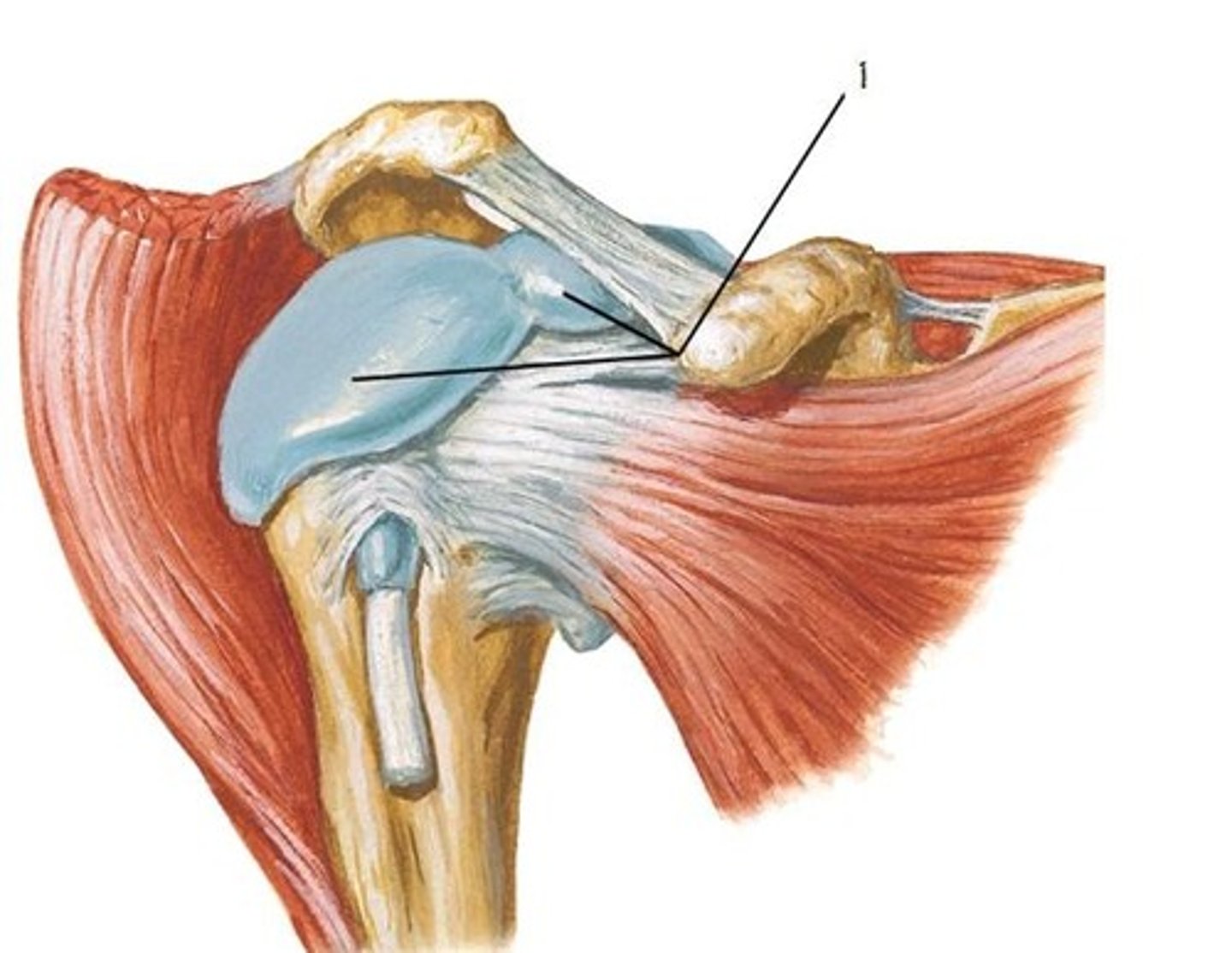
articulations of the glenohumeral joint
humeral head, glenoid fossa (only covers 1/2 of the humeral head) - components include glenoid labrum, fibrous capsule, and synovial membrane
ligaments of the glenohumeral joint
superior, middle, and inferior glenohumeral ligaments, transverse humeral ligament, coracohumeral ligament, coracoacromial ligament (accessory)
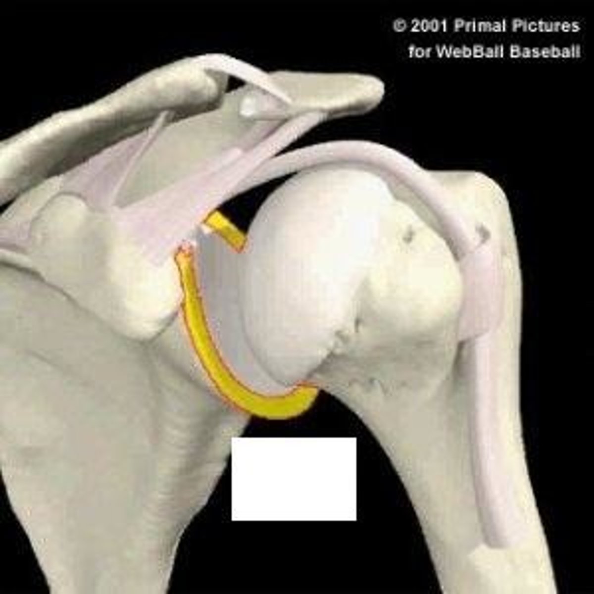
glenoid labrum
fibrocartilage ring that deepens glenoid cavity by 50%
static glenohumeral stabilizers
no contraction, provide structure and base for movement
- joint capsule/labrum
- joint cohesion (atmospheric pressure)
- geometry of humerus/glenoid
- ligaments: superior/middle/inferior GH ligaments, coracohumeral ligament, coracoacromial ligament
dynamic glenohumeral stabilizers
contracticle (muscles), produce movement at the girdle or joint
- stabilizers: SITS
- movers of the GH joint: pec major, lat. dorsi, LH biceps, deltoids, teres major
- movers of the shoulder girdle - serratus anterior, lat. dorsi, traps, rhomboids, levator scap.
tonic contraction (reflex)
special property of (the rotator cuff) muscles - low grade muscle contraction is always present while you are awake
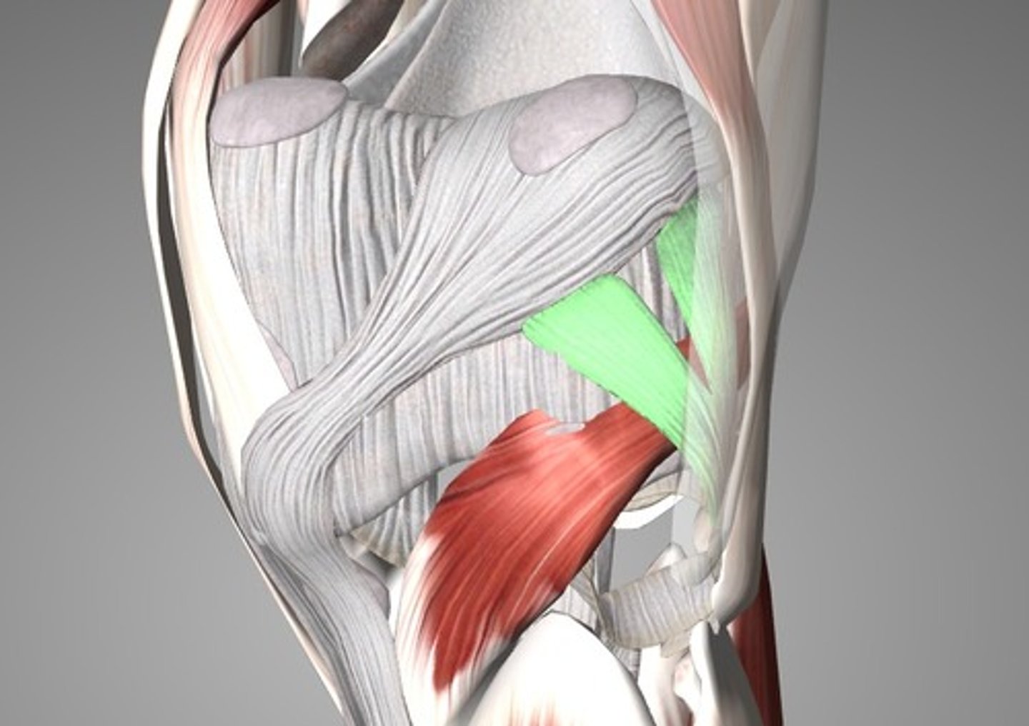
quadrangular/quadrilateral space boundaries
teres minor (superior), neck of humerus (lateral), teres major (inferior), triceps LH (medial)
quadrangular/quadrilateral space contents
axillary nerve, posterior circumflex humeral artery

triangular space boundaries
triceps LH (lateral), teres minor (superior), teres major (inferior)
triangular space contents
circumflex scapular artery
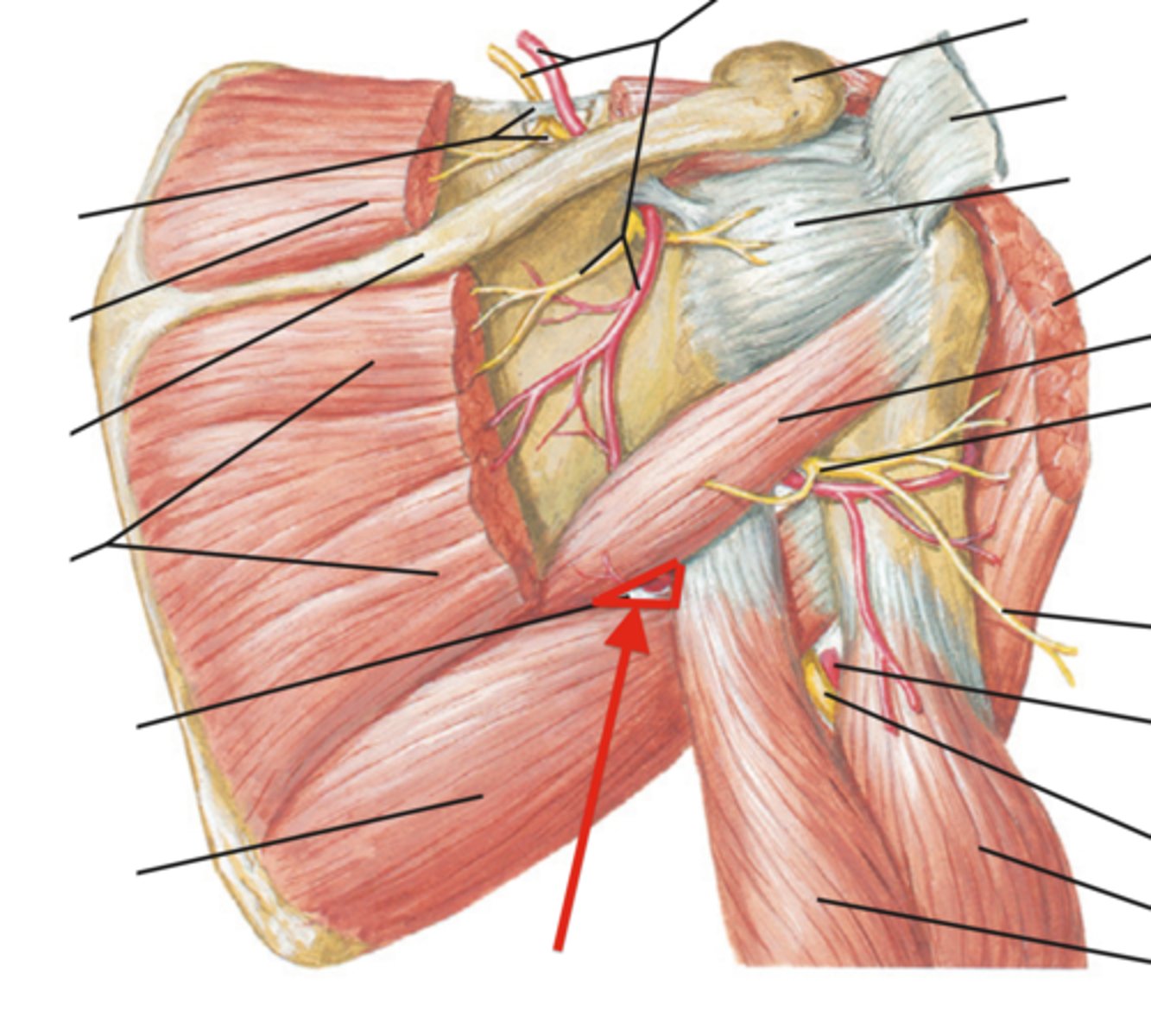
triangular interval boundaries
triceps LateralH (lateral), triceps LH (medial), teres major (superior)
triangular interval contents
deep/profunda brachial artery, radial nerve
bursa
fluid-filled sac made of synovial membrane - present in all major joints to allows for easy movement of one part of a joint over another (buffers friction) - when there is dysfunction, it produces more and fills with fluid and becomes inflamed --> causes pain
shoulder joint bursae (5)
subscapular, subacromial/subdeltoid, subcoracoid, supracoracoid
subscapular bursa
bursa between subscapularis m. and scapula - prevents friction the muscle and bone
subacromial/subdeltoid bursa
two interconnected bursae that share same reservoir of fluid that flows wherever inflammation is greatest - subacromial wedged in between acromion process and supraspinatus m., subdeltoid underneath deltoid m.
supracoracoid bursa
bursa underneath the coracoclavicular ligament
subcoracoid bursa
bursa between the glenoid fossa and subscapularis tendon
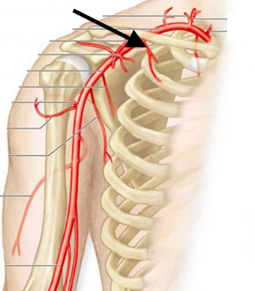
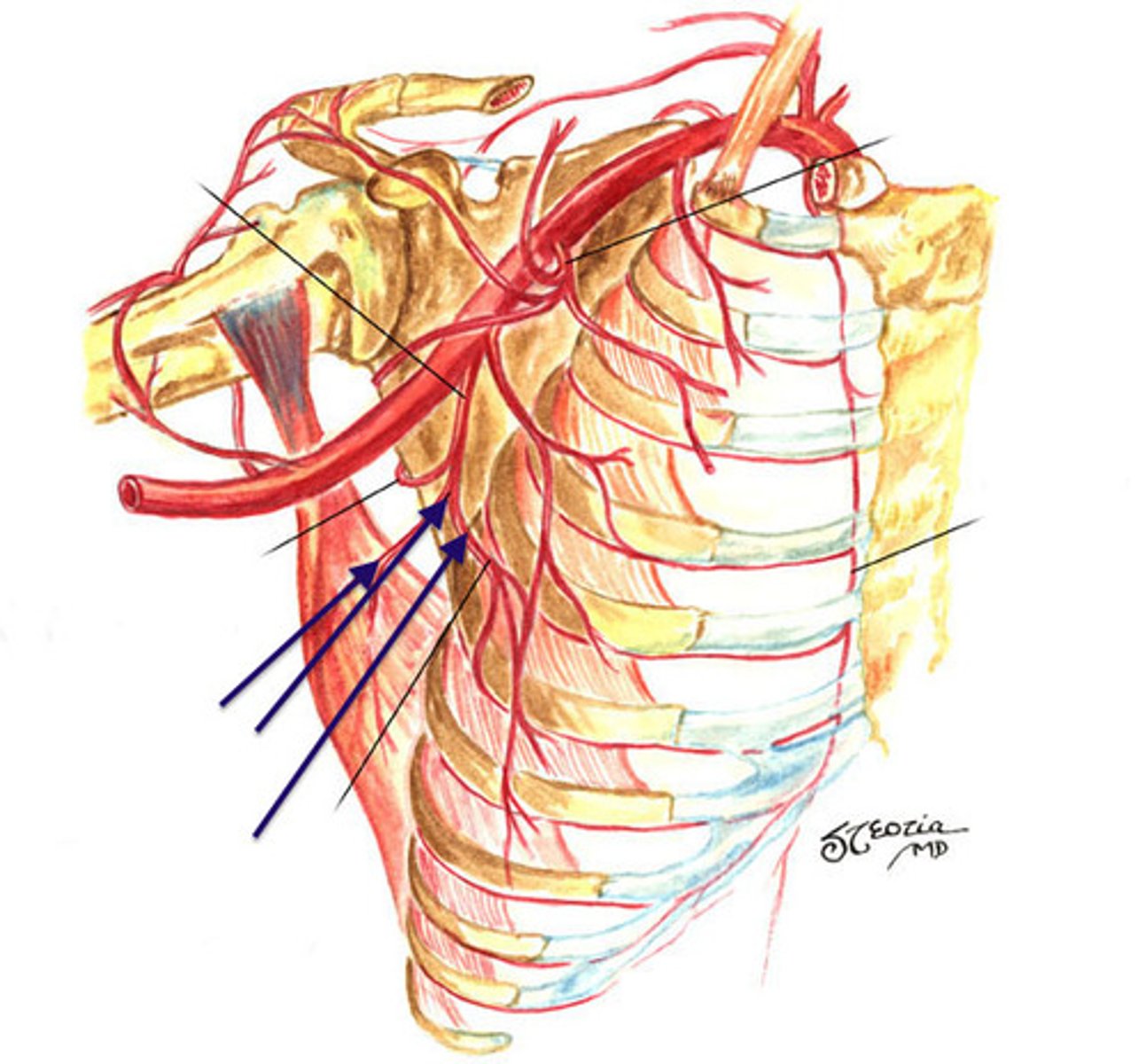
arteries of the posterior shoulder
circumflex scapular a., anterior and posterior circumflex humeral a., suprascapular a., dorsal scapular a. - functions as an anastomosis (collateral circulation)
subacromial bursitis
swelling of bursa --> coracoacromial ligament (ceiling of supraspinatus) causes more inflammation --> irritation/potential tearing of the supraspinatus muscle --> can lead to joint subluxation
Type 1 Shoulder Separation
AC ligament stretched --> bursa inflammation, AC laxity, pain
Type 2 Shoulder Separation
Partial rupture - tear of AC ligament - some integrity remains due to CC ligaments (but extra pressure is put on these ligaments)
Type 3 Shoulder Separation
Complete rupture - tear of AC and CC ligaments
Glenohumeral Subluxation
overhead work/overuse --> bursitis --> compression on arteries/nerves (b/c supraspinous fossa is "closed" by coracoacromial ligament) --> muscles become poorly innervated with less blood supply --> degradation of muscle --> no healthy movement of supraspinatus (most common SITS injury) --> no healthy movement of GH joint --> subluxation/dysfunction in UE
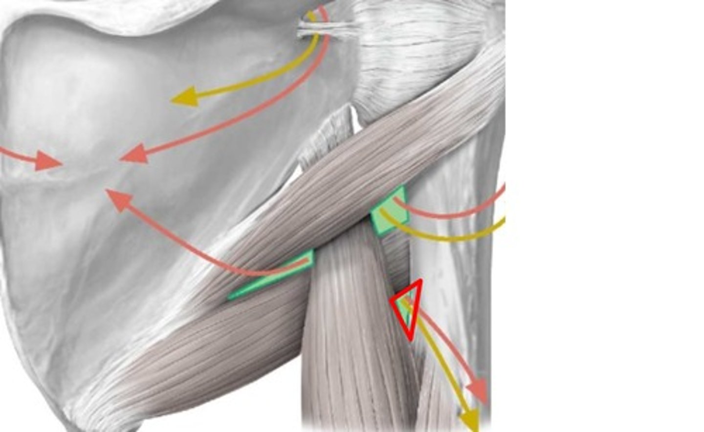
path of cephalic vein
back of hand --> anatomical snuffbox --> lateral forearm --> across bicep --> deltopectoral triangle --> cephalic opening (in costocoracoid membrane) --> combines with axillary vein
path of medial pectoral nerve
pierces pec minor and goes into pec major
costocoracoid membrane
in between ribs and coracoid process - covers the front of the pec minor, separating it from pec major - cephalic opening for thoracoacromial a., lateral pectoral n., and cephalic v.
path of thoracoacromial artery
branches off 2nd part of axillary a. --> comes out the cephalic opening of the costocoracoid membrane
path of lateral pectoral nerve
branches off lateral cord of brachial plexus --> comes out the cephalic opening of the costocoracoid membrane
boundaries of the axilla region
1st rib (apex), armpit (base), superior border of pec minor (anterior), subscapularis, teres major, latissimus dorsi (posterior)
contents of the axilla region
axillary artery (1st rib --> teres major), axillary vein, brachial plexus, axillary lymph nodes
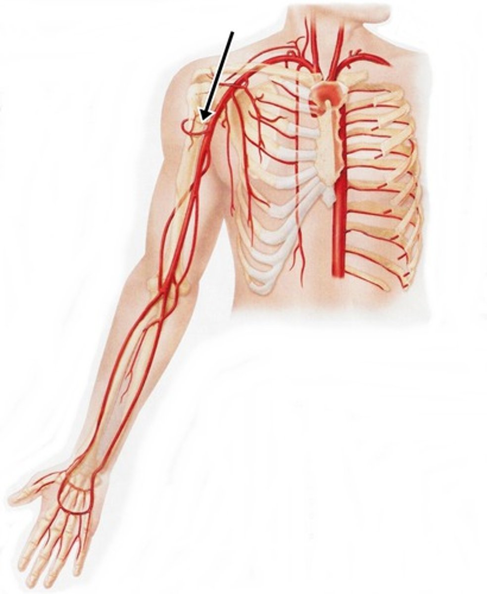
boundaries of the axillary artery
branch off subclavian a. --> 1st rib --> coracoid process (superior border) --> pec minor and teres major --> continues as brachial a.
supreme thoracic artery
branch off 1st part of axillary artery, goes through space between 1st and 2nd ribs
thoracoacromial artery
1st branch off 2nd part of axillary artery - most anterior, relatively large, has clavicular, acromial, deltoid, amd pectoral branches
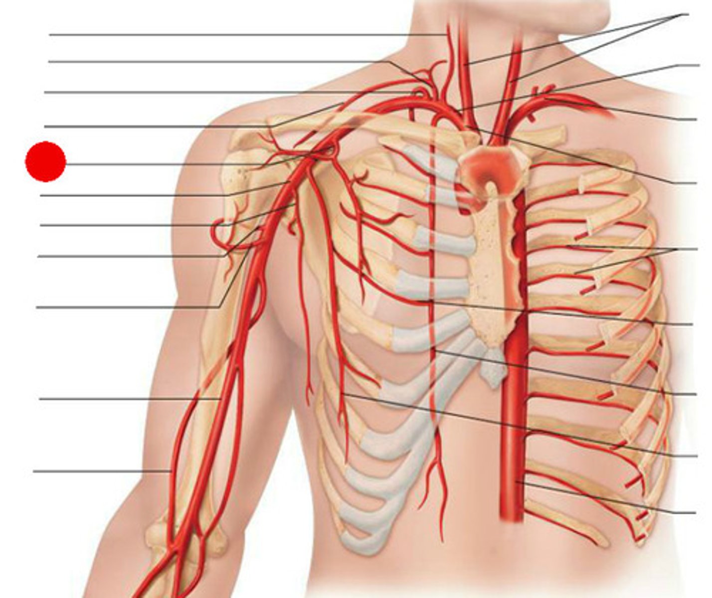
branches of the thoracoacromial artery
clavicular, acromial, deltoid, pectoral
lateral thoracic artery
2nd branch of 2nd part of axillary artery - supplies lateral thorax, serratus anterior
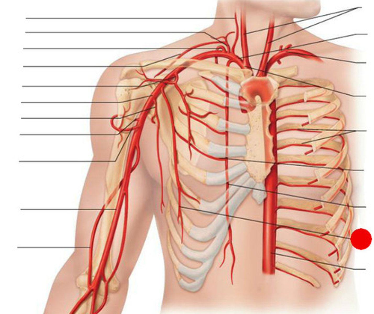
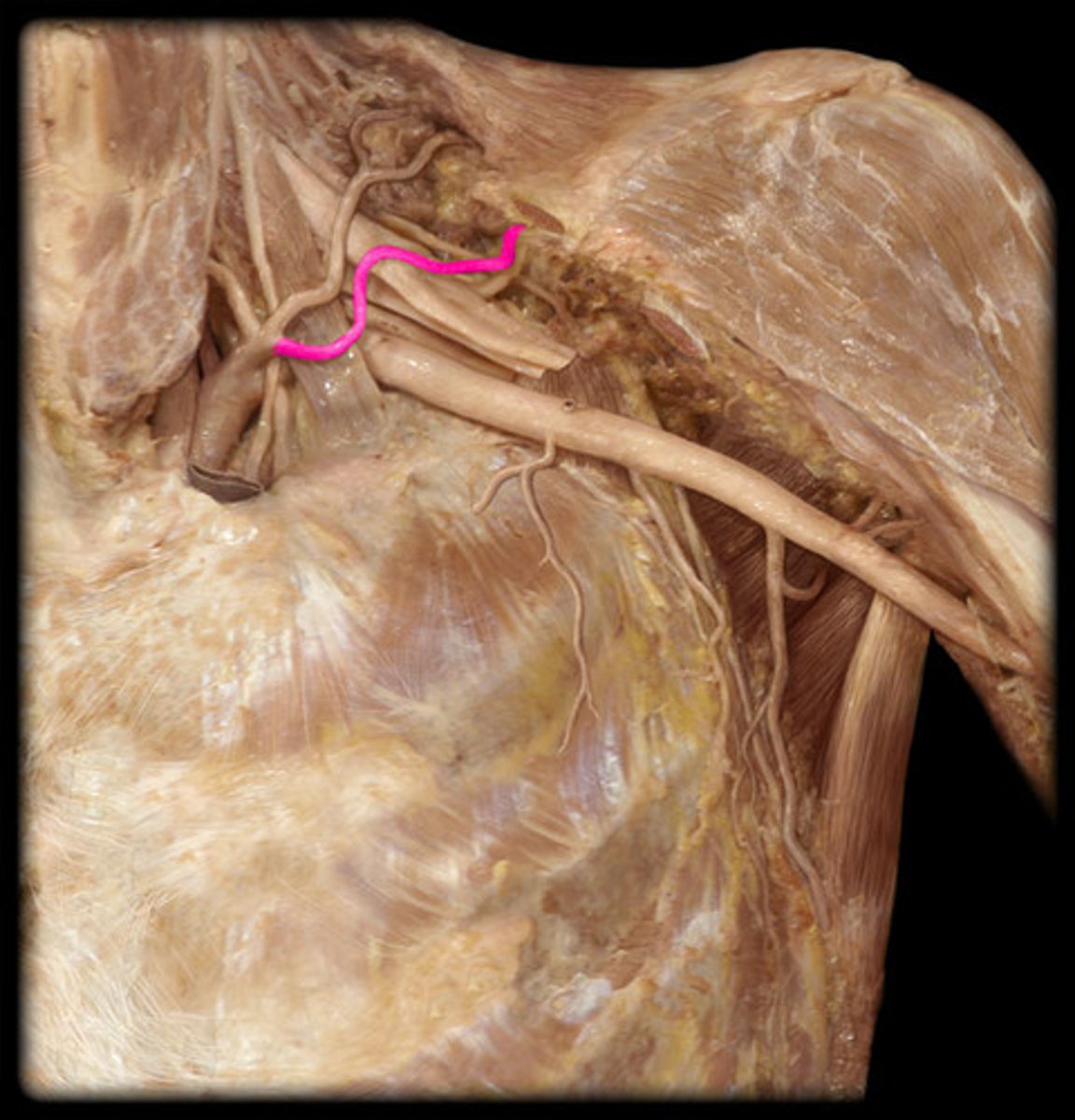
subscapular artery
1st branch off 3rd part of the axillary artery - sits on top of subscapularis --> branches into scapular circumflex a. (part of posterior shoulder anastomosis) and thoracodorsal a. (to lats)
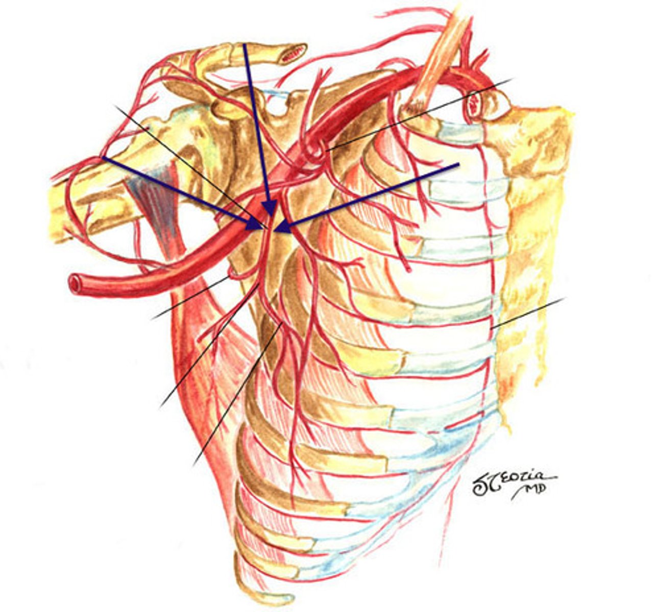
circumflex scapular artery
branches off subscapular artery (secondary branch off 3rd part of axillary a.) , participates in the arterial anastomosis of the posterior shoulder (found within the triangular space)
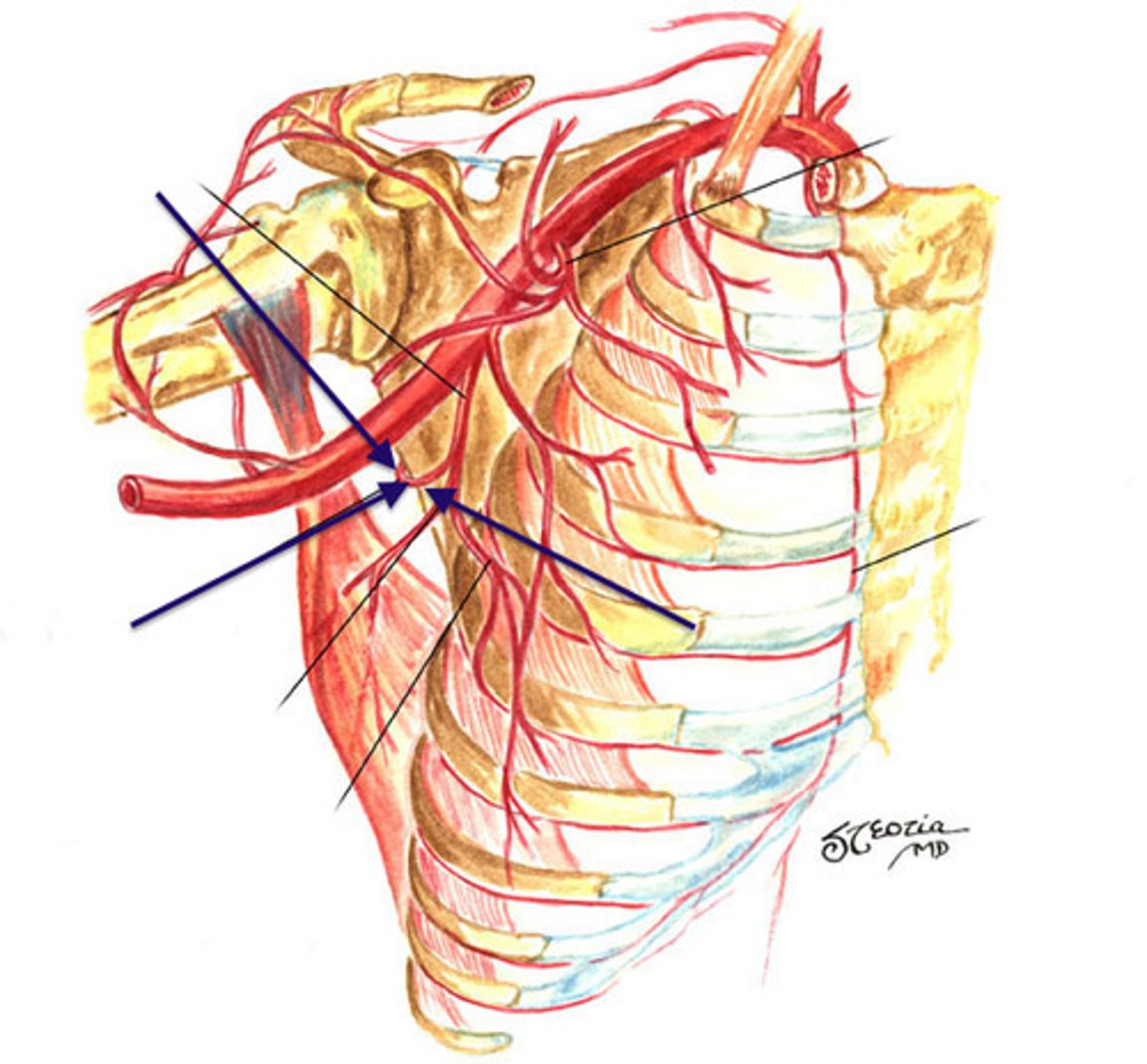
thoracodorsal artery
branches off the subscapular artery, (secondary branch off 3rd part of axillary a.) supplies blood to the latissimus dorsi
anterior and posterior circumflex humeral arteries
3rd branch off 3rd part of axillary artery - found in the quadrangular/quadrilateral space, participates in the arterial anastomosis of the posterior shoulder
supraclavicular branches of the brachial plexus
- dorsal scapular n.
- long thoracic n.
- nerve to subclavius n.
- suprascapular n.
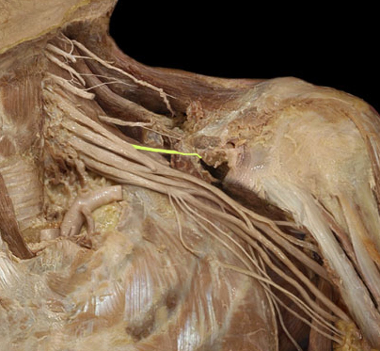
lateral cord
C5-C7 nerve roots --> anterior division of superior trunk and anterior division of middle trunk --> lateral pectoral n., musculocutaneous n., lateral root of median n.
medial cord
C8-T1 nerve roots --> anterior division of inferior trunk --> medial pectoral n., medial brachial cutaneous n., medial antebrachial cutaneous n., ulnar n., medial root of median n.
posterior cord
C5-T1 nerve roots --> posterior divisions of all 3 trunks --> upper, middle (thoracodorsal), and lower subscapular n., axillary n., radial n.
3 functions of nerves
motor, sensory (cutaneous), autonomic
path of axillary nerve
- branches off posterior cord
- through quadrangular space (w/ posterior circumflex humeral a.)
- splits into anterior and posterior branch
*motor innervation to deltoid and teres minor
**cutaneous innervation to regimental badge
damage to axillary nerve
- loss of shoulder abduction
- loss of sensation around regimental badge
(lateral rotation unaffected because infraspinatus takes over)
path of radial nerve
- branch off posterior cord
- through triangular interval (w/ deep brachial a.)
- through radial groove/sulcus down the lateral border of the humerus (*motor innervation to all heads to triceps and brachioradialis)
- pierce through intermuscular septum and passes through cubital fossa to land in anterior compartment of the forearm
- split into superficial branch and deep branch
- superficial branch lies underneath brachioradialis --> comes to surface and lands on the back of the hand (**cutaneous innervation to back of hand)
- deep branch pierces supinator (motor innervation to supinator and superficial extensors) --> runs to deep aspect of posterior forearm on interosseous membrane and becomes posterior interosseous n. (motor innervation to APL, EPL, EPB, EI) and ends at the level of the wrist
**cutaneous innervation to most of the posterior aspect of arm/lateral arm, thin strip down middle of posterior forearm, and dorsal/lateral hand (by the superficial branch)
damage to radial nerve
- sensory loss on the entire posterior aspect of the arm
- loss of extension of the elbow, wrist, and fingers
- loss of abduction of the thumb
- loss of supination
- wrist drop w/ flexed digits - ED cannot extend wrist/MCPs, short handle muscles cannot move fingers into full extension
musculocutaneous nerve pathway
- branch off lateral cord
- pierce the coracobrachialis (*motor innervation to coracobrachialis)
- lies in between biceps brachii and brachialis (*motor innervation to biceps brachii and brachialis)
- travels laterally, crosses cubital fossa, becomes superficial - called lateral antebrachial cutaneous n. (**cutaneous innervation to lateral aspect of forearm)
musculocutaneous nerve damage
- loss of elbow flexion
- sensory loss in lateral forearm
most common point of injury is when the nerve is pinched by an overdeveloped coracobrachialis
ulnar nerve pathway
- branch off medial cord
- no function in the arm, exposed down posteromedial aspect of the arm
- through cubital tunnel (*motor innervation to FCU)
- under FCU down medial forearm (*motor innervation to medial 1/2 of FDP)
- joins with ulnar artery and vein approx. halfway down forearm and becomes VAN (under FDP)
- pass OVER flexor retinaculum, between pisiform and hamate (Guyon's Canal) into hand and splits into superficial and deep branches
- superficial branch stays more on pinky side (**cutaneous innervation to skin of medial hand and medial 1 1/2 digits)
- deep branch crosses all deep bones of the hand (*motor innervation to dorsal/palmar interossei, adductor pollicis, deep fibers of FPB, hypothenar muscles)
cubital tunnel
created by the arcuate ligament (formed by 2 heads of flexor carpi ulnaris) and the medial epicondyle, ulnar nerve runs through (funny bone)
arcuate ligament
curved ligament formed by the 2 heads of the FCU, creates the cubital tunnel that the ulnar n. passes through
ulnar nerve damage
- loss of wrist adduction
- loss of some hand function
- loss of sensation to posterior/medial hand
median nerve pathway
- branch off medial and lateral cord
- travels down arm medial to bicep (w/ brachial a.) (***autonomic control of brachial a.)
- through cubital fossa
- pierce through 2 heads of pronator teres (*motor innervation of pronator teres)
- splits into median proper n. (same as median n.) and anterior interosseous n.
- median proper n. runs between FDS and FDP (motor innervation to FDS and superficial wrist flexors) --> passes UNDER flexor retinaculum through the carpal tunnel to the hand (motor innervation to lumbricals 1-2 and most thenar muscles) --> palmar branch over palmar carpal ligament **cutaneous innervation to digits 1,2,3, lateral half of 4
- anterior interosseous nerve goes deep to the anterior surface of the interosseous membrane (*motor innervation to lateral FDP, deep flexors, and pronator quadratus) and ends at pronator quadratus
median nerve damage
- loss of control of brachial a. --> cold hands
- loss of function in wrist and hand
- loss of sensation in posterior/lateral hand