ANAT 380 Exam Comprehensive Quiz Cards
1/206
There's no tags or description
Looks like no tags are added yet.
Name | Mastery | Learn | Test | Matching | Spaced | Call with Kai |
|---|
No analytics yet
Send a link to your students to track their progress
207 Terms
How many lobes make up the breast?
a) 5-10
b) 10-15
c) 15-20
d) 20-25
c)
each breast is divided into 15-20 lobes that contain the mammary glands
The lobes of the breast are divided by what structures?
a) radial septa
b) aponeuroses
c) tendinous sheaths
d) fibrous connective tissue
a)
lobes are separated by radial septa called suspensory ligaments of Cooper that help support the breast
What is the correct order of the layers of the pericardium from superficial to deep?
a) visceral serous, serous fluid, parietal serous, fibrous
b) parietal serous, serous fluid, visceral serous, fibrous
c) fibrous, parietal serous, serous fluid, visceral serous
d) visceral serous, fibrous, parietal serous, serous fluid
c)
the visceral serous (epicardium) layer covers the surface of the heart, the serous fluid is between the visceral and parietal serous pericardium, and the serous pericardium lines the fibrous pericardium.
The pericardium allows the heart to beat in a frictionless space.
This pleural recess is overlies the heart, is located in the anterior thorax between the costal and mediastinal parietal pleura, and is larger than its counterpart on the opposite side of the thorax
a) left costomediastinal recess
b) right costomediastinal recess
c) left costodiaphragmatic recess
d) right costodiaphragmatic recess
a)
Costomediastinal Recess
the right and left costomediastinal recesses are found between the anterior thorax, between the costal and mediastinal parietal pleura
the left costomeidastinal recess is larger than the right and overlies the heart
Costodiaphragmatic Recess
the costodiaphragmatic recesses are between the costal and diaphragmatic pleura, below the inferior part of the lungs
these recesses are filled with lung tissue only during deep inspiration
what is the name of the layer of the anterior abdominal wall that is deep to the transversus abdominus?
a) Camper’s fascia
b) investing fascia
c) external oblique
d) transversalis fascia
d)
Layers of the anterior abdominal wall:
Skin
superficial fascia
Camper’s fascia
membranous Scarpa’s fascia
Investing fascia
External oblique
Internal oblique
Transversus abdominus
Transversalis fascia
Extraperitoneal fat
Parietal peritoneum
mneumonic:
Some Creatures Say Intelligent Elephants Intend To Travel Every Plain
Which best describes the location of the superficial and deep inguinal ring? (in that order)
a) opening in the investing fascia superior to the pubic symphysis, invagination in the parietal peritoneum medial to the midpoint of the inguinal ligament
b) opening in the external oblique aponeurosis superior to the pubic tubercle, invagination in the transversalis fascia superior to the midpoint of the inguinal ligament
c) opening in the internal oblique superior to the ilium, invagination in the extraperitoneal fat lateral to the midpoint of the inguinal ligament
d) opening in the Camper’s fascia superior to the ischium, invagination in the investing fascia inferior to the inguinal ligament
b)
Which structure is found in the inguinal canal of both males and females?
a) ilioinguinal nerve
b) inguinal ligament
c) common iliac artery
d) pudendal artery
a)
What structures are contained in the spermatic cord?
a) ejaculatory duct, testicular nerve, urethra
b) vas deferens, testicular artery, pamoiniform plexus of veins
c) testicular vein, pudendal nerve, vas deferens
d) pampiniform plexus of veins, ejaculatory duct, urethra
b)
What structures are supplied by the ilioinguinal nerve? (males/females)
a) skin of lower scrotum, tip of penis, medial thigh/lower labia majora, mons pubis, medial thigh
b) skin of upper scrotum, prostatic urethra, lateral thigh/labia minora, labia majora, lateral thigh
c) skin of upper scrotum, root of penis, upper thigh/upper labia majora, mons pubis, upper thigh
d) skin of scrotum, prostate, lower thigh/labia minora, labia majora, lower thigh
c)
These large, double folds of peritoneum consisting of the greater (hanging below the stomach) and lesser (between the stomach and liver) portions, help to cushion the intestines and act as a protective barrier for infection and trauma of the underlying abdominal organs
a) mesenteries
b) omenta
c) rectovesicle pouch
d) tenae coli
b)
These double folds of peritoneum anchor the abdominal organs to the posterior abdominal wall, helping to keep them in place while still allowing for some mobility
a) epiploioc appendages
b) haustra
c) mesenteries
d) tenae coli
c)
which part of the duodenum is intraperitoneal?
a) superior
b) descending
c) horizontal
d) ascending
a)
this structure is a fold of peritoneum that divides the lobes of the liver
a) annular ligament
b) ligamentum flavum
c) falciform ligament
d) suspensory ligament
c)
the body of the gallbladder projects posteriorly and narrows to the neck, which is continuous with which duct?
a) bile duct
b) cystic duct
c) main pancreatic duct
d) ampulla of vater
b)
which structures make up the portal triad?
a) common bile duct, hepatic artery, renal artery
b) cystic duct, hepatic artery, renal vein
c) common bile duct, main pancreatic duct, cystic duct
d) hepatic artery proper, hepatic portal vein, common bile duct
d)
Which of the following statements is completely true about the path of the ureters?
a) The right ureter has to travel farther to reach the bladder, the ureters pass anterior to the psoas and the bifurcation of the common iliac artery, in females the ureters cross superior to the uterine artery and anterior to the vas deferens in males
b) The left ureter has to travel farther to reach the bladder, the ureters pass anterior to the psoas and the bifurcation of the common iliac artery and vein, in females the ureters cross inferior to the uterine aretery in females and posterior to the vas deferens in males
c) the left ureter has to travel farther to reach the bladder, the ureters pass posterior to the psoas and bifurcation of the uterine artery, in females the ureters cross inferior to the uterine aretery in females and posterior to the vas deferens in males
d) The right ureter has to travel farther to reach the bladder, the ureters pass posterior to the psoas and the bifurcation of the common iliac artery, in females the ureters cross superior to the uterine artery and anterior to the vas deferens in males
b)
the sigmoid colon is becomes continuous with the rectum at what level of the vertebral column?
a) S1
b) S2
c) S3
d) S4
b)
As the vas deferens travel superior to the epididymus into the abdominal cavity towards the posterior surface of the bladder, it meets the seminal vesicle to form which structure?
a) ejaculatory duct
b) spermatic cord
c) prostatic urethra
d) Cowper’s gland
a)
Which best describes the borders of the perineum? (anterior/lateral/posterior)
a) pubic symphysis/ishcial tuberosities/sacral canal
b) pelvic outlet/ischium/sacral canal
c) pelvic inlet/ilium/coccyx
d) pubic symphysis/ischial tuberosities/coccyx
d)
which structures are contained in the superficial perineal pouch?
a) muscle, skin, external genitalia
b) skeletal muscle, urethral sphincter, deep transverse perineal muscles
c) urogenital diaphragm, urethral sphincter, external genetalia
d) skin, urethral sphincter, urogenital diaphragm
a)
which area of the scalp is highly vascularized and innervated, contains hair follicles, and may bleed profusely if lacerated?
a) skin
b) connective tissue (dense)
c) aponeuroses
d) loose connective tissue
e) pericranium/periosteum
b)
which salivary gland is the second largest, can be palpated in the neck medial to the lower border of the mandible, and exits through Wharton’s duct into the sublingual papillae behind the lower incisors?
a) parotid gland
b) sublingual gland
c) submandibular gland
d) Stenson’s duct
c)
which border of the middle ear is a thin bone that separates the tympanic cavity from the internal carotid artery and the upper wall is incomplete due to an opening for the eustachian tube?
a) lateral border
b) medial border
c) posterior border
d) anterior border
e) roof
f) floor
d)
Lateral border: tympanic membrane
Medial border: separates middle from inner ear
Posterior border: thin bone separating tympanic cavity from mastoid air cells of temporal bone
Roof: tegmen tympani separates middle ear from cranial fossa
Floor: jugular wall separates middle ear from internal jugular vein
Anterior border: thin bone separates tympanic cavity from internal carotid artery
Inferiorly, the nasopharynx communicates with this structure, which can be closed by elevation of the soft palate during swallowing
a) choanae
b) pharyngeal isthmus
c) piriform recesses
d) palantine tonsils
b)
The anterior palatoglossal arch and the posterior pharyngeal arch contain the palantine tonsil between them and are located on either side of this structure
a) nasopharynx
b) oropharynx
c) laryngopharynx
d) thoracic esophagus
b)
the thyroid gland consists of left and right lateral lobes with an isthmus that connects them. It sits on the anterior surface of the trachea, just inferior to which structure?
a) oropharynx
b) laryngopharynx
c) larynx
d) cervical esophagus
c)
This muscle is formed by the merging of the iliacus and the psoas major, inserts on the lesser trochanter of the femur, and is a muscle of the posterior abdominal wall
a) quadratus lumborum
b) coccygeus
c) adductor magnus
d) iliopsoas
d)
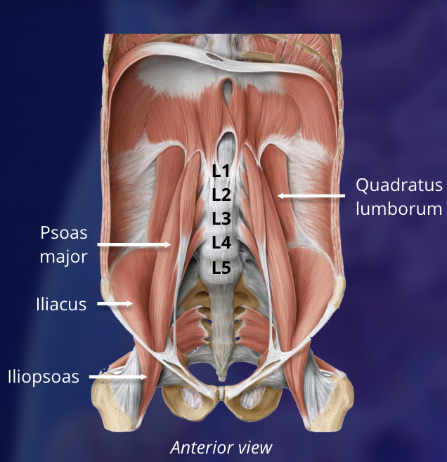
this muscle is a muscle of the posterior abdominal wall, stabilizes the spine, enables lateral flexion, lumbar extension, and pelvic hiking. it originates on the iliac crest and lumbar vertebrae and inserts onto rib 12
a) quadratus lumborum
b) coccygeus
c) iliacus
d) adductor magnus
a)
This is a broad muscle group situated on either side of the pelvis and is the main muscle of the pelvic floor
a) coccygeus
b) levator ani
c) puborectal sling
d) ischiocavernous muscles
b)
this muscle makes up the posterior portion of the pelvic floor
a) coccygeus
b) bulbospongiosus muscles
c) puborectal sling
d) ischiocavernous muscles
a)
this muscle is formed by fibres of the levator ani and forms the anorectal flexure to keep the rectum closed until defecation
a) inferior gemellus
b) superior gemellus
c) puborectal sling
d) ischiocavernous muscles
c)
this fibromuscular mass at the centre of the perineum acts as an attachment site for muscles of the perineum, helps strengthen the pelvic floor, is larger in females, and is also known as the central tendon of the perineum
a) vestibular bulb
b) perineal body
c) urogenital hiatus
d) pubic symphysis
b)
this lateral rotator attaches to the lateral part of the sacrum, leaves the pelvic cavity through the greater sciatic foramen, and attaches on the femur
a) piriformis
b) obturator internus
c) superior & inferior gemellus
d) quadratus femoris
e) obturator externis
a)
this lateral rotator originates in the obturator membrane of the pelvis, its tendon exits through the lesser sciatic foramen to insert on the femur, and the pudendal canal is formed within its membrane to supply the structures of the perineum
a) piriformis
b) obturator internus
c) superior & inferior gemellus
d) quadratus femoris
e) obturator externis
b)
this lateral rotator(s) run above and below the obturator internus
a) piriformis
b) obturator internus
c) superior & inferior gemellus
d) quadratus femoris
e) obturator externis
c)
this lateral rotator runs from the outer surface of the obturator membrane to the femur and is better visualized from an anterior view
a) piriformis
b) obturator internus
c) superior & inferior gemellus
d) quadratus femoris
e) obturator externis
e)
this lateral rotator runs from the ischial tuberosity to the femur
a) piriformis
b) obturator internus
c) superior & inferior gemellus
d) quadratus femoris
e) obturator externis
d)
these ligaments are oriented in a spiral fashion around the hip joint so they tighten during extension and go slack during flexion, and hold the head of the femur in the acetabulum
a) ligamentum teres
b) pectineus
c) fibrous joint capsule
d) acetabular labrum
c)
this is a ring of fibrocartilage that deepens the acetabulum to improve the stability of the hip joint
a) ligamentum teres
b) pectineus
c) fibrous joint capsule
d) acetabular labrum
d)
this acts as a secondary stabilizer of the hip joint, supplementing the work of the capsular ligament
a) ligamentum teres
b) pectineus
c) fibrous joint capsule
d) acetabular labrum
a)
this muscle is an adductor that aids in adduction of the thigh and internal hip rotation
a) adductor brevis
b) adductor longus
c) gracilis
d) adductor magnus
e) pectineus
e)
this muscle is an adductor that is deep to the adductor longus and pectineus and adducts, flexes, and rotates the hip
a) adductor brevis
b) adductor longus
c) gracilis
d) adductor magnus
e) pectineus
a)
this muscle is an adductor muscle that adducts the hip primarily and originates from the pubic body and inserts onto the linea aspera of the femur
a) adductor brevis
b) adductor longus
c) gracilis
d) adductor magnus
e) pectineus
b)
this is a long, thin, superficial adductor muscle that acts ant both the hip and knee joints to adduct the thigh, flex the leg, and internally rotate the knee
a) adductor brevis
b) adductor longus
c) gracilis
d) adductor magnus
e) pectineus
c)
this is the deepest and largest adductor muscle that inserts along the length of the linea aspera of the femur, rotates the thigh and medially rotates the thigh at the hip joint, and forms the adductor hiatus
a) adductor brevis
b) adductor longus
c) gracilis
d) adductor magnus
e) pectineus
d)
this is the most medial of the 3 hamstring muscles. it originates on the ischial tuberosity and inserts on the medial tibial condyle
a) semitendinosus
b) semimembranosus
c) biceps femoris
d) popliteus
b)
this muscle is the middle of the 3 hamstring muscles. it originates on the ischial tuberosity and inserts on the proximal medial tibia
a) semitendinosus
b) semimembranosus
c) biceps femoris
d) popliteus
a)
this is the most lateral of the 3 hamstring muscles and has two heads (long head and short head) that insert on the fibular head, crural fascia, and lateral condyle of the tibia
a) semitendinosus
b) semimembranosus
c) biceps femoris
d) popliteus
c)
the femoral triangle has a superior boundary, floor, and roof. these are the:
a) ilioinguinal ligament, psoas major, quadriceps tendon
b) inguinal ligament, iliopsoas and pectineus, fascia lata
c) external oblique, iliacus, quadriceps tendon
d) external oblique, pectineus, iliopsoas
b)
this knee ligament is broader and joins the distal femur to the proximal tibia
a) ACL
b) PCL
c) MCL
d) LCL
e) patellar ligament
c)
this knee ligament is cord like and joins the distal femur to the head of the fibula
a) ACL
b) PCL
c) MCL
d) LCL
e) patellar ligament
d)
this knee ligament prevents anterior dislocation of the tibia on the fixed femur
a) ACL
b) PCL
c) MCL
d) LCL
e) patellar ligament
a)
this knee ligament prevents posterior dislocation of the tibia on the fixed femur
a) ACL
b) PCL
c) MCL
d) LCL
e) patellar ligament
b)
this is the deepest muscle of the posterior compartment of the lower leg. it originates on the lateral femur and inserts on the posterior surface of the tibia
a) medial head of gastrocnemius
b) lateral head of gastrocnemius
c) popliteus
d) soleus
c)
this muscle of the posterior compartment of the lower leg has two heads (medial and lateral) that originate on the medial and lateral sides of the femur and insert on the calcaneus bone, thus crossing two joints
a) soleus
b) gastrocnemius
c) plantaris
d) tibialis posterior
b)
this muscle is a long, thin muscle in the posterior compartment of the leg that originates on the posterolateral part of the femur and inserts on the calcaneus
a) soleus
b) tibialis anterior
c) tibialis posterior
d) plantaris
d)
these structures are contained in the popliteal fossa
a) common peroneal nerve, tibial artery, tibial vein
b) popliteal nerve, tibial artery, tibial vein
c) popliteal artery and vein, common peroneal nerve, tibial nerve
d) tibial nerve, popliteal artery and vein, fibular nerve
c)
these are the borders of the popliteal fossa
superior, medial: semimembranosus
superior, lateral: biceps femoris
inferior, medial: medial head of gastrocnemius
inferior, lateral: lateral head of gastrocnemius AND _________
a) pectineus
b) plantaris
c) piriformis
d) flexor pollicis longus
b)
this tarsal bone articulates with the first metatarsal
a) calcaneus
b) medial cuneiform
c) cuboid
d) intermediate cuneiform
b)
this is formed from 5 fused vertebrae and is located at the terminal part of the vertebral column where it forms the posterior aspect of the bony pelvis
a) sacral cornua
b) sacral foramina
c) sacrum
d) promontory of the sacrum
c)
this broad, triangular muscle has multiple points of origin and acts to extend, adduct, and internally rotated the humerus
a) trapezius
b) rhomboid minor
c) rhomboid major
d) latissimus dorsi
e) levator scapulae
d)
this muscle of the upper back elevates, upwardly rotates, retracts, and depresses the scapula. it also extends the neck
a) trapezius
b) rhomboid minor
c) rhomboid major
d) latissimus dorsi
e) levator scapulae
a)
this muscle downwardly rotates the scapula
a) trapezius
b) rhomboid minor
c) rhomboid major
d) latissimus dorsi
e) levator scapulae
b)
this muscle retracts, downwardly rotates, and stabilizes the scapula
a) trapezius
b) rhomboid minor
c) rhomboid major
d) latissimus dorsi
e) levator scapulae
c)
this muscle elevates and downwardly rotates the scapula
a) trapezius
b) rhomboid minor
c) rhomboid major
d) latissimus dorsi
e) levator scapulae
e)
this is the most lateral, longest, and widest of the three spinal erectors and is divided into 3 divisions (cervicis, thoracis, lumborum)
a) spinalis
b) longissimus
c) iliocostalis
c)
the posterior wall of this pyramidal shaped structure is made up of the subscapularis, latissimus dorsi, and teres major muscles
a) cubital fossa
b) popliteal fossa
c) femoral triangle
d) axilla
d)
this ligament of the pectoral girdle reinforces the acromioclavicular joint and supports the superior surface of the shoulder. it is located between the acromion process of the scapula and the clavicle
a) coracoacromial ligament
b) acromioclavicular ligament
c) coracoclavicular ligament
d) glenohumeral ligaments
b)
this ligament of the pectoral girdle connects the acromion and coracoid processes of the scapula and forms a ‘vault’ that prevents displacement of the humeral head
a) coracoacromial ligament
b) acromioclavicular ligament
c) coracoclavicular ligament
d) glenohumeral ligaments
a)
this ligament of the pectoral girdle is the main stabilizer of the acromioclavicular joint as it anchors the clavicle to the coracoid process of the scapula
a) coracoacromial ligament
b) acromioclavicular ligament
c) coracoclavicular ligament
d) glenohumeral ligaments
c)
these are the largest ligaments of the pectoral girdle, and stabilize the glenohumeral joint especially during adduction of the arm
a) coracoacromial ligament
b) acromioclavicular ligament
c) coracoclavicular ligament
d) glenohumeral ligaments
d)
the glenoid labrum, rotator cuff muscles, long head of biceps brachii, and scapulohumeral muscles act as what?
a) rotator cuff muscles
b) muscles of the pectoral girdle
c) stabilizers of the glenohumeral joint
d) scapulohumeral muscles
c)
the infraspinatus, supraspinatus, teres minor, and subscapularis act as what?
a) rotator cuff muscles
b) muscles of the pectoral girdle
c) stabilizers of the glenohumeral joint
d) scapulohumeral muscles
a)
the deltoids, teres major, and the rotator cuff muscles act as what?
a) rotator cuff muscles
b) muscles of the pectoral girdle
c) stabilizers of the glenohumeral joint
d) scapulohumeral muscles
d)
the subacromial and subdeltoid bursa do not protect from friction with what structure?
a) coracoclavicular ligament
b) coracoacromial ligament
c) acromioclavicular ligament
d) supraspinatus tendon
c)
this muscle is a scapulohumeral muscle that extends and medially rotates the humerus
a) anterior deltoid
b) teres major
c) teres minor
c) subscapularis
b)
this muscle is both a rotator cuff muscle and scapulohumeral muscle that inserts on the greater tubercle of the humerus and initiates abduction of the humerus
a) supraspinatus
b) infraspinatus
c) subscapularis
d) teres minor
c)
these three muscles make up the anterior compartment of the arm
a) anconeus, biceps brachii, brachialis
b) biceps brachii, coracobrachialis, brachialis
c) biceps brachii, brachioradialis, pronator teres
d) anconeus, brachioradialis, pronator teres
b)
the cubital fossa is a trianglular depression on the anterior surface of the elbow joint containing the biceps brachii tendon, brachial artery, and median nerve. the lateral border is the brachioradialis muscle and the medial border is the _____
a) anconeus
b) brachialis
c) pronator teres
d) biceps brachii
c)
this forms a fibrous joint that spans the space between the radius and ulna, and divides the forearm into posterior and anterior compartments
a) proximal radioulnar joint
b) annular ligament
c) interossesous membrane
d) distal radioulnar joint
c)
the flexor digitorum profundus, flexor pollicis longus, and pronator quadratus make up which compartment of the arm?
a) posterior compartment of the arm
b) posterior compartment of the forearm
c) superficial muscles of the anterior compartment of the forearm
d) intermediate muscles of the anterior compartment of the forearm
e) deep muscles of the anterior compartment of the forearm
e)
this compartment of the arm contains the extensor digitorum, extends digits 2-5, and extends the hand at the wrist
a) posterior compartment of the arm
b) posterior compartment of the forearm
c) superficial muscles of the anterior compartment of the forearm
d) intermediate muscles of the anterior compartment of the forearm
e) deep muscles of the anterior compartment of the forearm
b)
this joint is a biaxial, synovial, ellipsodial (condyloid) joint formed between the distal end of the radius and the proximal row of carpal bones with the exception of the psiform
a) radiocarpal joint
b) intercarpal joints
c) midcarpal joints
d) carpal tunnel
a)
these intrinsic muscles of the hand are responsible for finger abduction
a) hypothenar muscles
b) thenar muscles
c) dorsal interossei
d) palmar interossei
c)
these intrinsic muscles of the hand are responsible for movements of the thumb
a) hypothenar muscles
b) thenar muscles
c) dorsal interossei
d) palmar interossei
b)
these intrinsic muscles of the hand are responsible for movements of the 5th digit
a) hypothenar muscles
b) thenar muscles
c) dorsal interossei
d) palmar interossei
a)
these intrinsic muscles of the hand are responsible for adduction of the fingers
a) hypothenar muscles
b) thenar muscles
c) dorsal interossei
d) palmar interossei
d)
these muscles depress the eye with the pupil in the midline
a) lateral rectus and medial rectus
b) inferior rectus and superior oblique
c) superior rectus and inferior oblique
d) inferior rectus and inferior oblique
b)
the temporalis, masseter, and medial pterygoid are responsible for which action
a) elevation
b) depression
c) lateral movemnt
d) protraction (protrusion)
e) retraction
a)
the temporalis on its own is responsible for which action
a) elevation
b) depression
c) lateral movemnt
d) protraction (protrusion)
e) retraction
e)
the medial and lateral pterygoids are responsible for which action
a) elevation
b) depression
c) lateral movement
d) protraction (protrusion)
e) retraction
c)
gravity and the relaxation of muscles is responsible for which action
a) elevation
b) depression
c) lateral movemnt
d) protraction (protrusion)
e) retraction
b)
the lateral pterygoid on its own is responsible for which action
a) elevation
b) depression
c) lateral movemnt
d) protraction (protrusion)
e) retraction
d)
this is formed by the lower part of the inferior constrictor muscles of the pharynx
a) lower esophageal sphincter
b) upper esophageal sphincter
c) larynx
d) vocal folds
b)
this structure is located in the back of the larynx and has the primary function of closing the vocal folds during swallowing and secondary function of changing tension of the vocal folds during sound production
a) cricothyroid ligament
b) artenoid cartilage
c) thyroartenoid muscle
d) cricothyroid muscle
b)
the upper margin of this structure forms the true vocal fold (vocal ligament)
a) cricothyroid ligament
b) artenoid cartilage
c) thyroartenoid muscle
d) cricothyroid muscle
a)
this intrinsic muscle of the larynx that attaches to the anterolateral part of the arytenoid cartilage receives motor innervation from the recurrent laryngeal nerve to relax the vocal ligament and produce a softer voice
a) cricothyroid ligament
b) artenoid cartilage
c) thyroartenoid muscle
d) cricothyroid muscle
c)
this intrinsic muscle of the larynx is innervated by the external laryngeal nerve and stretches and tenses the vocal ligaments for the production of loud speech
a) cricothyroid ligament
b) artenoid cartilage
c) thyroartenoid muscle
d) cricothyroid muscle
d)
this superficial muscle of the neck runs from the upper two ribsto the lower margin of the mandible to tense the skin of the neck and aid in facial expression
a) sternocleidomastoid
b) scalenes
c) platysma
d) levator scapulae
c)
this paired superficial muscle of the neck innervated by the accessory nerve originates on the manubrium of the sternum and medial end of the clavicle to insert inferiorly on the mastoid process. the two muscles together flex the head and neck while individually will tilt the head toward the shoulder on the same side, rotating the head to turn the face to the opposite side
a) sternocleidomastoid
b) scalenes
c) platysma
d) levator scapulae
a)