psyc367- neuroscience: test 1
1/82
Earn XP
Description and Tags
human neuroscience 🧠
Name | Mastery | Learn | Test | Matching | Spaced | Call with Kai |
|---|
No analytics yet
Send a link to your students to track their progress
83 Terms
cutaneous mechanoreceptors
receptors in the skin that open ion channels in response to physical distortion (stimuli)
nociceptors
receptors on the skin that respond specifically to noxious or harmful stimuli
agonist
something that mimics the effects of a neurotransmitter
antagonist
something that blocks or reduces the effects of a neurotransmitter
dura mater
“hard mother”!!- outermost meninge layer which is the thickest and strongest
pia mater
“tender mother”!!- meninge that sits tight against the brain (think seran wrap)
afferent
information that travels to the central nervous system, otherwise known as sensory information
efferent
information that travels away from the central nervous system, otherwise known as motor information
voltage-gated ion channels are located primarily at which part of the neuron
the axon hillock
which of the following is not a way that neurotransmitters are cleaned up from the synapse
they are inactivated or degraded by enyzmes
they float away
they are recycled by the presynaptic cell
they are absorbed by the postsynaptic cell
they are absorbed by the postsynaptic cell.
what creates the necessary environment for neuronal communication
electrochemical gradient
which cell is not found in the central nervous system
neurons
astrocytes
microglia
schwann cells
schwann cells
what are parts of the brain that are dense with axons
white matter
what is the period of time during which it is impossible for another action potential to fire
absolute refractory period
if you were sitting in your chair and fidgeting with your pen, which tract would be sending the info. from the CNS to your muscles
dorsolateral corticospinal tract
what brain region contributes to pain perception through initial emotion identification
insula
what ion is responsible for neurotransmitter release from vesicles
calcium
which cell is responsible for maintaining the physical structure of the brain/creating the blood brain barrier
astrocytes
what part of the brain is the central “relay station”
thalamus
what brain region is responsible for the storage of sensory information
secondary somatosensory cortex
dendrites
“tree”- recieve information
soma/cell body
contains organelles and nucleus
axon
sends information
axon hillock
gatekeeper of information
axon terminal
end of the axon, contains vesicles w/ neurotransmitters
myelin
increases speed of signal/protects the axon
nodes of ranvier
recharge/refuel stations for signal
synapse
gap between the axon terminal and the dendrites of the next neuron
step 1 of action potential
stimulus: ligand-gated ion channels open (Na+ flows in)
step 2 of action potential
huge depolarization: voltage-gated ion channels open (Na+ flows in)
step 3 of action potential
repolarization: voltage-gated K+ channels open (slowly), flow out
step 4 of action potential
hyperpolarization: undershoot of negative charge (more negative than at rest) due to slow K+ channels
last step of action potential
return to rest: -70mV – Na+/K+ pump (3 Na+ out, 2 K+ in)
what are the 3 main divisions of the brain
brainstem, cerebrum, and cerebellum
what are the subdivisions under the brainstem
medulla, pons, and midbrain
what are the subdivisions of the cerebrum (the cortex)
frontal, temporal, occipital, & parietal lobe
function of the frontal cortex
action lob
function of the temporal lobe
hearing/processing
function of the occipital lobe
vision
function of the parietal lobe
somatosensory
function of the cerebellum
fine tuner, especially for movement
characteristics of small molecule
stored/made at the axon terminals
1 type per cell
small vesicles
fast release
ex. amino acids, monoamines
characteristics of a peptide
stored/made in the soma
multiple types per cell
dense core vesicles
slower(er) release
ex. substance P, neuropeptide Y, dynorphin
what would happen: the ion channels on the cutaneous mechanoreceptors do not function
not going to get sensation at all (not even making it to the spine!)
what would happen: the dorsal column is severed
no signal will make it to the brain (end in the spine) – no perception. reflexes still intact
what would happen: the connection between the VPN and the S1 is damaged
no perception of touch but yes reflexes
what would happen: the PPC is nonfunctional
perception–semi-intact. inhibited understanding of orientation and movement
Ramon y Cajal
first neuroscientists, discovered the neuron
relative refractory period
a new impulse can be generated, but only by a stronger-than-normal (suprathreshold) stimulus
absolute refractory period
interval immediately following an action potential during which a second action potential cannot be initiated, regardless of the stimulus intensity
ligand-gated voltage channels
open when a specific chemical attaches. located @ dendrites
voltage gated ion channels
open in response to a specific charge. located @ axon hillock
excitatory post synaptic potential (EPSP)
depolarization of the next neuron- caused by Na+
inhibitory post synaptic potential (IPSP)
hyperpolarization of the next neuron- caused by Cl- or K+
propogration
progression of the action potential down the axon
electroencephalogram (EEG)
nonivasive method to record electrical activity in the brain
pros of EEG’s
non-ivasive
multipurpose
relatively cheap
cons of EEG’s
many obstructions (hair, layers, etc.)
not comfortable
depth of reading- only records outside of the brain
accuracy isn’t 100%
layers of the brain
bone- skull & vertebrae column
duramater
meninges
arachnoid layer
subarachnoid lymphatic like membrane
pia mater
brain
cerebral spinal fluid
flow around arachnoid layer and SLLYM
function: makes brain float, delivers nutrients, and removes waste
central sulcus
seperates frontal and parietal lobes
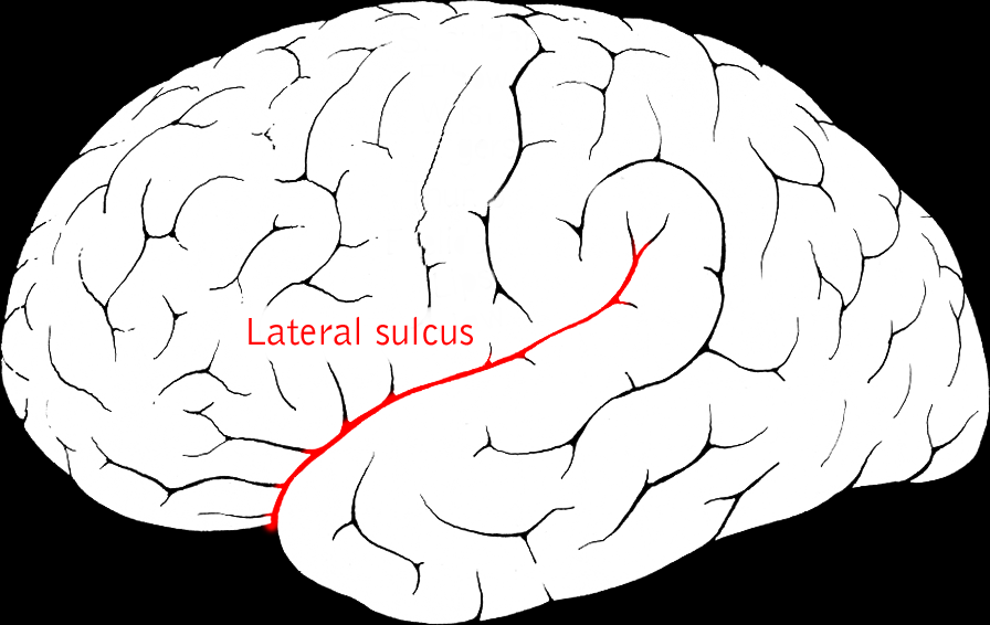
lateral fissure
separates temporal from frontal and occipital lobes
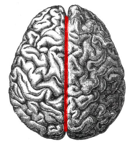
longitudinal fissure
separates hemispheres
gray matter
areas of brain dens with cell bodies and dendrites

white matter
communication highways- areas of brain dense with axons

outermost layer
higher order thinking
subcortical layer
basic level cognition

commissure
white matter connecting left/right hemispheres

functional MRI (fMRI)
detects changes in blood/oxygen levels as an indirect measure of activity
helps in finding functional connectivity
structural MRI
anatomic information
fMRI pros
noninvasive
functional- where is energy being used/required
3d image
subcortical images
fMRI cons
EXPENSIVE
must hold fully still
blood/oxygen level is an indirect measure- room for error
what neurotransmitters can not excite AND inhibit
glutamate- only excitatory
GABA- only inhibitory
glycine- only inhibitory
ionotrophic receptors
neurotransmitter attaches to receptors @ active sight to open ion channel
receptor and ion channel are 1
excitatory OR inhibitory
1:1 NT to open ion channel
metabotrophic receptor
neurotransmitter attaches to receptor @ active sight to activate a G-protein
receptor and ion channel are not the same
excitatory or inhibitory
1 NT has multiple effects
sensation
detection of stimuli by specialized receptors
afferent
info going AT the CNS- sensory!!
efferent
info that exits the CNS- motor!!
what is process of sensation
stimulus
cutaneous mechanoreceptor
through dorsal root of spinal nerve
SPLIT in dorsal horn- main axon goes into dorsal column (reflex)
second axon goes up dorsal column
1ST ORDER NEURON- @ medulla- info crosses into opposite side (contralateral)
2ND ORDER NEURON- @ thalamus- organized in ventral posterior nucleus
when does perception begin
3RD ORDER NEURON- @ primary somatosensory cortex (s1)
primary somatosensory cortex (s1)
initial identification of sensory information
secondary somatosensory cortex (s2)
storage, processing, & retention of sensory info- remembering what feels like what!
posterior parietal cortex (PPC)
gives orientation and movement information- such as grabbing water in the dark