APE I-21. Enfermedades degenerativas y desmielinizantes SNC
1/28
There's no tags or description
Looks like no tags are added yet.
Name | Mastery | Learn | Test | Matching | Spaced | Call with Kai |
|---|
No analytics yet
Send a link to your students to track their progress
29 Terms
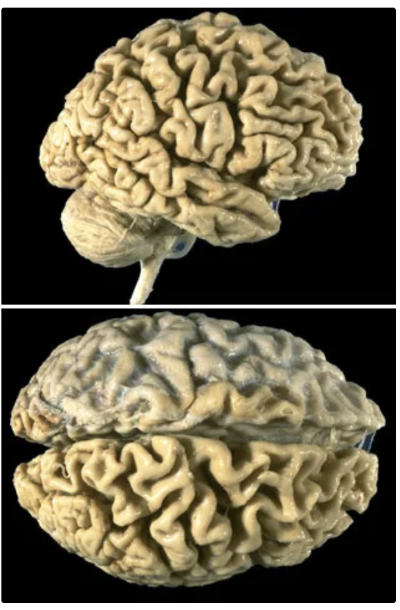
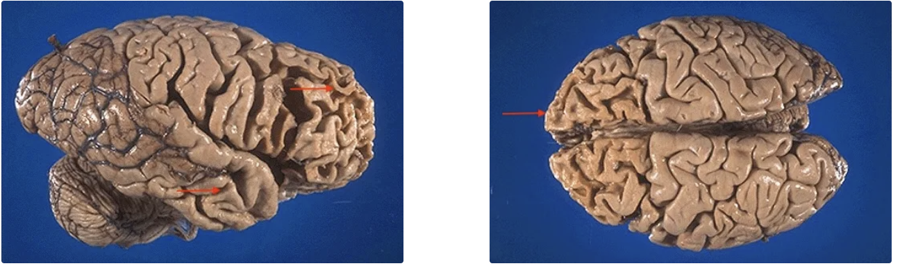
Atrofia cerebral
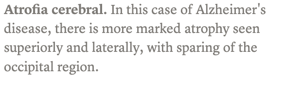
Hidrocefalia ex vacuo.
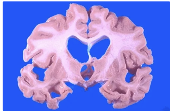
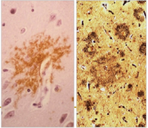
Placa senil clásica
Placa senil difusa
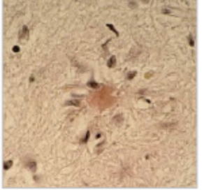
Placa senil quemada
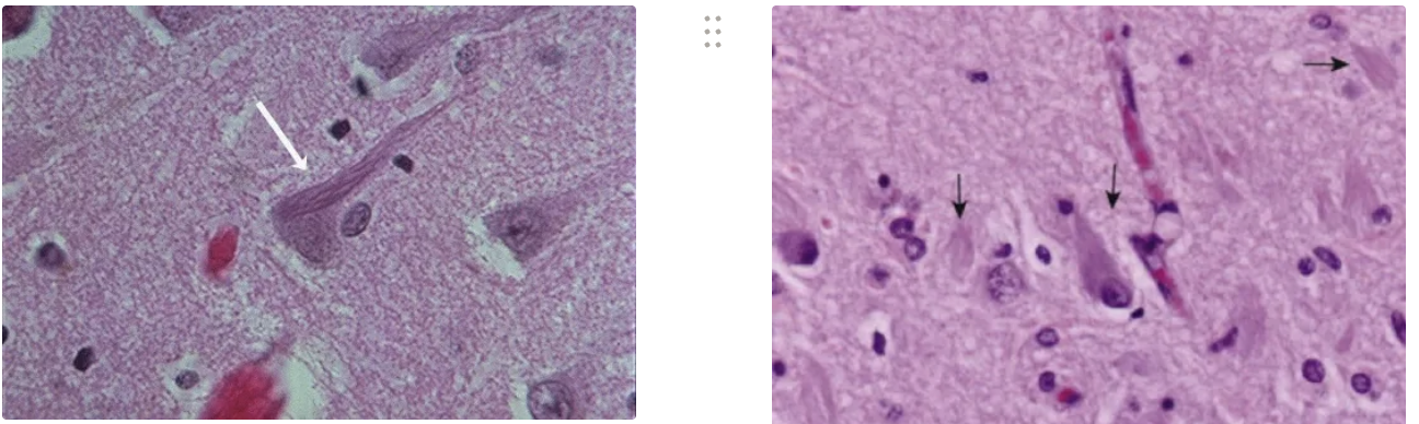
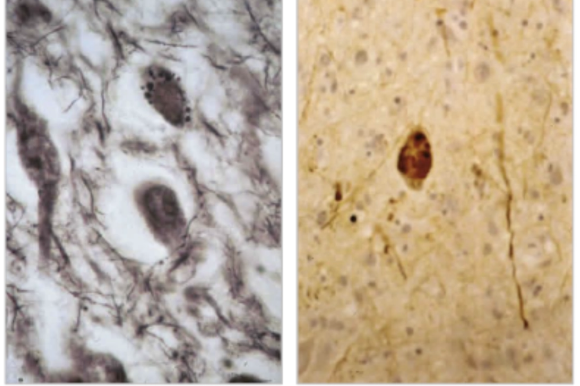
Ovillos neurofibrilares
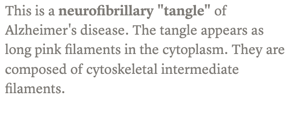
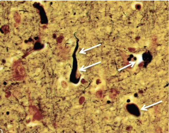
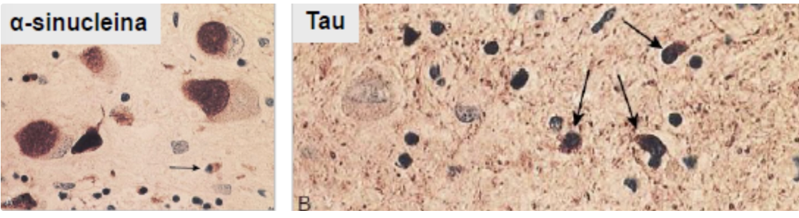
Tau-containing neurofibrillary tangle (black) within a neuron.
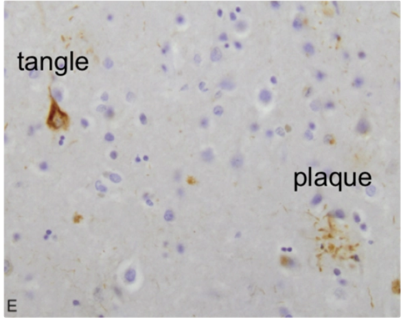
Image shows accumulation of proteins in Alzheimer disease, a tangle and a plaque.
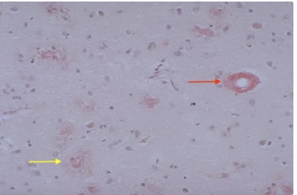
Neuritic plaques (yellow) with Alzheimer's disease are seen here. They have an amyloid core as seen here with Congo red stain. Small peripheral cerebral arteries (red; angiopatía amiloide).
Angiopatía amiloide
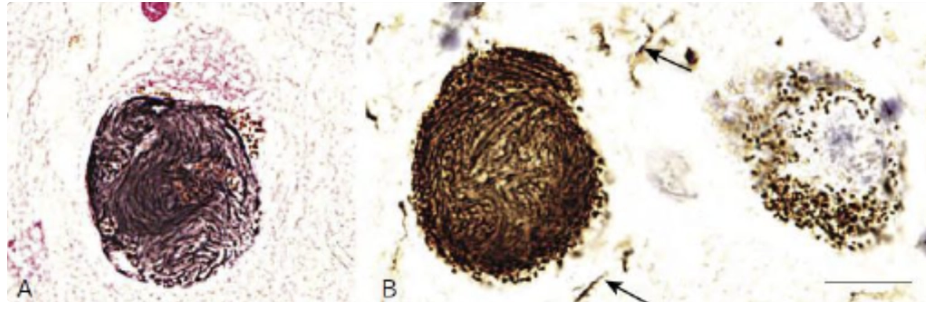
Degeneración granulovacuolar en neuronas de tamaño medio vistas con impregnación argéntica (A) e inmunotinción para neurofilamentos fosforilados (B
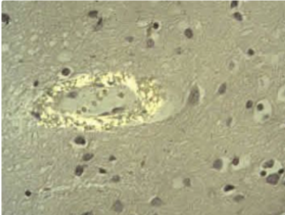
Cuerpos de Hiramo
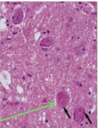
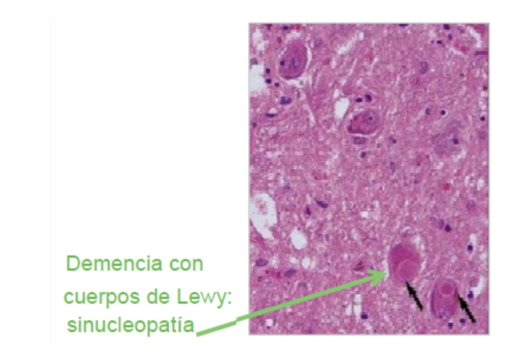
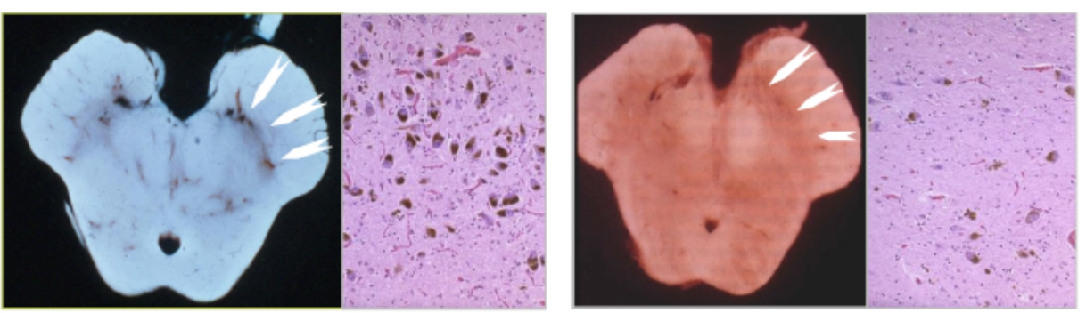
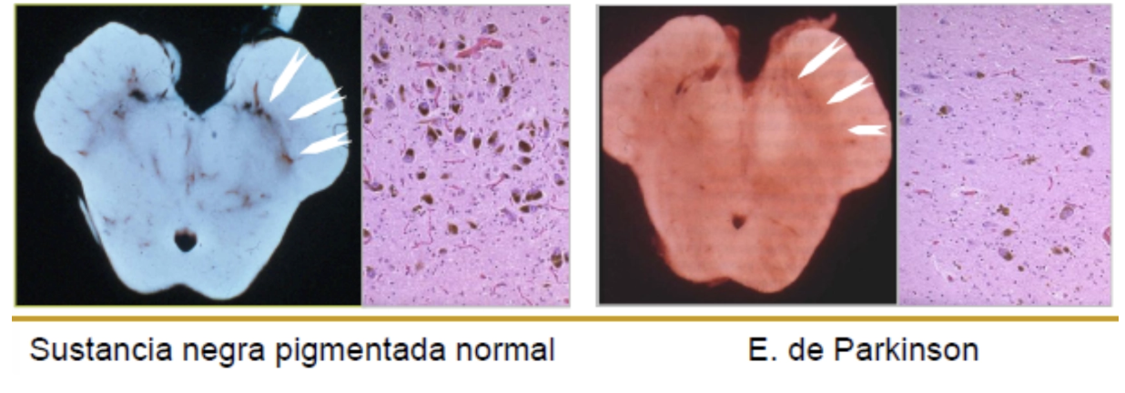
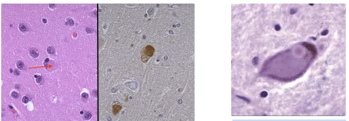
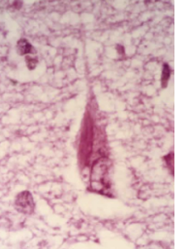
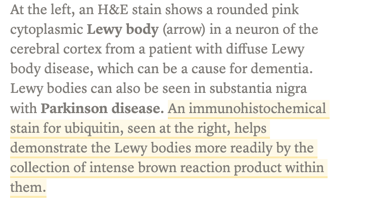
Cuerpo de Lewy en sustancia negra.
ovillos neurofibrilares globosos de Tau 4R en neuronas y glía
Atrofia de Múltiples Sistemas (AMS): Depósitos: Tau, α-sinucleína, ubicuitina, αβ-cristalina
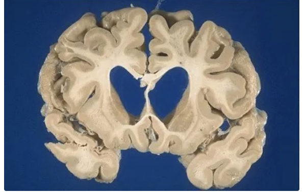
Enfermedad de Huntington (EH)
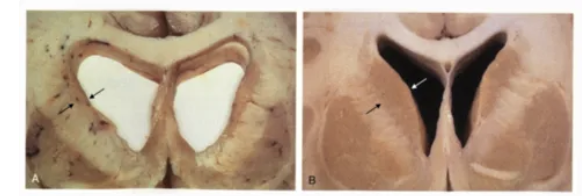
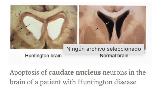
Enfermedad de Huntington (EH) → pérdida neuronal en caudado y putamen con agregados de ubicuitina + gliosis fibrilar
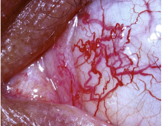
Ataxia-Telangiectasia: Telangiectasia en conjuntiva
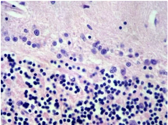
Ataxia-Telangiectasia: Cerebelo: Pérdida de células de Purkinje.
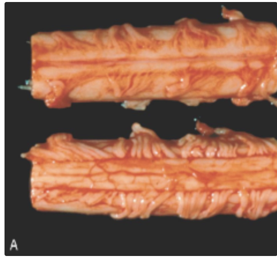
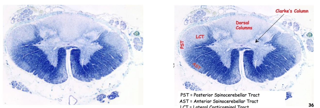
ELA (Esclerosis Lateral Amiotrófica):
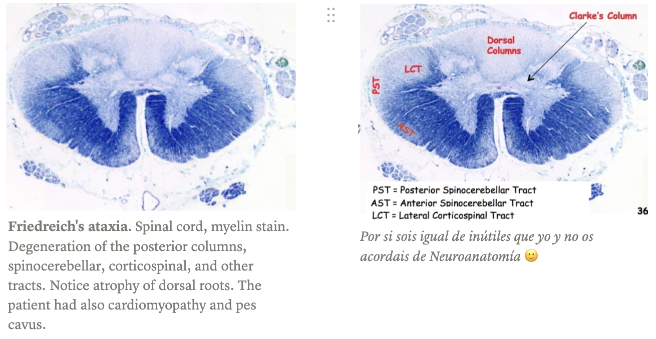
The top one shows thin atrophic ventral nerve roots. The bottom one shows normal dorsal nerve roots.
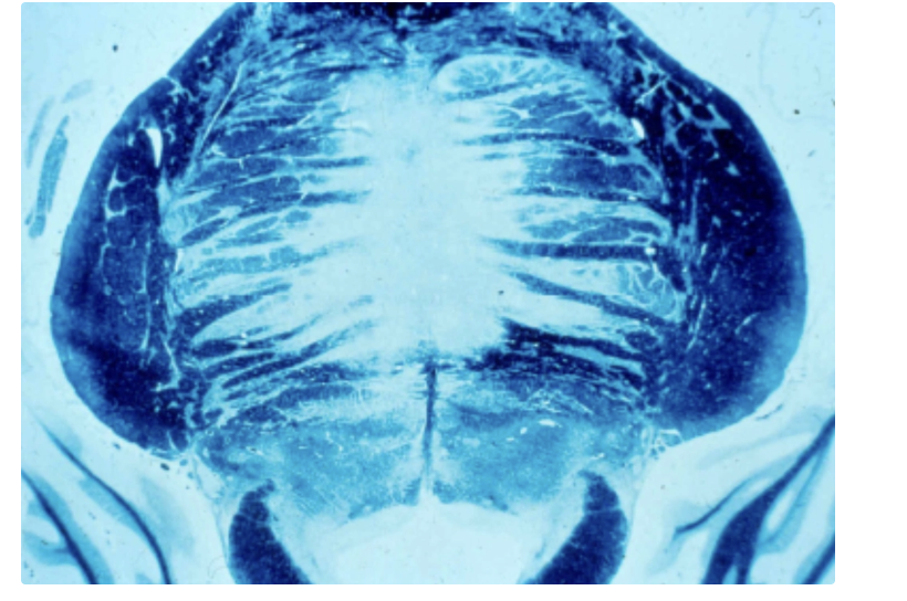
Mielinolisis Centropontina