Lecture 7: Cartilage tissue
1/83
There's no tags or description
Looks like no tags are added yet.
Name | Mastery | Learn | Test | Matching | Spaced | Call with Kai |
|---|
No analytics yet
Send a link to your students to track their progress
84 Terms
A specialized type of connective tissue.
What type of connective tissue is cartilage?
High concentrations of glycosaminoglycans (GAGs) and proteoglycans.
What is the composition of the extracellular matrix (ECM) in cartilage?
No, cartilage is avascular.
Does cartilage have a vascular supply?***
By diffusion from the surrounding perichondrium (connective tissue).
How do nutrients reach cartilage cells?
No, cartilage has no innervation.
Does cartilage have innervation?
It forms the framework for soft tissues such as the respiratory tract, ears, and nose.
What framework does cartilage provide in the body?
Provides cushioning and sliding regions with high resiliency and a smooth, lubricated surface.
What role does cartilage play within joints?
Its high resiliency and smooth, lubricated surface allow easy movement of bones.
How does cartilage facilitate bone movements?
It guides development of long bones before and after birth as a cartilaginous model in the fetus and infants.
What developmental role does cartilage serve in long bones?
Because of its high content of bound water.
Why does cartilage function as a shock absorber?
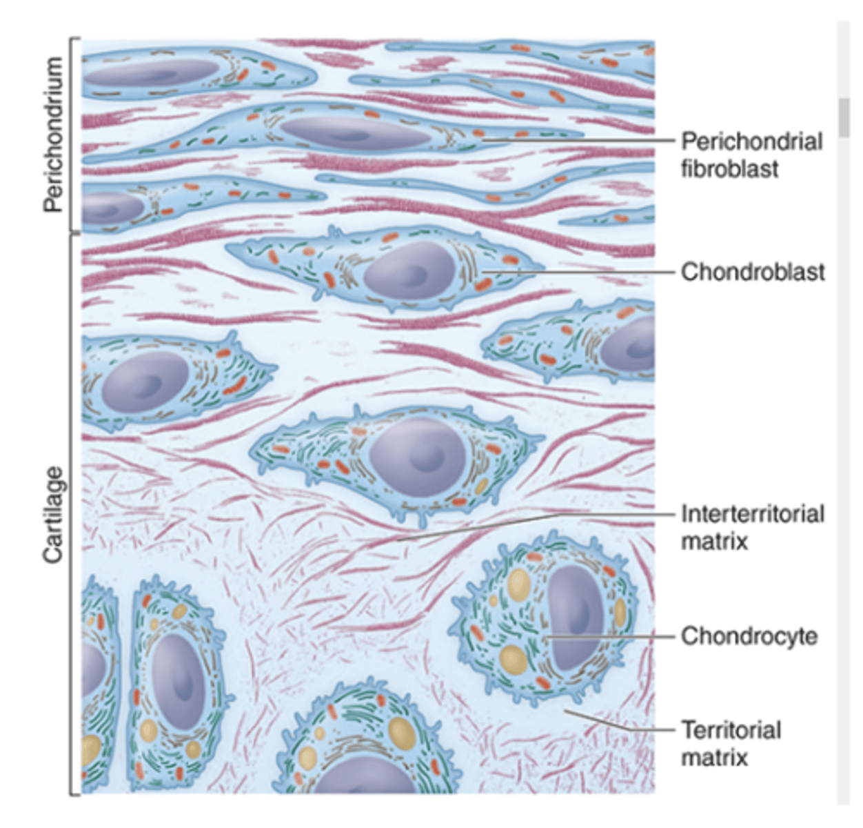
The perichondrium (dense irregular connective tissue).
What surrounds cartilage and provides support?

More than 95% of the tissue.
What percentage of cartilage is made up of extracellular matrix (ECM)?

Type II collagen.
What is the primary collagen type in cartilage ECM?***
Hyaluronan and sulfated GAGs.
What glycosaminoglycans (GAGs) are found in cartilage ECM? (2)

1. Chondroblasts: synthesize ECM components
2. Chondrocytes: synthesize and maintain ECM components (within lacunae)
What cells are found in cartilage, and what are their functions? (2)

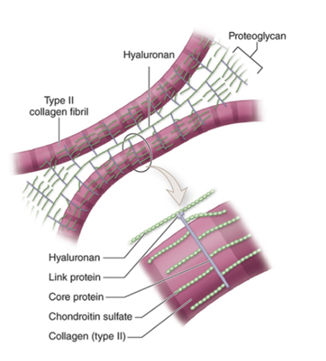
•Type II collagen fibrils
What type of collagen fibrils are found in the ECM of cartilage?***

1. Hyaluronan
2. Sulfated GAGs: chondroitin sulfate
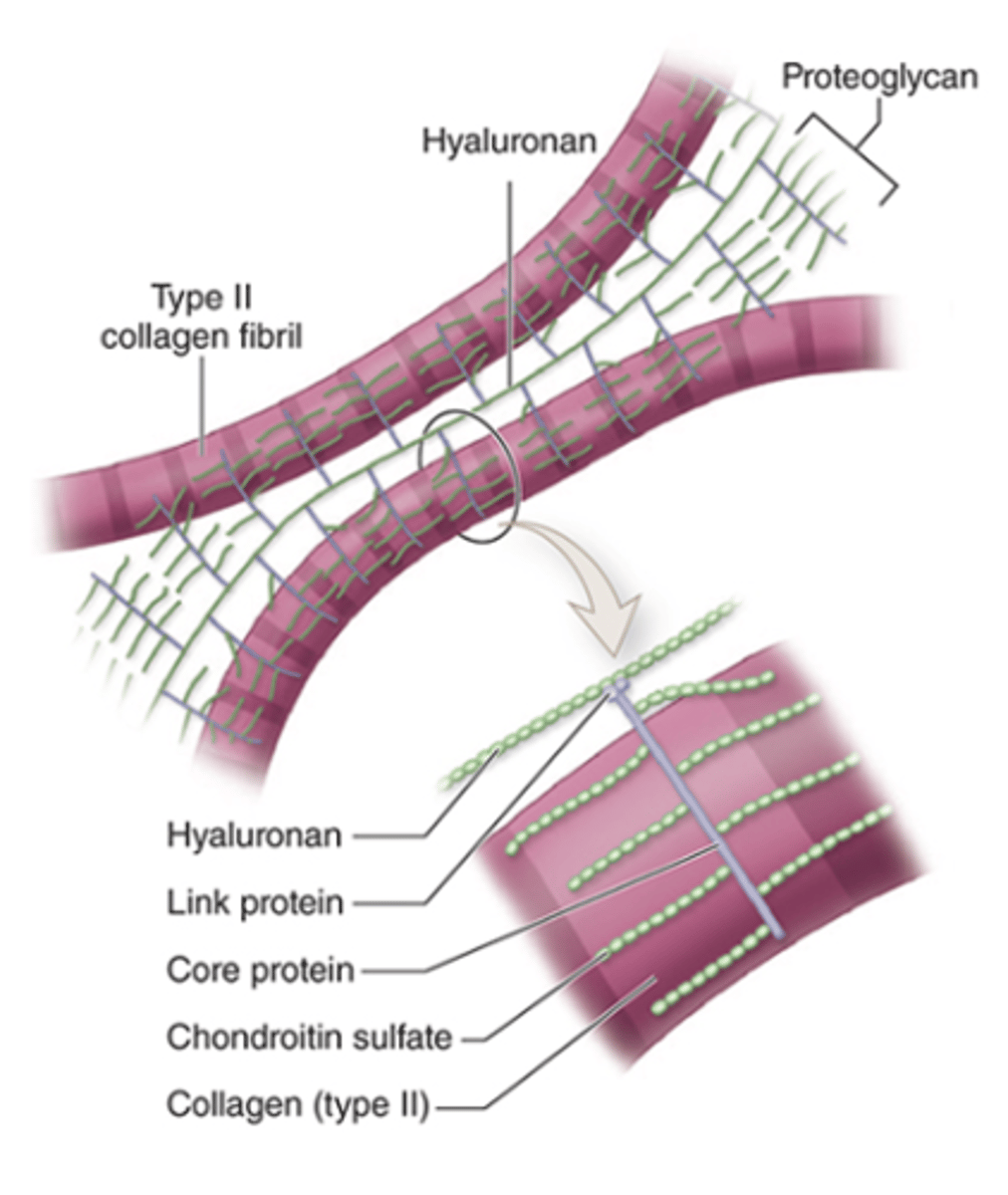
What glycosaminoglycans (GAGs) are present in the cartilage ECM?*** (2)

•Core protein and side chains of chondroitin sulfate
•Link proteins attach them to hyaluronan
What is the basic structure of proteoglycans in cartilage ECM?

bound by electrostatic bonds
How are Type II collagen and proteoglycan components held together in the ECM?
1. Hyaline cartilage
2. Elastic cartilage
3. Fibrocartilage
What are the three types of cartilage based on variations in the ECM?
Dense connective tissue.
What type of connective tissue makes up the perichondrium?
Surrounds cartilage, provides an interface between cartilage and other tissues, and supplies vascular support.
What is the main function of the perichondrium?

Yes, it has a small neural component.
Does the perichondrium contain nerves?

1. Articular cartilage (hyaline)
2. Epiphyseal cartilage (hyaline)
3. Fibrocartilage
Which types of cartilage lack a perichondrium? (3)

Glass
What does "hyalos" mean in hyaline cartilage?
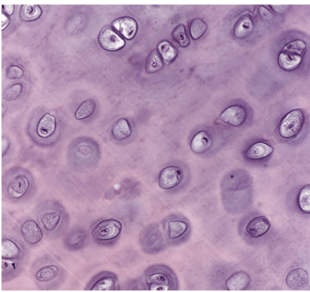
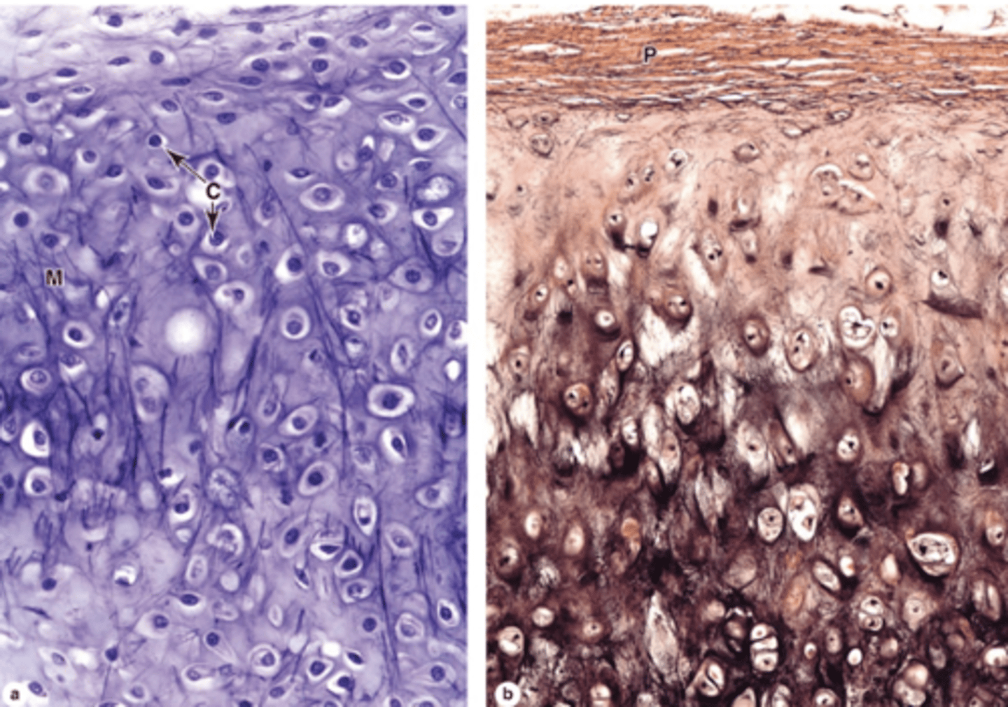
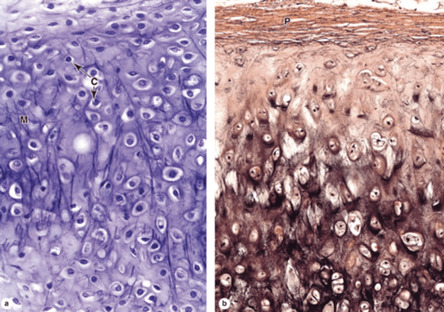
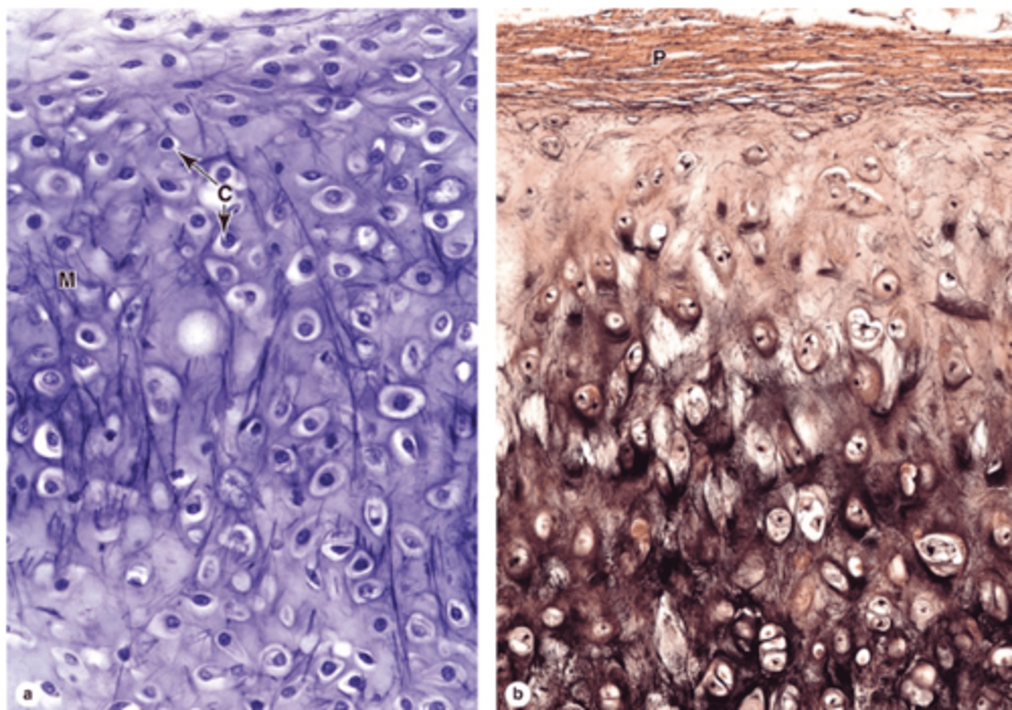
Hyaline cartilage
Which type of cartilage is the most common in the body? ***

Homogeneous and semitransparent.
How does hyaline cartilage appear under the microscope

1. Articular surfaces of diarthroses
2. Walls of respiratory passages
3. Ventral ends of ribs
4. Epiphyseal plates
5. Temporary fetal skeleton
Where is hyaline cartilage commonly found? (5)

About 40%.
What percentage of hyaline cartilage ECM is collagen?

Proteoglycans and structural glycoproteins.
What forms the hydrated gel in hyaline cartilage ECM?
Type II collagen (often barely discernible).
What is the main type of collagen in hyaline cartilage?

Due to the high content of proteoglycans.
Why does hyaline cartilage stain basophilic?

A proteoglycan with side chains of chondroitin sulfate and keratan sulfate, covalently bound to long polymers of hyaluronan.
What is aggrecan and what is its structure?

They are bound to type II collagen.
How are proteoglycan complexes organized in the ECM?

60%–80%, bound to proteoglycans.
What proportion of ECM in fresh hyaline cartilage is water?

Chondronectin (a multiadhesive protein).
What protein mediates the adherence of chondrocytes to ECM components?

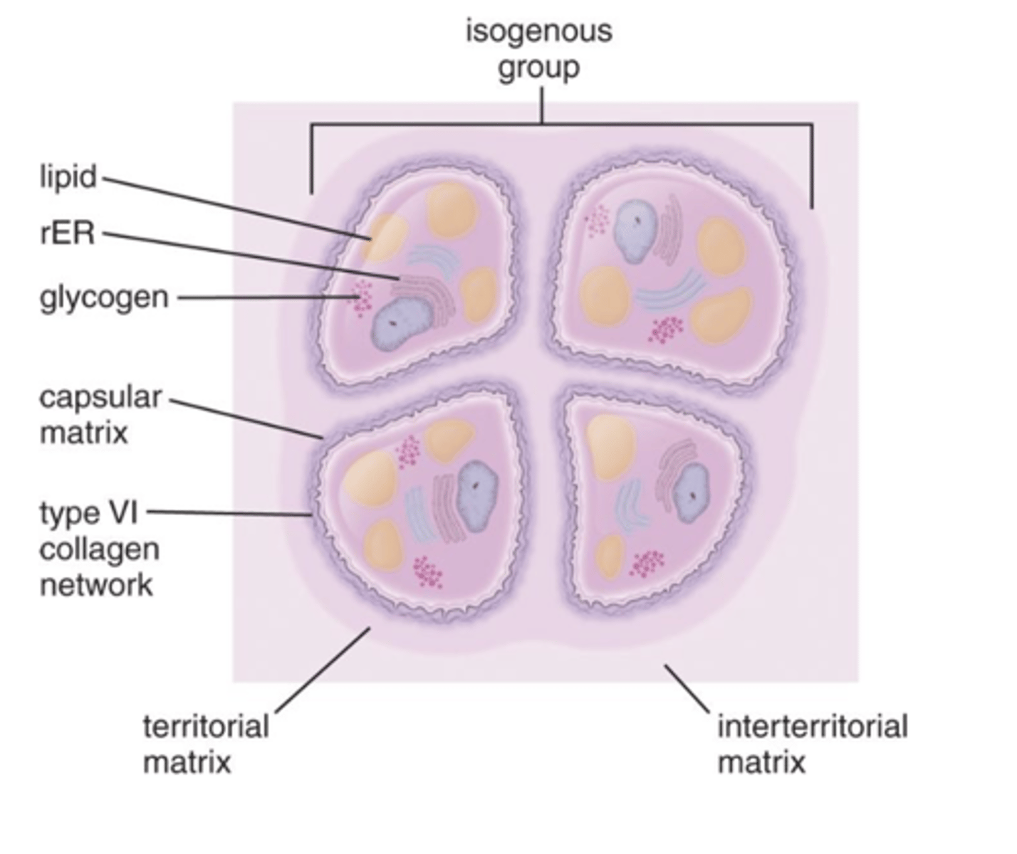
- Densely staining matrix surrounding the cell
- Highest concentration of proteoglycans
- Contains type VI collagen
What is the capsular matrix in cartilage ECM?
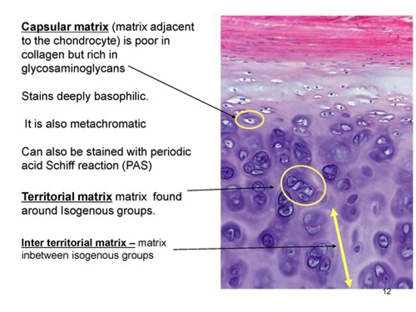
- intensely basophilic
- More GAGs than collagen (mainly type II)
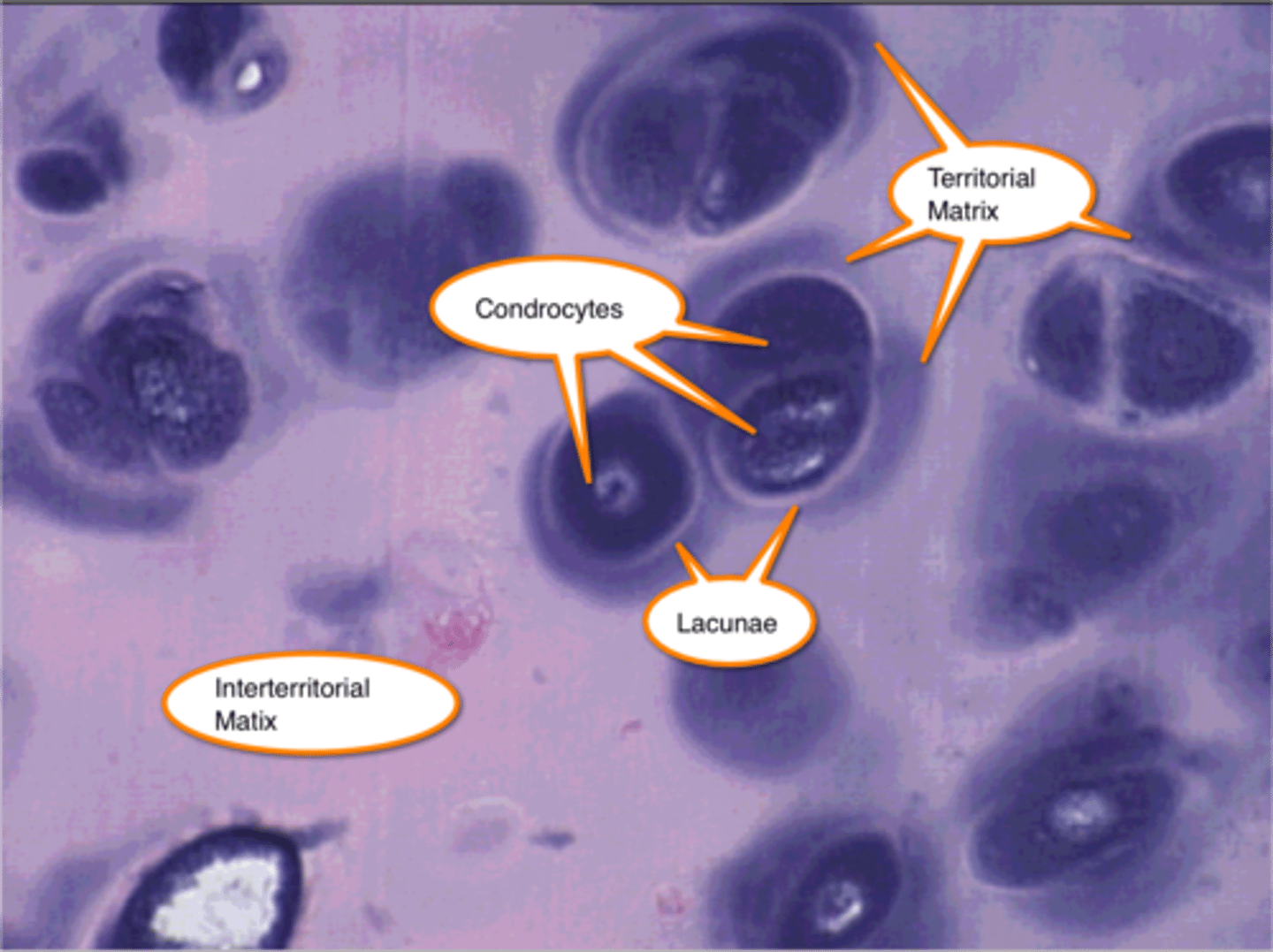
- Surrounds isogenous groups of chondrocytes
What is the territorial matrix in cartilage ECM?
- Weaker basophilia
- Less GAG content
- Found between groups of chondrocytes
What is the interterritorial matrix in cartilage ECM?
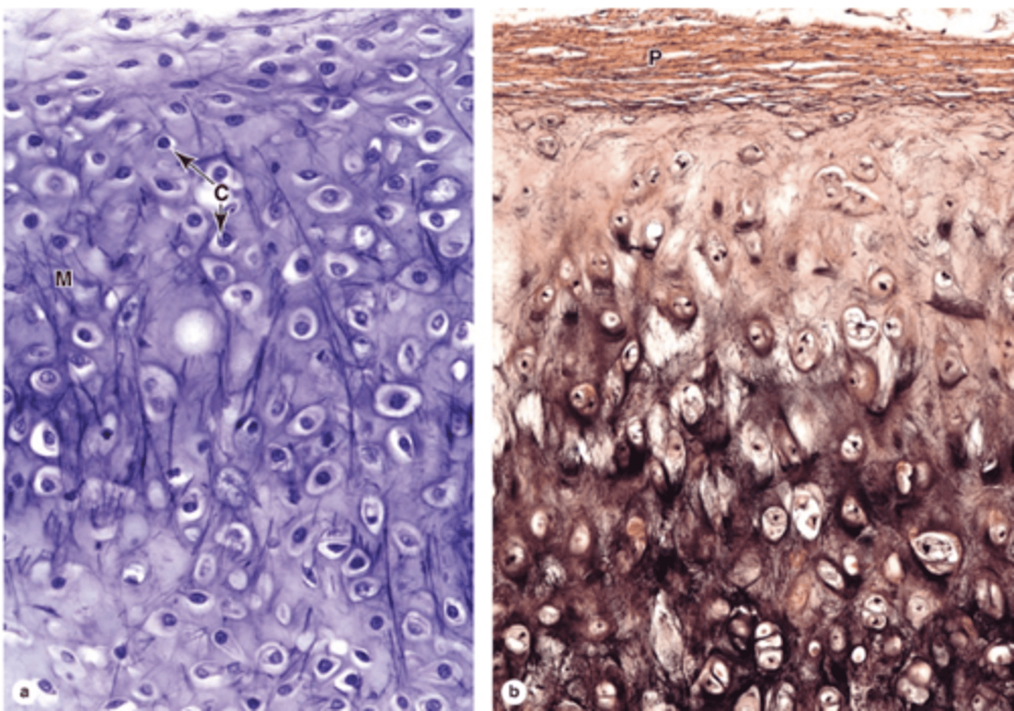
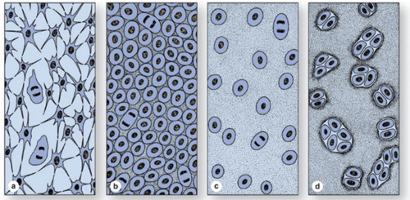
Young chondrocytes located peripherally
Elliptic in shape with long axes parallel to the surface
What are chondroblasts and where are they located?
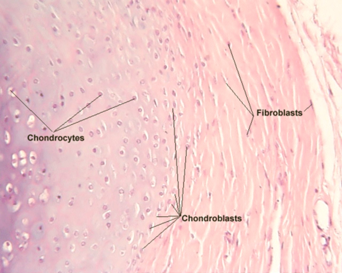
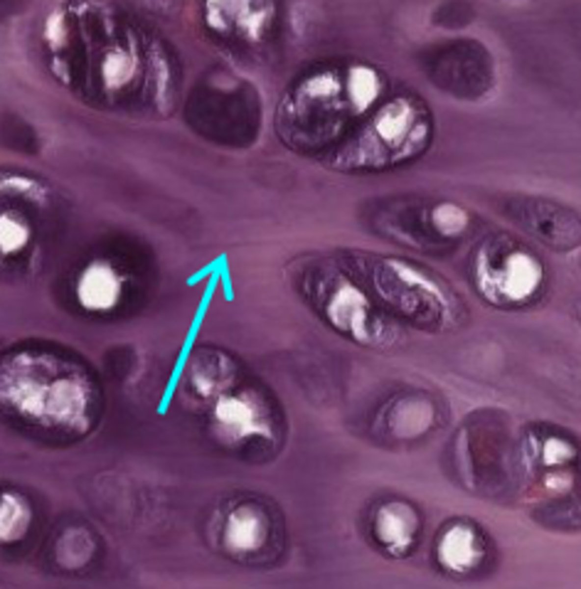
Mature cartilage cells located deeper in cartilage
Reside within lacunae
What are chondrocytes and where are they found?
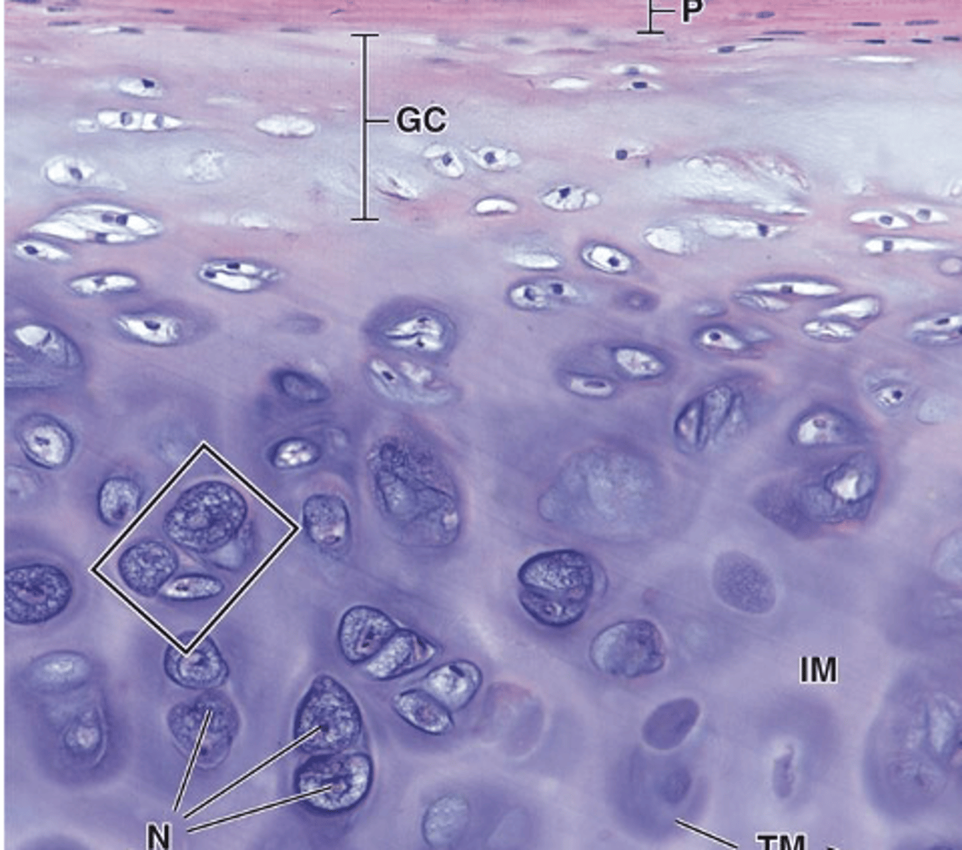
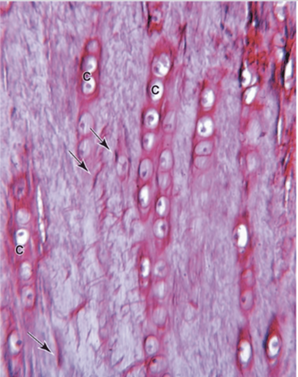
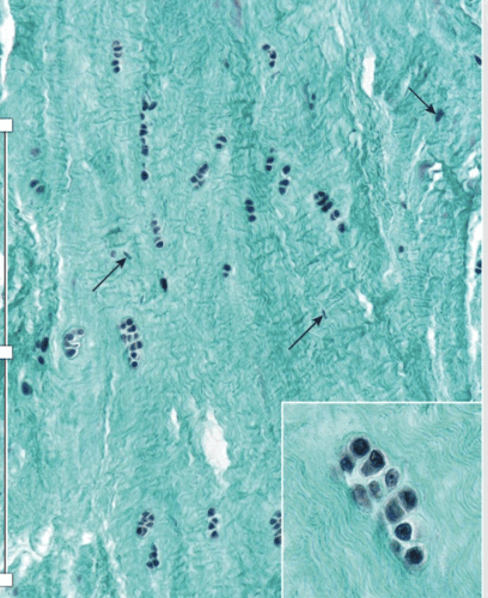
- A cluster of up to eight chondrocytes formed by mitosis of a single chondroblast
- ECM synthesis separates them into individual lacunae
What is an isogenous group?
Anaerobic glycolysis.
What is the main type of metabolism in chondrocytes***?
Somatomedins (insulin-like growth factors) from the liver.
What stimulates chondrocytes to synthesize ECM?
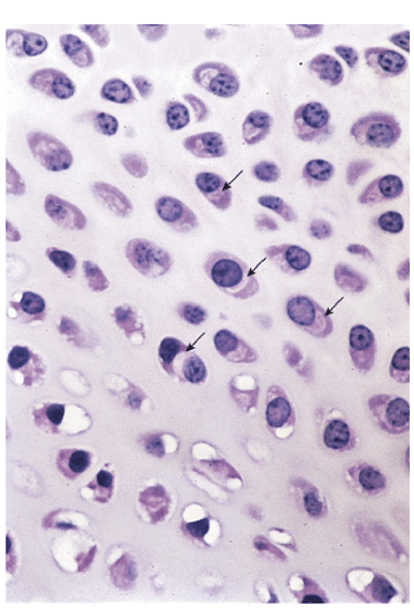
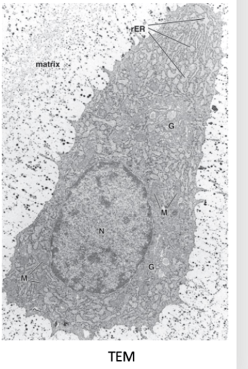
- Cytoplasmic basophilia (active protein synthesis, RER)
- Clear areas representing large Golgi apparatus
- Euchromatic nucleus (active)
How does an active chondrocyte appear under light microscopy? (3)
1. Rough endoplasmic reticulum (RER)
2. Large Golgi apparatus
3. Secretory granules and vesicles
4. Cytoskeleton: intermediate filaments, microtubules, and actin filaments
5. Eccentric euchromatic nucleus
What organelles and structures are visible in chondrocytes under TEM? (5)
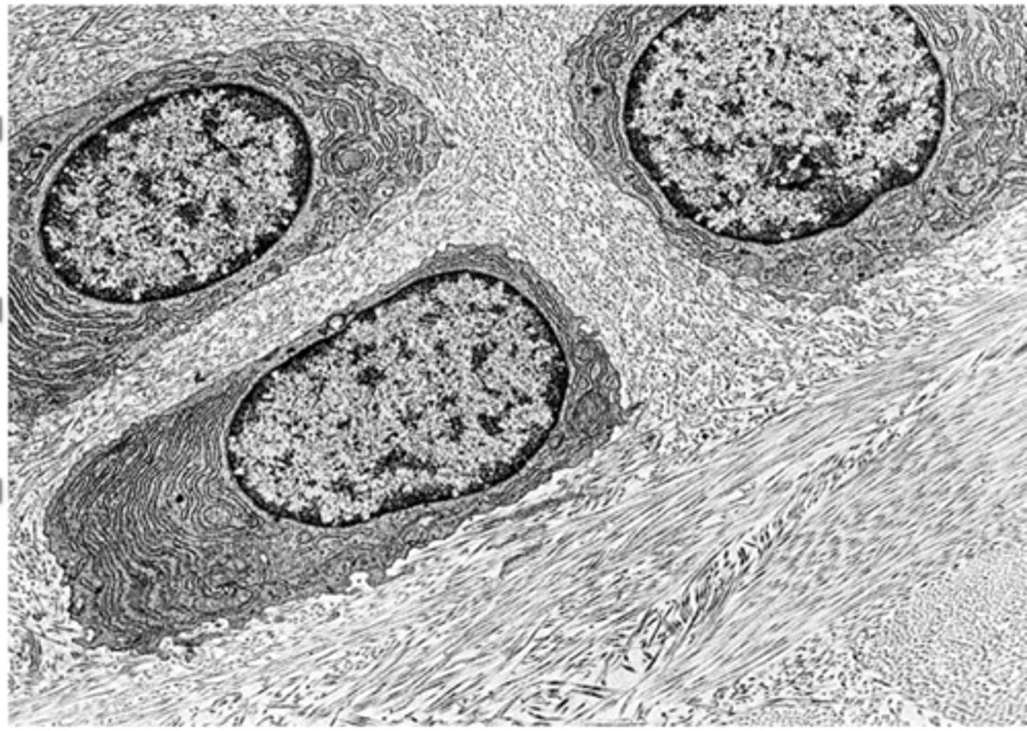
•Chondrocytes of fibrocartilage
•Abundant RER
•Euchromatic, eccentric nucleus
•Prominent Golgi apparatus
•Collagen fibers around the cells
How do active chondrocytes in growing cartilage appear under TEM?
All hyaline cartilage surfaces except articular cartilage and epiphyseal cartilage.
Which hyaline cartilage surfaces are covered by perichondrium?
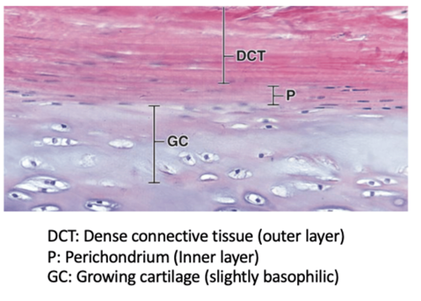
Two layers: an outer fibrous layer and an inner cellular layer.
How many layers does the perichondrium have in actively growing cartilage?

Type I collagen and fibroblasts.
What is the composition of the outer fibrous layer of the perichondrium? (2)

next to cartilage matrix with mesenchymal stem cells
- Contains mesenchymal stem cells that can divide and differentiate into chondroblasts → chondrocytes
What is the composition and function of the inner cellular layer of the perichondrium?

Mesenchymal stem cells → chondroblasts → chondrocytes.
What is the sequence of differentiation from stem cells in the perichondrium?
Similar structure but with an abundant, unevenly distributed network of elastic fibers.
How does elastic cartilage compare to hyaline cartilage?
Type II collagen fibers.
What fibers form the meshwork in elastic cartilage ECM?
Highly flexible
What is a key functional property of elastic cartilage?
1. Auricle of ear
2. Walls of external auditory canal
3. Auditory tubes
4. Epiglottis
5. Upper respiratory tract
Where is elastic cartilage found? (5)
Yes, perichondrium is always present
Does elastic cartilage have a perichondrium?

Hyaline cartilage and dense regular connective tissue.
What tissues is fibrocartilage a combination of? (2)

Provides cushioning and support for bones.
What is the main function of fibrocartilage?
1. Intervertebral discs
2. Attachment of some ligaments
3. Pubic symphysis
Where is fibrocartilage commonly found? (3)
Single or aligned in isogenous aggregates.
How are chondrocytes arranged in fibrocartilage?

Type I collagen and the usual components of ECM.
What do fibrocartilage chondrocytes synthesize?

- Separate areas with chondrocytes and hyaline matrix
- Provide extra tensile strength
What do regions with fibrocartilage that contain fibroblasts and type I collagen do? (2)
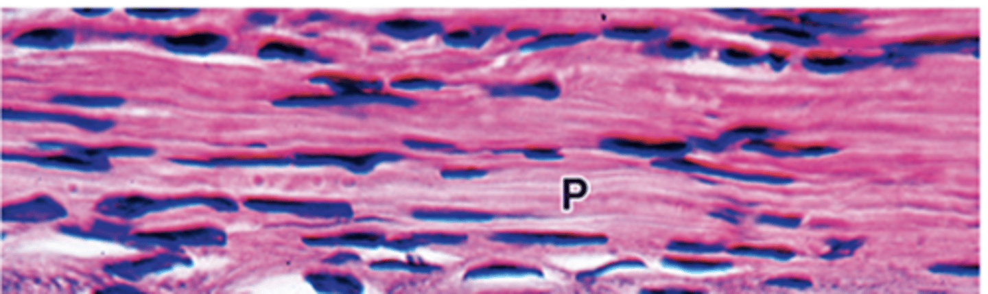
Acidophilic due to high type I collagen content.
- less proteoglycans
How does the fibrocartilage matrix stain and why?
NO PERICHONDRIUM
Does fibrocartilage have a perichondrium?***
a) Embryonic mesenchymal cells retract their processes, round up, and rapidly multiply
b) New chondroblasts become densely packed
c) Synthesis and swelling of ECM separates the cells (now chondrocytes)
d) Mitosis of single chondrocytes leads to formation of isogenic groups
What are the steps of cartilage formation (condrogenesis)? (4)
- Chondroblasts differentiate from progenitor perichondrial cells
- Synthesis of matrix contributes to cartilage growth
- More important during postnatal development
- Never occurs in articular cartilage
What is appositional growth in cartilage?
- Mitosis of pre-existing chondrocytes
- Synthesis of matrix contributes to cartilage growth
- Important for increasing the length of long bones
- Seen in articular cartilage
What is interstitial growth in cartilage?
Interstitial growth
What type of cartilage growth is seen in articular cartilage?
Slow and incomplete, except in young children
How effective is cartilage repair in general?
From perichondrial cells invading the injured tissue, but this is very limited.
Where does new cartilage come from during repair?
A scar of dense connective tissue.
What often forms in damaged cartilage areas instead of new cartilage?
Due to avascularity and slow metabolism.
Why is cartilage repair difficult and incomplete?
Hyaline cartilage
Which type of cartilage is most prone to calcification?
1. Portion of articular cartilage in contact with bone tissue (growing and adult bones)
2. During endochondral ossification in growth
3. As part of the aging process in adult hyaline cartilage
What determines the sites of cartilage calcification? (3)
a lengthy process
How long does cartilage calcification take?
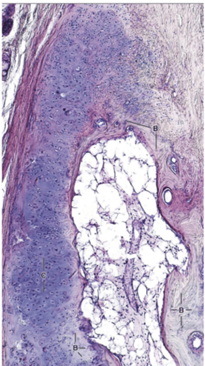
It is eventually replaced by bone.
What always happens to calcified cartilage?

The calcified matrix impedes diffusion of nutrients.
How does calcification affect nutrient diffusion in cartilage?

Chondrocytes swell and die.
What happens to chondrocytes during calcification?
It is removed and replaced by bone.
What happens to the calcified matrix after chondrocytes die?
Special connective
What type of tissue is cartilage?
Fibrocartilage
What type of cartilage has abundant type I collagen fibers?
by diffusion
How do Chondrocytes receive their nutrients
Hyaline cartilage
Osteoarthritis is a degenerative disease that limits the movement of synovial joints by affecting the articular cartilage, which is made up of ________