6.3 Skeletal muscles are stimulated to contract by nerves and act as effectors
1/9
There's no tags or description
Looks like no tags are added yet.
Name | Mastery | Learn | Test | Matching | Spaced | Call with Kai |
|---|
No analytics yet
Send a link to your students to track their progress
10 Terms
Describe how muscles work
work in antagonistic pairs → pull in opposite directions e.g. biceps / triceps
→ one muscle contracts (agonist), pulling on bone / producing force
→ one muscle relaxes (antagonist)
skeleton is incompressible so muscle can transmit force to bone
Advantage - the second muscle required to reverse movement caused by the first (muscles can only pull) and contraction of both muscles help maintain pressure
Describe the gross and microscopic structure of skeletal muscle
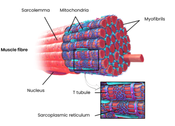
made of many bundles of muscle fibres (cells) packaged together
attached to bones by tendons
muscle fibres contain:
→ sarcolemma (cell membrane) which folds inwards (invagination) to form transverse (T) tubules
→ sarcoplasm (cytoplasm)
→ multiple nuclei
→ many myofibrils
→ sarcoplasmic reticulum (endoplasmic reticulum)
→ many mitochondria
Describe the ultrastructure of a myofibril
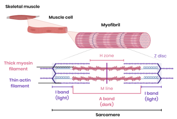
made of two types of long protein filaments arranged in parallel
→ myosin - thick filament
→ actin - thin filament
arranged in functional units called sarcomeres
→ ends - Z-line / disc
→ middle - M -line
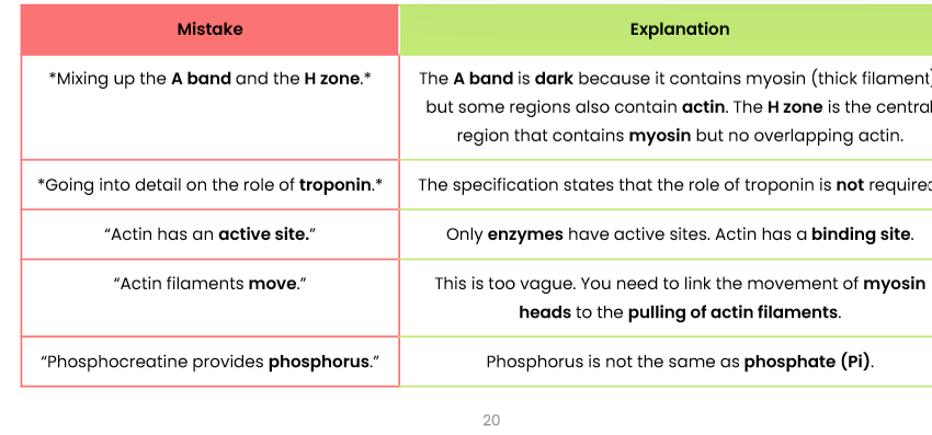
→ H zone - contains only myosin
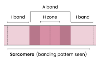
Explain the banding pattern to be seen in myofibrils
I-bands - light bands containing only thin actin filaments
A-bands - dark bands containing thick myosin filaments (and some actin filaments)
→ H zone contains only myosin
→ darkest region contains overlapping actin and myosin
Give an overview of muscle contraction
myosin heads slide actin along myosin causing the sarcomere to contract
simultaneous contraction of many sarcomeres causes myofibrils and muscle fibres to contract
when sarcomeres contract (shorten)…
→ H zones get shorter
→ I bands get shorter
→ A bands stay the same
→ Z lines get closer
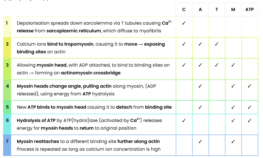
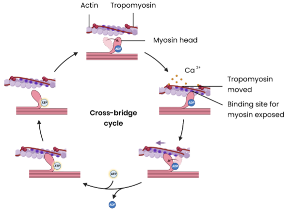
Describe the roles of actin (A), myosin (M), calcium ions (C), tropomyosin (T) and ATP in myofibril contraction
Action potentials travels deep into the muscle fibre through system of T tubules that are extensions of the sarcolemma and branch throughout the sarcoplasm
Tubules are in contact with the sarcoplasmic reticulum which has actively transported calcium ions from the cytoplasm of the muscle leading to a low calcium ions concentration in the sarcoplasm (at rest)
Action potential opens the calcium ion protein channels on the sarcoplasmic reticulum membrane and calcium ions diffuse into the sarcoplasm down their electrochemical gradient
Calcium ions cause the tropomysoin molecules that were blocking the binding sites on the actin filaments to pull away
ADP molecules attached to the myosin heads mean that they are in a state to bind to the actin filament and form an actin-myosin cross bridge
Once attached to the actin filament, the myosin heads change their angle, pulling the actin filaments along as they do so and releasing a molecule of ADP (power stroke)
An ATP molecule attaches to each myosin head, causing it to become detached from the actin filament
Calcium ions then activate the enzyme ATPase which hydrolyses the ATP to ADP and Pi. The hydrolysis of ATP to ADP provides energy for the myosin heads to return to its original position
The myosin head, once more with an attached ADP molecule, then reattaches itself further along the actin filament and the cycle is repeated as long as the concentration of calcium ions in the myofibril remains high.
What happens during muscle relaxation?
Ca2+ actively transported back into the endoplasmic reticulum using energy from ATP
tropomyosin moves back to block myosin binding site on actin again → no actinomyosin cross bridges
Describe the role of phosphocreatine in muscle contraction
a source of inorganic phosphate (Pi) → rapidly phosphorylates ADP to regenerate ATP
→ ADP + phosphocreatine → ATP + creatine
runs out after a few seconds → used in short bursts of vigorous exercise
anaerobic and alactic
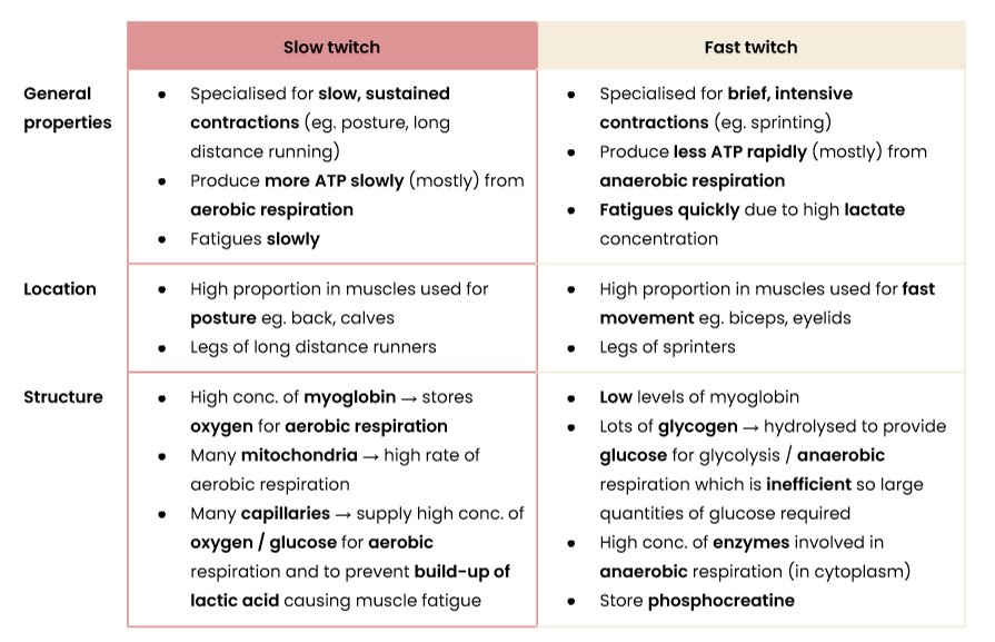
Compare the structure, location and general properties of slow and fast skeletal muscle fibres
Exam Insights: common mistakes