Day 1
1/53
There's no tags or description
Looks like no tags are added yet.
Name | Mastery | Learn | Test | Matching | Spaced | Call with Kai |
|---|
No analytics yet
Send a link to your students to track their progress
54 Terms
Example of unconditioned reflex
Patellar (knee-jerk) reflex
Example of conditioned reflex
Pavlov’s salivary reflex
Unconditioned reflexes are ______ , inherited responses that do not require prior learning. They possess a ________ ______ ___ and are triggered by an _______ stimulus.
In contrast, conditioned reflexes are acquired during life through learning and experience. They are ________, ________, and depend on the formation of new neural connections in the cerebral cortex. They are _________, individual, and depend on the formation of ___ ________ _____________ in the ________ ______. Conditioned reflexes arise when a neutral stimulus is repeatedly associated with a stimulus that naturally evokes a response.
innate, temporary reflex arc, adequate
Temporary, individual, new neural connections, cerebral cortex,
4 ways reflexes are categorized depending on reflex arc
Somato-somatic
Somato-viceral
Vicero-somatic
Vicero-viceral
Somato-somatic reflex
Origin/arise from :
Affect :
Example :
Somato-somatic reflex
Origin/arise from :
Affect :
Example :
Exteroceptors, skeletal muscles, plantar reflex
Somato-visceral reflex
Origin/arise from :
Affect :
Example :
Exteroceptors, smooth muscles + glands, oculocardiac reflex
Vicero-somatic reflex
Origin/arise from :
Affect :
Example :
interoceptors, skeletal muscles, abdominal wall rigidity
Vicero-visceral reflex
Origin/arise from :
Affect :
Example :
interceptors, smooth muscles and glands, pressor reflex.
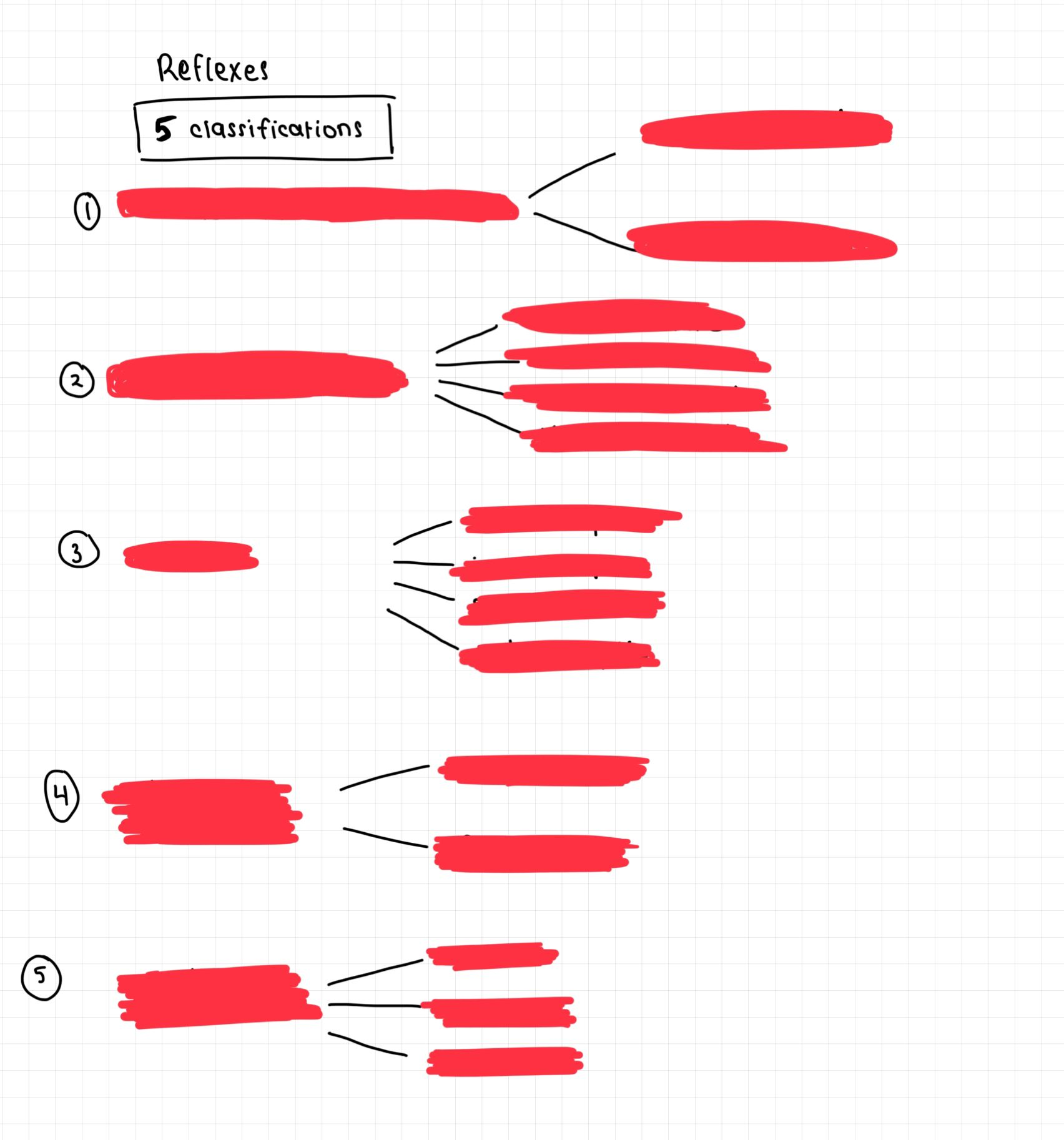
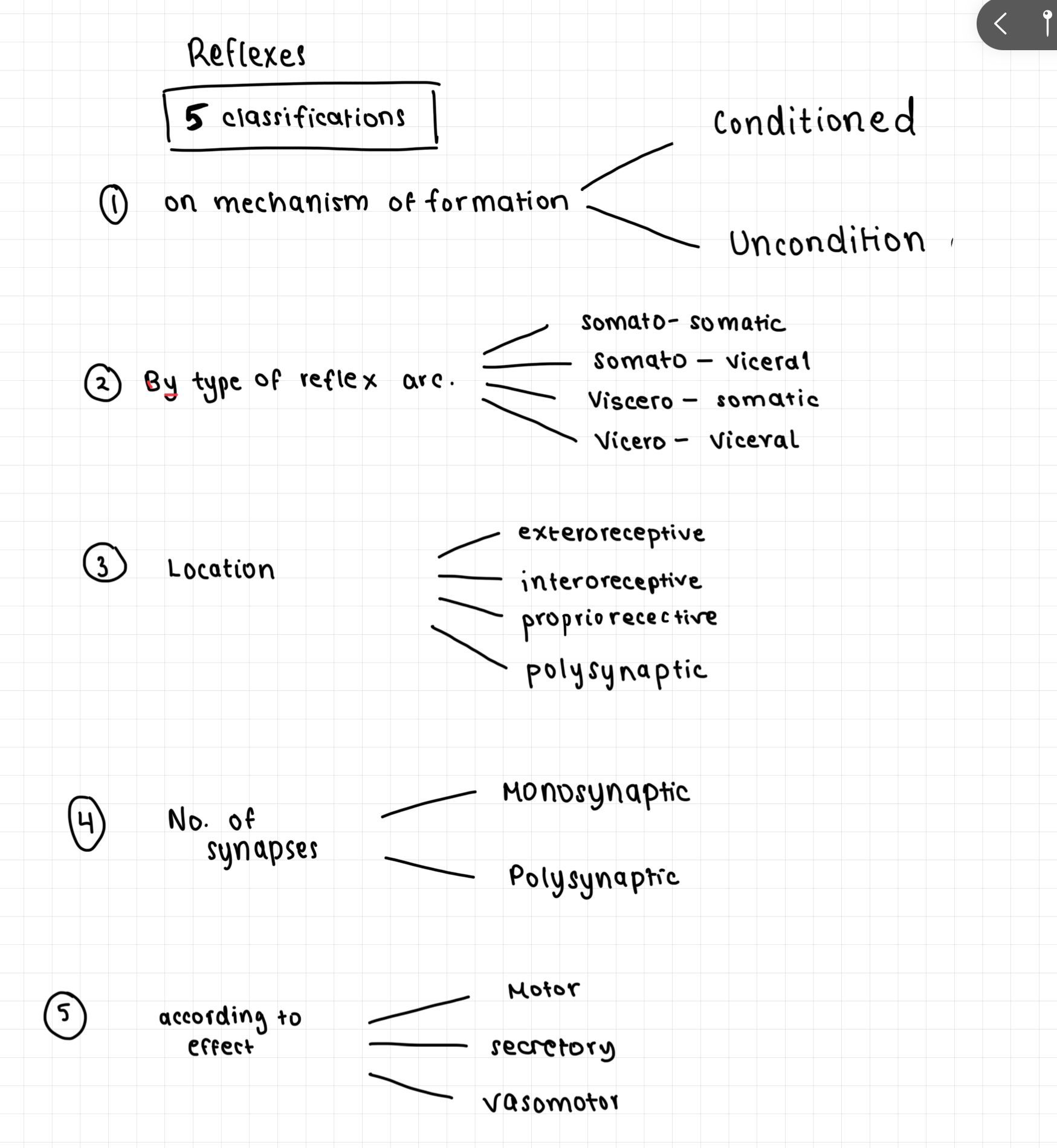
According to receptor location, reflexes can be classified as ______________, _______________ or _________________ (give their locations)
exteroceptive - inner ear
interoceptive - internal organs
proprioceptive - muscular joints
Classification according to number of synapses
Monosynaptic
Polysynaptic —> involving one or more interneurons
3 reflexes according to effect:
Motor
Secretory
Vasomotor
A nerve centre is a collection of ____________ within the CNS where information is __________, _________, and _____________. Nerve centres are rich in __________ _________ and are responsible for controlling specific physiological functions.
Examples include the :
Respiratory and cardiovascular centres -
Vasomotor centre -
Defecation centre -
Sensory centre -
Motor centre -
Visual centre -
Auditory centre -
A nerve centre is a collection of interneurons within the CNS where information is received, processed, and integrated. Nerve centres are rich in chemical synapses and are responsible for controlling specific physiological functions.
Examples include the :
Respiratory and cardiovascular centres - medulla oblongata and pons
Vasomotor centre - medulla
Defecation centre - lumbosacral spinal cord
Sensory centre - parietal cortex
Motor centre - frontal lobe
Visual centre - occipital lobe and midbrain
Auditory centre - temporal lobe and midbrain.
Role of lysosomes
Vesicular structure formed by Golgi apparatus
Contains digestive enzymes hydrolases which degrade proteins
Role of peroxisomes
Contain digestive enzyme oxidase
Breaks down H2O2 (harmful to cell) into H2O by catalases
It degrades fatty acids
What are the different types of cell junctions
Junction | Main Function | Cytoskeleton Attached |
|---|---|---|
1. Tight junction (Zonula occludens) | Seals adjacent cells together, preventing leakage of substances between cells | Actin filaments |
2. Adherens junction (Zonula adherens) | Strong cell-to-cell adhesion; joins actin filaments of neighboring cells | Actin filaments |
3. Desmosome (Macula adherens) | Strong mechanical attachment between cells; resists stretching forces Found in epithelia (e.g. skin) | Intermediate filaments (keratin) |
4. Hemidesmosome | Anchors epithelial cells to the basal lamina (basement membrane) | Intermediate filaments (keratin) |
5. Gap junction (Nexus) | Intercellular channels allows passage btw. cells of ions and small molecules | No major cytoskeletal attachment |
Tight junctions: Prevent paracellular movement of substances.
Adherens junctions: Connect actin cytoskeletons of adjacent cells.
Desmosomes: Connect intermediate filaments of adjacent cells.
Hemidesmosomes: Connect intermediate filaments to the basal lamina.
Gap junctions: Allow passage of ions and small molecules between neighboring cells.
What are the types of signaling (intercellular)
Endocrine signaling - secretes substances into the bloodstream and affects the target cells in different parts of the body
Paracrine signaling - secretes substances that act on adjacent cells
Autocrine signaling - cell affects itself
Juxtacrine signaling - contact dependent à a ligand on one surface binds to a receptor on the other cell surface
Is cell membrane polar or non-polar? Why?
POLAR
induces electric potential difference permeable to several different ions
Depends on conc. inside and outside of memb.
What are the 3 states of membrane for osmosis?
Isotonic - normal conc. of solute and water
Hypotonic - Little solute and big water conc —> bursts
Hypertonic - conc. of solute is too high —> shrinks
What are the 2 types of hormonal regulation ?
Hormonal
not-well addressed
slow
hormones released form endocrine and exocrine glands
long-term response
Neural
well-addressed
fast
based on reflexes of CNS and ANS
give short term response
What are the 2 feedback systems ?
Negative feedback
Response opposes the original stimulus and returns variable toward its normal value
Characteristics:
Most common homeostatic mechanism
Stabilizes the internal environment
Reduces deviation from the set point
Examples
Blood glucose regulation
↑ Glucose → insulin release → ↓ glucose
Thermoregulation
↑ Temperature → sweating
↓ Temperature → shivering
Blood pressure regulation
↑ Blood pressure → vasodilation → ↓ pressure
Positive feedback
This amplifies the original stimulus and causes further deviation from the normal value
Characteristics:
Less common
Continues until a specific event is completed
Examples
Childbirth
Cervical stretching → oxytocin release → stronger contractions → more stretching
Blood clotting
Activated clotting factors stimulate activation of additional clotting factors
Action Potential Generation
Depolarization opens sodium channels → further depolarization
What are the 3 basic components of the homeostatic regulatory system?
1. Receptor (Sensor)
Detects changes in the internal or external environment.
Examples:
Thermoreceptors —> detect changes in temp. of internal + external environment, vasodilation/vasoconstriction
Chemoreceptors —> chemical changes in blood and fluids
Baroreceptors
2. Control Center (Integrating Center)
Receives information from receptors
Compares it with the normal value (set point)
Determines the appropriate response
Examples:
Hypothalamus
Brainstem
Endocrine glands
3. Effector
Produces the corrective response.
Examples:
Muscles
Glands
Blood vessels
Homeostatic Sequence
Stimulus → Receptor → Control Center → Effector → Response
Homeostatic regulatory systems
Skeletal system
Supports posture and movement
Mineral reservoir (Ca 2+ and phosphate)
Blood cell formation
Cardiovascular system
Transport of nutrients, gases and hormones
Regulate BP
Lymphatic system
Returns excess tissue fluid
Immunity
Fat absorption
Respiratory system
Digestive system
Urinary system
Excretes waste
Regulates water and electrolyte balance
Reproductive system
Maintains reproductive function
Produces sex hormones
Immune system
Muscular system
Produces movement
Generates heat thru contraction
Integumentary system
Protects against pathogens
Regulated temp
Maintains temp and ion balance
Levels of physiological regulation
Intracellular level —> gene expression, enzyme activity, metabolism, osmotic balance
Tissue level
Organ level
Organ system level
Organismal level
What are the types of regulation
Proportional regulation
The response is proportional to the size of the change.
Example: CO₂ and breathing
Normal CO₂ → normal breathing
Slightly ↑ CO₂ → slightly faster breathing
Very ↑ CO₂ → much faster breathing
Differential regulation
The body responds to the rate of change, not just the value itself
Example: Baroreceptor reflex
Imagine blood pressure falls:
Case 1
BP drops from 120 → 110 mmHg slowly over hours.
The body doesn't panic.
Case 2
BP drops from 120 → 80 mmHg in 5 seconds.
The body reacts immediately:
↑ Heart rate
↑ Vasoconstriction
The actual BP value matters less than how quickly it changed.
Memory: Differential = speed of change.
Integral regulation
The body takes into account the accumulated error over time.
Example: Blood volume
Suppose you lose a little blood.
For 5 minutes:
Not a huge response.
For several hours:
Kidneys retain water.
ADH secretion increases.
Aldosterone increases.
The body is responding because the deviation has been present for a long time.
What is the difference btw. irritability and excitability?
Irritability
Ability of the cell to react to stimuli from external or internal environment by increase or decrease of its activity
Stimuli may be mechanical, chemical, electrical, or thermal
Stimuli differ in intensity, duration and rate of application
Excitability
Ability of specialised cells to respond to change in environment (irritation)
Does this by changing their membrane potential and generating an action potential when the stimulus reaches a threshold value
What are the excitable tissues ?
Motor neuron —> longest to cause action potential
Skeletal muscle —> shortest to cause action potential
Heart muscle —> contraction of muscle cells
Endocrine cells —> produces and releases hormones
What is membrane potential ?
The electrical difference between the inside and outside of the cell membrane
Voltage diff. across cell memb. due to differences in ion conc.
What is resting memb. potential ?
Electrochemical gradient that exists when cell is not transmitting a signal
Typically -70mV in neurons
Muscle cells may have values btw. -70 to -90 mV
This means inside of cell = negatively charged relative to outside
Action potential process/steps
Put Pic From Ipad !!!!!!!!!!!!!!
Resting state
Memb. potential -70mV
To every 3 Na+ flowing out of cell, 2 k+ flow inside
More +ve charges outside, memb. potential is -ve
Depolarisation
If stimulus reaches threshold of -55 mV = action potential
Voltage-gated channels open = Na+ flow inside
POSITIVE FEEDBACK - more voltage-gated channels open leading to memb. potential rising to 30 mV
Repolarisation
Sodium channels close
Voltage gated potassium channels open —> K+ out cell
Voltage-gated channels for K+ open causing flow of K+ out of cell
Memb. potential becomes -ve
Hyperpolarisation
As the K+ voltage-gated channels take a long time to close
membrane may become temporarily more negative than the resting potential, producing a phase called hyperpolarization
What are the different types of ion channels and pumps?
Voltage-gated channels
Open/close in response to changes in memb. potential
Ligand-gated channels
Open/close in response to binding of a specific molecule
Ion pumps
Use energy (ATP)
actively transport ions against conc. gradient (Na+/k+ ATPase pump)
embedded in cell memb., help to maintain and alter ionic balance
How can excitability be measured ? What are the parameters?
Hoorweg-Weiss strength-duration relationship
Rheobase
The minimum stimulus intensity capable of producing excitation.
Utilization Time
The minimum duration required for a rheobase-strength stimulus to excite tissue.
Chronaxie
The minimum duration required for a stimulus with twice the rheobase intensity to produce excitation.
Liability
The maximum number of impulses a tissue can transmit per second.
What are the 2 diff. types of synapses?
Electrical
Consist of gap junctions that create v. narrow space of abt. 2.5 nm btw. adjacent cells
Signal transmission depends on VOLTAGE GATED ion channels —> open + close depending on memb. potential
Allow direct passage of ions from one cell to another = extremely rapid transmission w. virtually no synaptic delay
Conduction = BIDIRECTIONAL (impulse travels 2 ways)
Mainly muscles cells —> synchronized activity is required
Depolarisation = increase in memb. potential
Higher conduction velocity, greater no. of cells can be excited simultaneously
Chemical
Most common in nervous system.
Characterized by synaptic cleft of 20-30nm btw. presynaptic + postsynaptic cells
UNIDIRECTIONAL
neurotransmitters released from presynaptic neuron and act ONLY on receptors of postsynaptic memb.
Physiological delay of - 0.3-0.5 ms
Chemical synapses exhibit plasticity —> strength can increase or decrease depending on activity
Can be excitatory or inhibitory
According to location —> conventional synapses (axodendritic, axosomatic, and axoaxonic), which are generally excitatory, and non-conventional synapses (dendrodendritic, dendroaxonic, and dendrosomatic)
Transmission only occurs thru ligand-gated ion channels —> open when a neurotransmitter binds to its receptor, allows specific ions to pass thru memb. and alter excitability of postsynaptic cell
Types of postsynaptic channel receptors
Ionotropic
Metabotropic
Types of ionotropic receptors
N-cholinoreceptors
Glutamate
Glycine
GABA receptors
Types of metabotropic
G protein-coupled receptors —>
Alpha and Beta adrenoreceptors
M-cholinoreceptors
Inhibitory postsynaptic potentials are due to:
GABA
Glycine
Serotonin
Excitatory postsynaptic potentials are due to:
Glutamate
norepinephrine
Epinephrine
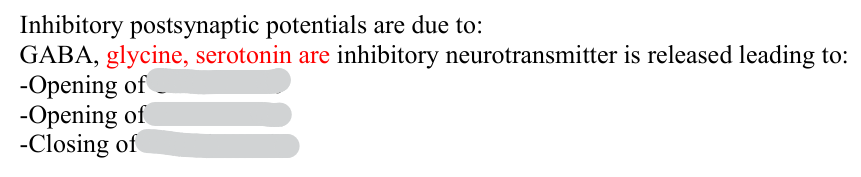
Cl- channels
K+ channels
Na+ channels
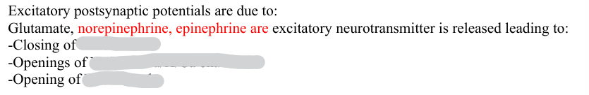
K+ channels
Na/K or Na/K/Ca channels
Na channels
Types of reflexes according to mechanism of formation ?
Unconditioned
Conditioned
What are unconditioned reflexes
Innate, inherited responses that don’t require learning
Permanent reflex arc
need adequate stimulus
What are conditioned reflexes?
acquired during life through life via learning and experience
No fixed reflex arc
Temporary reflexes
elicited by stimuli that acquire signal significance through repeated association with another stimulus.
Types of reflexes according to the characteristics of the reflex arc
Monosynaptic reflexes - patellar reflex
Polysynaptic reflexes - withdrawal reflex
Spinal reflexes - stretch reflex, withdrawal reflex
Cranial reflex - pupillary light reflex, gag reflex
somato-somatic —> plantar reflex
somato-visceral --> oculocardial reflex
viscero-somatic —> abdominal wall rigidity
viscero-visceral —> pressor reflex
Nerve centers -
________ __ ______ in the CNS responsible for __________ and _________ information, and coordinating specific physiological functions or behaviors
Examples: ________ _____ (regulating vital functions), ____________ (endocrine and autonomic regulation), _____ ____ ____ _____ (reflex control)
Clusters of neurons in the CNS responsible for processing and integrating information, and coordinating specific physiological functions or behaviors
Examples: brainstem nuclei (regulating vital functions), hypothalamus (endocrine and autonomic regulation), spinal cord gray matter (reflex control)
What are the 4 nerve centre types
Sensory centre
Motor centre
Autonomic centre
Integrative centre
Which part of the brain is responsible for regulation of motor centres ?
Cerebral cortex
Cerebellum
Basal ganglia
Which part of the brain is responsible for regulation of autonomic centres ?
Medulla oblongata and hypothalamus
Which part of the brain is responsible for regulation of integrative centre?
Hippocampus
Prefrontal cortex
Thalamus
Cerebral Blood Flow
Brain requires a constant supply of oxygen and glucose to function properly, supplied by the cerebral blood flow:
Arterial supply: provided mainly by the_________ _______ and _______ ________, forming the ______ __ ____ at the base of the brain
Venous drainage: ______ veins empty into _____ ______ _______, which drain into the _______ ______ veins
Blood-brain barrier: _______ __________ of cerebral blood vessels, restricting the passage of certain substances between the bloodstream and the brain
Autoregulation: maintenance of ______ cerebral blood flow despite fluctuations in blood pressure or metabolic demands
Brain requires a constant supply of oxygen and glucose to function properly, supplied by the cerebral blood flow:
Arterial supply: provided mainly by the internal carotid and vertebral arteries, forming the Circle of Willis at the base of the brain
Venous drainage: cerebral veins empty into dural venous sinuses, which drain into the internal jugular veins
Blood-brain barrier: selective permeability of cerebral blood vessels, restricting the passage of certain substances between the bloodstream and the brain
Autoregulation: maintenance of constant cerebral blood flow despite fluctuations in blood pressure or metabolic demands
Cerebrospinal fluid
_______ fluid that circulates the _________ of the brain, _____ _____ of the spinal cord and ___________ _____
Function : __________ , protecting the brain and spinal cord and maintains ___ ____________, removes metabolic waste
Produced by : ________ _______ within _________ of the brain ( __________ cells)
Absorbed into bloodstream via ________ ___________ along the dural venous sinuses
___ mL of CSF is produced per ___
At any-time we have ____ mL of CSF circulating
CSF can be obtained by _______ _________
Cerebrospinal fluid
Colorless fluid that circulates the ventricles of the brain, centre canal of the spinal cord and subarachnoid space
Function : cushioning, protecting the brain and spinal cord and maintains ion concentrations, removes metabolic waste
Produced by : Choroid plexuses within ventricles of the brain (ependymal cells)
Absorbed into bloodstream via arachnoid granulations along the dural venous sinuses
500 mL of CSF is produced per day
At any-time we have 150 mL of CSF circulating
CSF can be obtained by lumbar puncture