processing the environment - MCAT
 Call Kai
 Learn
Learn Practice Test
 Spaced Repetition
 Match
Match Flashcards
 Knowt Play
1/32

Earn XP
33 Terms
Visual cues
features that the brain uses to organize and interpret sensory information
Binocular cues
visual cues that require both eyes working together
Retinal disparity: brain calculates difference between what left & right eye see, bigger difference = closer object
Convergence: eyeballs turn inward when you focus on very close object; eyes converge more = closer object
Monocular cues
visual cues that only require one eye — Raw Raw Raw SLIT
Relative size
Relative height
Relative motion / Motion parallax
Shading and Contour
Linear perspective
Interposition
Texture gradient
Weber’s Law
The change needed to notice a difference in a stimulus is always a constant proportion of the original stimulus’s intensity
K = ΔI / I
K = constant specific to sense being measured, “Weber fraction”
ΔI = just-noticeable difference
I = initial intensity
Absolute threshold of sensation
minimum intensity required to notice stimulus 50% of the time
influenced by psychological state, expectations, experience, motivation, alertness, etc.
stimuli with lower intensities than ATS are subliminal
Somatosensation
Body’s sensory system for:
Temperature — Thermoception
Pain — Nociception
Pressure — Mechanoreception
Movement/position — Proprioception
Sensory receptor adaptation types
3 types
categorize chemoreceptors, nociceptors and thermoceptors
Differ based on what happens to firing rate when exposed to continuous unchanging stimulus
Fast-adapting receptors fire intensely when stimulus begins, then quickly level off to nothing (eg. thermoceptors)
Slow-adapting receptors fire rapidly when stimulus begins, then slowly level off, continuing to fire at steady low rate. (eg. nociceptors)
Non-adapting receptors fire steadily, unchanging as the stimulus itself (eg. chemoreceptors)
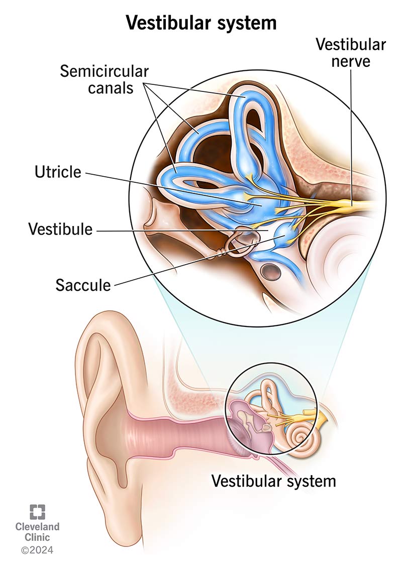
Vestibular system
Sensory network in inner ear that controls balance and spacial orientation
Semicircular canals (anterior, posterior and lateral) containing endolymph that shits w/ movement
detect rotational movement & angular acceleration
Otolith organs (Utricle & Saccule) containing calcium crystals attached to hair cells in gel
detect linear acceleration, gravitational pull and head tilt
Signal detection theory
explains how we make decisions under uncertainty
Detection of signal depends not only on intensity but also on background distractions (noise) and observer’s psychological state
4 outcomes: Hit (true positive), Miss (false negative), False alarm (true positive), Correct rejection (true negative)
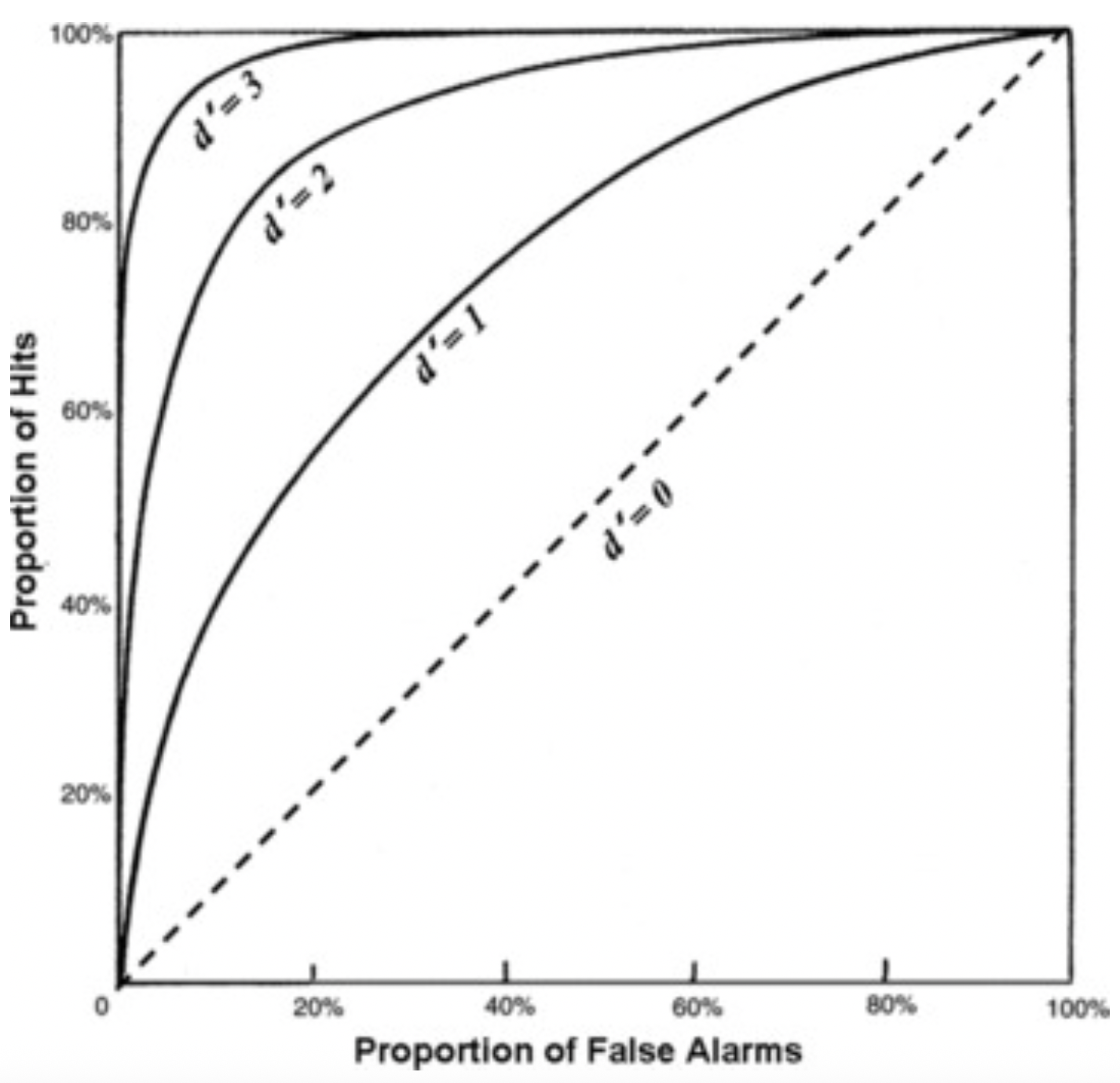
d’ (sensitivity): represents the strength of signal relative to background noise. Higher d’ → easier to distinguish signal → noise and signal distributions are further apart
C or β (Criterion / Bias): represents the observer's psychological threshold or strategy, based on expectations and the consequences of their choices.
Liberal Strategy: (lower C) The observer says "yes" to almost everything to ensure they never miss a signal. This increases hits but also leads to more false alarms.
Conservative Strategy: (higher C) The observer only says "yes" if they are absolutely certain. This reduces false alarms but results in more misses.

Receiver-Operating Characteristic (ROC) Curve
Illustrates trade-off between sensitivity and specificity
true positive on y axis, false positive on x
Bottom-up vs. Top-down processing
Stimulus influences perception, data-driven
vs.
Expectations and prior knowledge influence perception
Gestalt principles
Explain how our brains naturally organize visual elements into organized patterns and recognizable shapes
Succ Fat PP, Chew Colossal Cok
Similarity: group similar objects together
Figure-Ground: ability to separate into figure (closer, distinct shape) and ground (background, indistinct)
Proximity: group close objects together
Pragnanz: reduce reality to simplest form
Continuity: lines are seen as following smoothest path
Common Fate: group together objects moving in same direction
Closure: objects grouped together are seen as a whole
The Gestalt principles of grouping hold that …
… the brain processes the whole rather than the sum of its parts
Aβ vs. Aδ fibers
Large, thickly myelinated. Quickly transmit touch, pressure, vibration
vs.
Small, thinly myelinated. Transmit temperature and pain slower (though still fast. Slow / dull or aching pain is transmitted by C fibers)
Receptive field
region of sensory space where stimulus modifies firing activity of neuron
ig it’s the scope of sensory nerve’s sensitivity
eg. small region of visual field where light hits photoreceptors
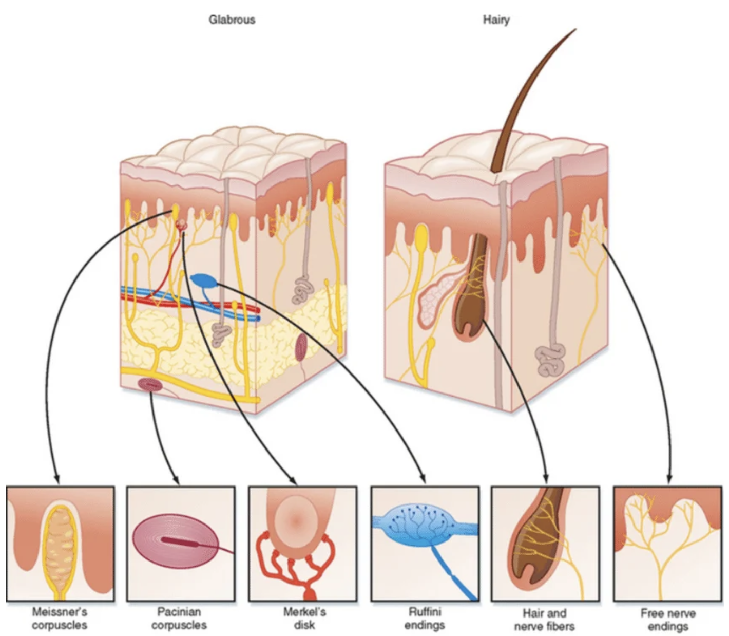
5 types of mechanoreceptors
“How’s My Pretty Mechano Receptor”
Hair Follicle Receptor
Meissner’s Corpuscle
Pacinian Corpuscle
Merkel’s Disc
Ruffini’s Corpuscle
All work by allowing influx of Na+ one way or another
Hair Follicle Receptor
structure/function
location
type of touch
When hair bends, opening created to receptor, allowing influx of Na+
located in reticular dermis
senses changing light pressure on hairy skin (no way)
large receptive field
senses changing pressure because area around hair is rich in collagen, which quickly fills in the gap and blocks Na+
Meissner’s Corpuscle
Looks like a beehive
Changing light pressure displaces disc, movement allows influx of Na+
located in papillary dermis
only mechanoreceptor to be absent in hairy skin (only works in glabrous skin)
small receptive field
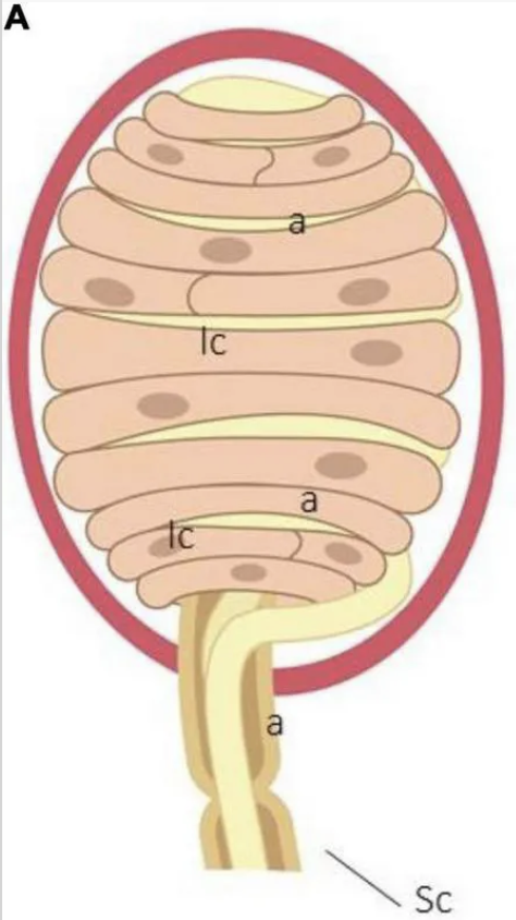
Pacinian’s corpuscle
Looks like an onion and has the rings to boot
Changing deep pressure spins rings, allowing influx of Na+
Located in hypodermis
large receptive field
Merkel’s disc
Modified epithelial cell which contains vesicles filled with peptides
Pressure causes vesicles to burst and release peptides, which bind to receptor and trigger influx of Na+
located in stratum basale — papillary dermis, senses sustained light pressure
small receptive field

Ruffini’s Corpuscle
looks kinda like a mushroom
Senses sustained deep touch
located in reticular dermis
Full of collagen, deep touch stretches collagen allowing influx of Na+
Large receptive field

Draw a labeled diagram of the auditory system

Sensory adaptation & amplification
Adaptation = downregulation
eg. getting used to a smell — sensory neurons firing rate decreases
Amplification = upregulation
eg. single ray of light in the dark activates one cell which goes on to activate many more cells so it is detectable
Define / differentiate between Proprioception, Kinesthesia, Vestibular sense and Somatosensation
Proprioception: sense of position
Kinesthesia: sense of movement
Vestibular sense: sense of balance
Somatosensation: senses of touch + pain + temperature + position
Docking theory and Vibrational theory of olfaction
Odor molecules act as ligands and bind to specific GPCRs in nasal epithelium, and its shape/properties dock into receptor active site to trigger response
vs.
Nose acts as spectroscope to detect vibrational frequencies of odorant molecules emitted when they bind
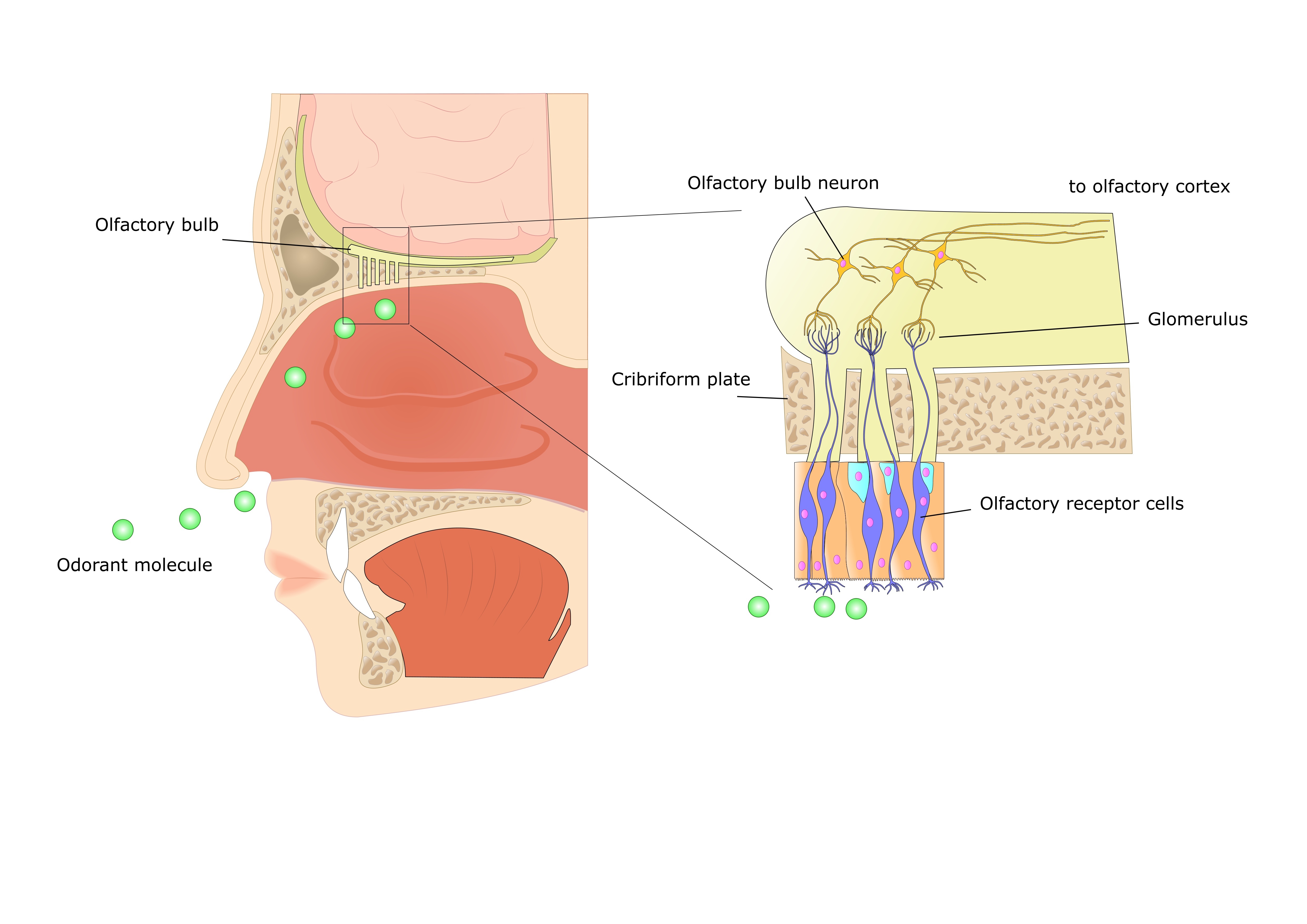
Describe / diagram how we smell things
Odor particles get inhaled up the nasal passage and bind to GPCRs on the dendrites of olfactory sensory neurons
G proteins activate ion channels which depolarize OSNs so they fire action potential and activate mitral/tufted cells in the olfactory bulb (meeting of OSN axon terminals and mitral cell dendrites is called a glomerulus)
signal travels from olfactory bulb → amygdala → piriform cortex → orbitofrontal cortex
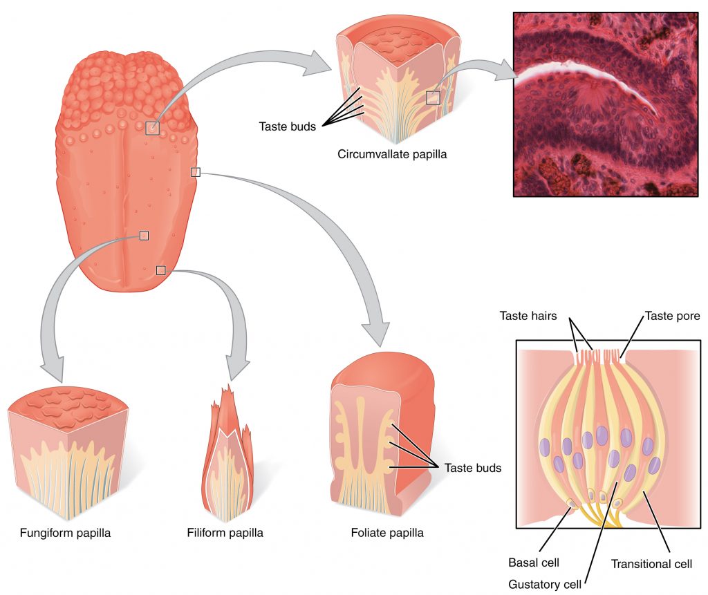
Different types of tongue papillae
location
function
Fungiform — front
Foliate — sides
Circumvallate — back
Filiform — all, no taste buds
5 flavors and how they are detected
Bitter, Salty, Sweet, Sour, Umami (glutamate)
Every taste bud can detect all 5 flavors, though within the taste buds there are many sensory cells specific to 1 flavor each
Ipsilateral vs. Contralateral
Ipsilateral = right brain controls right side of body, left brain controls left side of body
Contralateral = right brain controls left side of body, and vice versa
States of consciousness
Alertness — beta waves (12-30 Hz)
Daydreaming / light meditation — alpha waves (8-13 Hz)
Drowsiness / deep meditation — theta waves (4-7 Hz)
Sleep
Sleep stages
Non-REM 1 = N1: b/t awake and asleep, start of theta waves and hypnogenic hallucinations
N2: more theta waves, K complexes and sleep spindles
N3: delta waves, sleepwalking/talking, difficult to wake up
REM: active mind, paralyzed body. Most of one’s vivid, ‘structured’ dreams occur here
Sleep recommendations by age
4-11 months: 12 hrs / night
3-5 years: 10 hrs
6-13 years: 9 hrs
14-18 years: 8-10 hrs
18-64: 7-9 hrs
65+: 7-8 hrs
Freud’s theory of dreams vs. Activation-synthesis hypothesis
Sigma Freud thought dreams are the product of unconscious desire
What actually happens in dream = manifest content
The hidden meaning of the dream = latent content
The Activation-synthesis hypothesis suggests that dreams are just a biological byproduct
While we sleep, pons of brainstem fires spontaneous bursts of electrical energy that stimulate areas of brain for movement, emotion and sensation
Cerebral cortex attempts to organize signals into logical ‘story’ by cross-referencing stored memories and emotions