Pons
1/40
There's no tags or description
Looks like no tags are added yet.
Name | Mastery | Learn | Test | Matching | Spaced | Call with Kai |
|---|
No analytics yet
Send a link to your students to track their progress
41 Terms
Describe the Dev. of the Pons
Derived from?
Location
Differentiate the dev. of Alar vs Basal Plates
Describe formation of Basilar Pons
What will axons of these nuclei form?
Pons Development:
derived from:
rhombencephalon (primary brain vesicle)
later as part of metencephalon along w/ cerebellum.
Location:
pontine flexure btw metencephalon and myelencephalon.
Alar vs Basal:
Alar: sensory-related nuclei associated w/ CNs
CN sensory nuclei develop/remain in dorsal and lateral position in the pons
Basal: midline and dorsal motor components of pontine cranial nerve nuclei
Formation of Basilar Pons:
pontine nuclei migrate from part of alar plate → ventrally + medially → basilar pons.
Axons of these nuclei will form:
middle cerebellar peduncle that wraps around the brainstem in a lateral position
Describe the two major divisions within the pons
Location within pons?
Components? (3) (3)
Pontine Tegmentum:
Dorsal (and medial) portion
Components:
Cont. of medullary reticular formation/ associated tracts
Pontine CN nuclei
Ascending sensory tracts
Basilar Pons
Ventral (and lateral) portion
Components:
Cortically derived axons
corticobulbar (includes corticopontine)
Pontine nuclei
Projections → cerebellum = middle cerebellar peduncle
Describe the PMJ
Level of What?
Nuclei Overlaps?
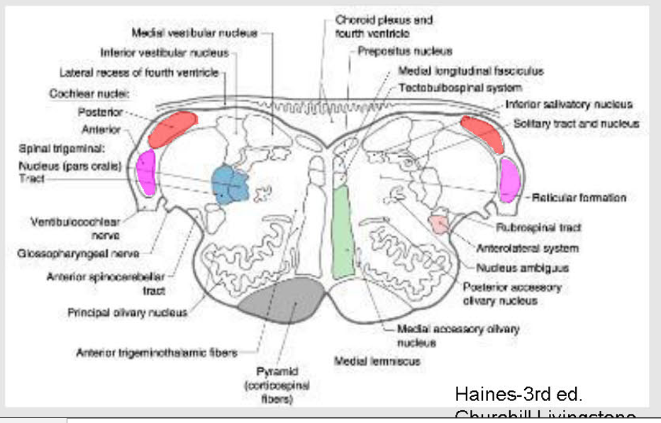
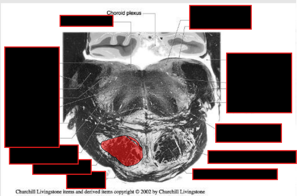
Ponto Medullary Junction (PMJ):
Level Of:
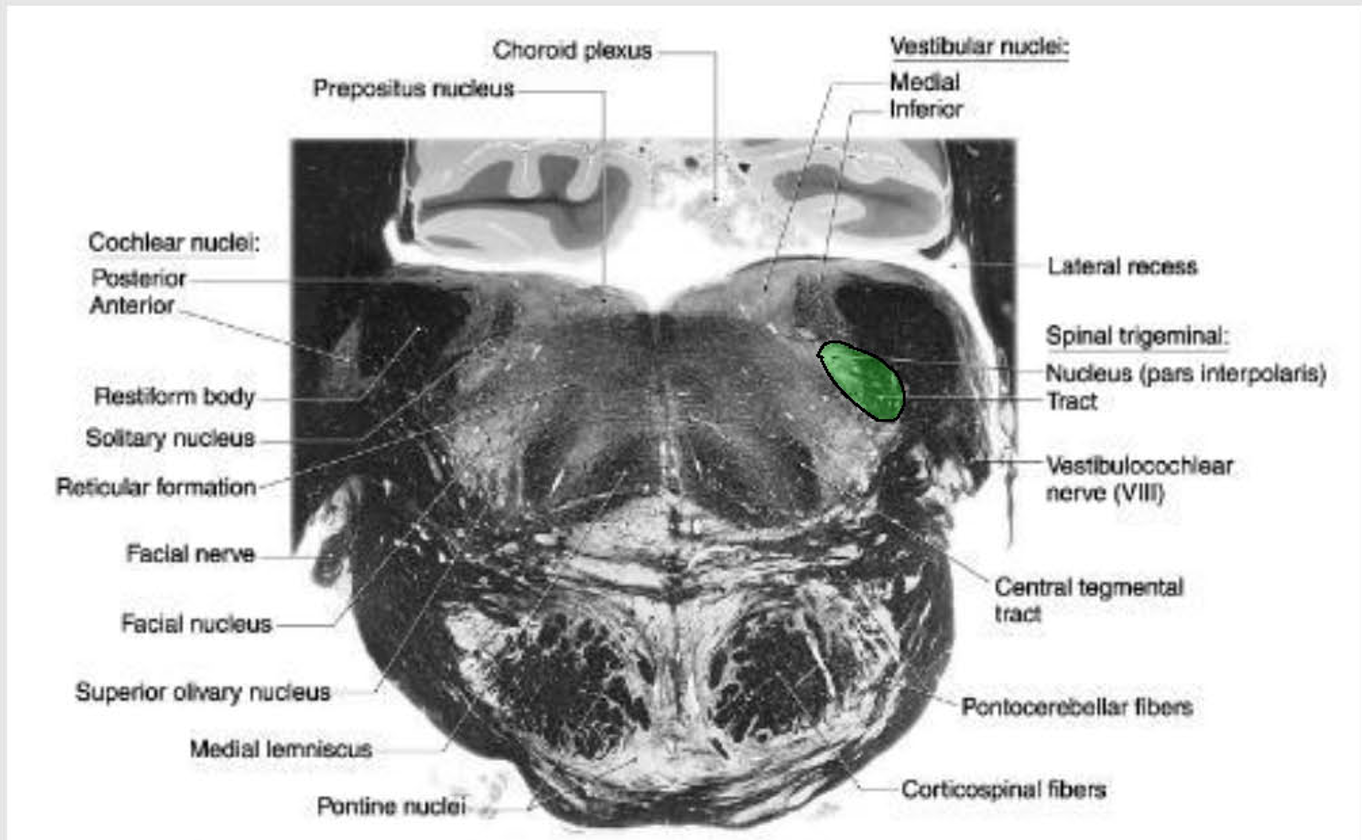
CN VIII (SSA) - Cochlear Component
nuclei overlap between the pons and medulla, and in some cases is classified as lying in both.
Describe PMJ-CN VIII - Cochlear Component
Origins
Function
Synapse @
Pathway?
What happens to the Info in Contra Lat. Lemniscus?
Describe the Accessory Auditory Nuclei in Pons:
Function?
List/Function
Describe the Lat. Lemniscus:
Travel up in what position?
Clinical Importance?
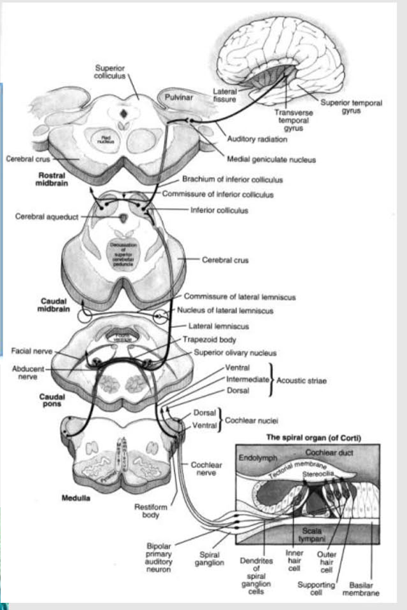
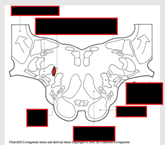
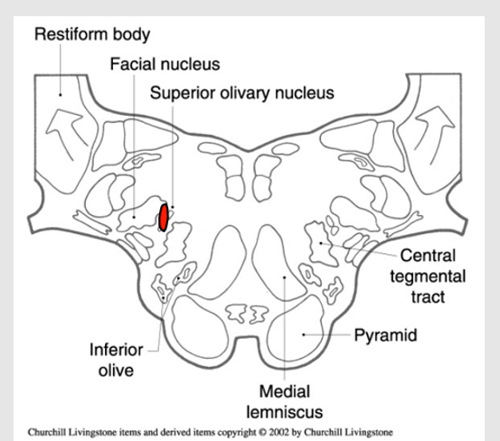
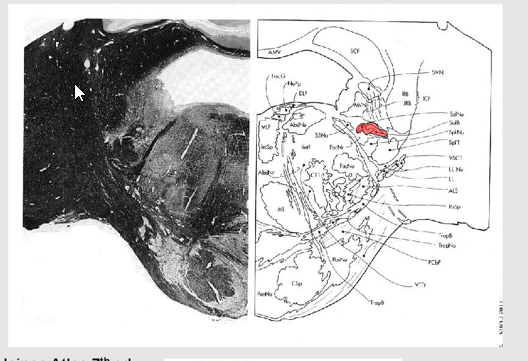
PMJ-CN VIII - Cochlear Component
Origin: spiral ganglia of the cochlea
Function: carry auditory information to the brainstem
Synapse @ Brainstem:
Dorsal/Ventral Cochlear Nucleus
Pathway:
Dorsal/Ventral Cochlear Nuelci → Lateral Lemniscus (primary ascending auditory pathway)
Axons Leave nuclei in dorsal, intermediate and ventral acoustic stria → Cross @ more rostral levels (there is small ipsilateral component)
Trapezoid Body = region in brainstem where axons cross
Info in Contra Lat. Lemniscus:
Ascends to:
Inferior Colliculus
Medial geniculate body (thalamus)
Cortex (temporal, transverse gyri of Heschel)
Some Axons → accessory auditory nuclei adj. to tract
Accessory Auditory Nuclei in Pons:
Main Function = Sound processing; particularly dampening and localization
Primary Ones:
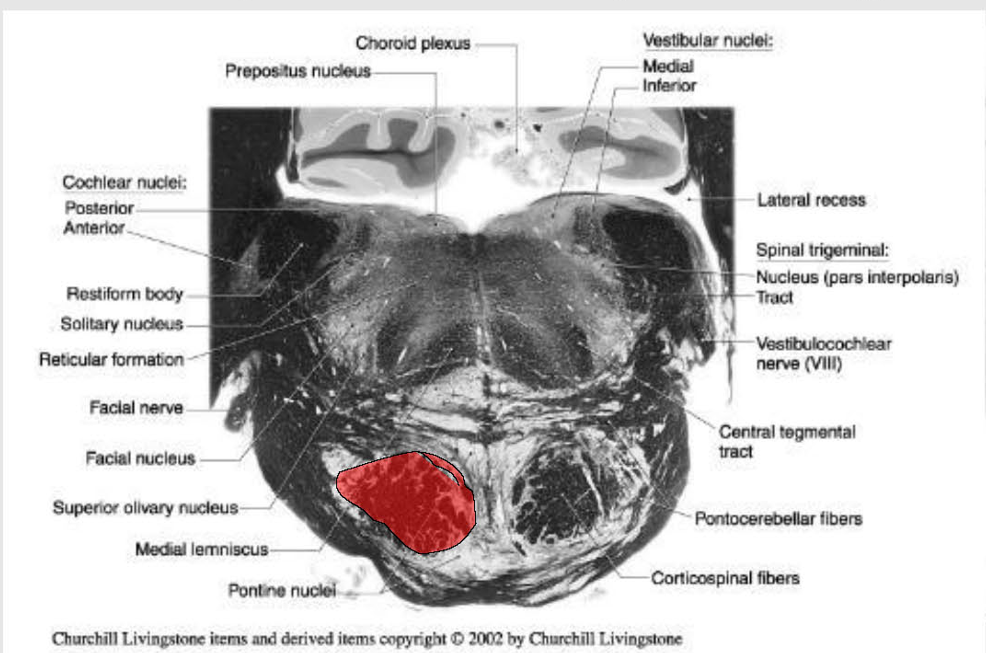
Superior olivary nuclear complex
sound localization- reflex sound dampening
via stapedius muscle (CN VII)and tensor tympanic (CN V)
Nucleus of trapezoid body
localization of sounds
Nucleus of lateral lemniscus
sound localization
acoustic reflexes (not as well defined)
Lateral Lemniscus Notes:
Travels up brainstem in dorsolateral position
There is some Ipsilateral Components
→ ipsilateral lesion of tract in pons/midbrain → no noticeable unilateral hearing loss
EXCEPTION: unless the nerve or cochlear nuclei are involved.
Draw out the Cochlear component pathway
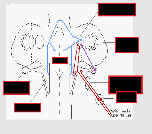
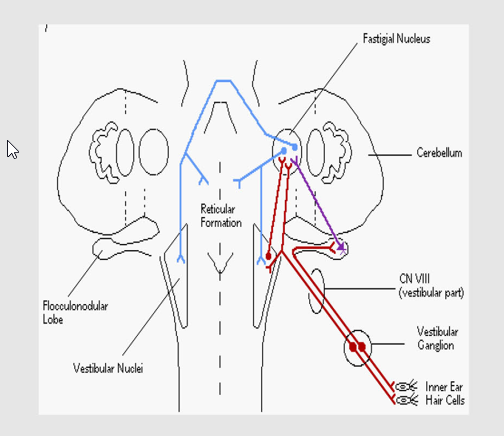
Describe PMJ-CN VIII - Vestibular Component
Origins
Function
Innervates? (4)
Central Nuclei projections? (3)
List and describe function of the 4 CN VIII - Vestibular Related Pathways
PMJ-CN VIII - Vestibular Component
Origins: vestibular ganglion;
Function:
Transmits afferent information from vestibular apparatus
Innervates:
four nuclei in dorsal and lateral part of the PMJ
Medial Vestibular Nucleus
Lateral Vestibular Nucleus (Deiter’s nucleus)
Superior Vestibular Nucleus
Inferior Vestibular Nucleus
Central Nuclei Projections:
→ Cerebellum via juxtarestiform body (balance coordination)
Some direct connections to:
flocculonodular lobe
deep cerebellar nuclei.
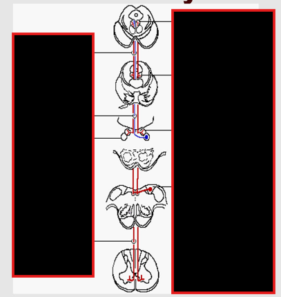
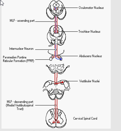
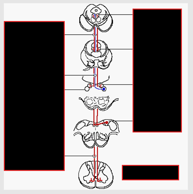
CN VIII - Vestibular Related Pathways
Oculomotor nuclei:
via medial longitudinal fasciculus (MLF)-
Thalamus:
via MLF:
→ cortex to give conscious perception of equilibrium and orientation.
From SC:
Lateral vestibulospinal tract
from lateral vestibular nucleus (Deiter's nucleus),
Function: excitatory to extensor spinal alpha motor neurons.
Medial vestibulospinal tract (a.k.a. MLF)
Descends in Spinal Cord
Function:
Inhibitory to neck and upper trunk muscles
Contributes to head- righting reflexes in relation to vision, etc
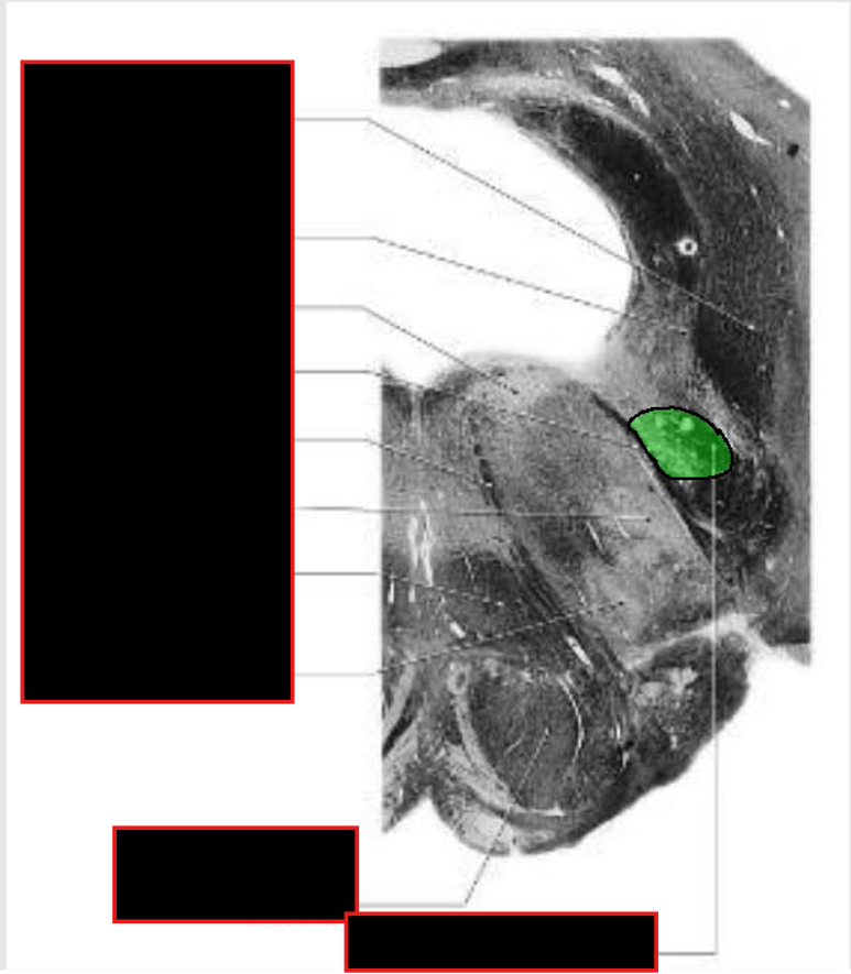
What is rostral to nuclei of CN VIII
rostral to nuclei of CN VIII @ PMJ:
caudal pons contains nuclei for Cranial Nerves VI/VII
exit from nuclei in ventral and slightly caudal direction to exit @ PMJ
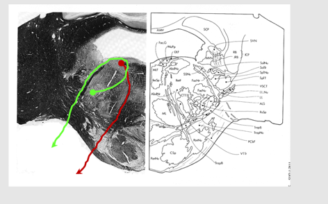
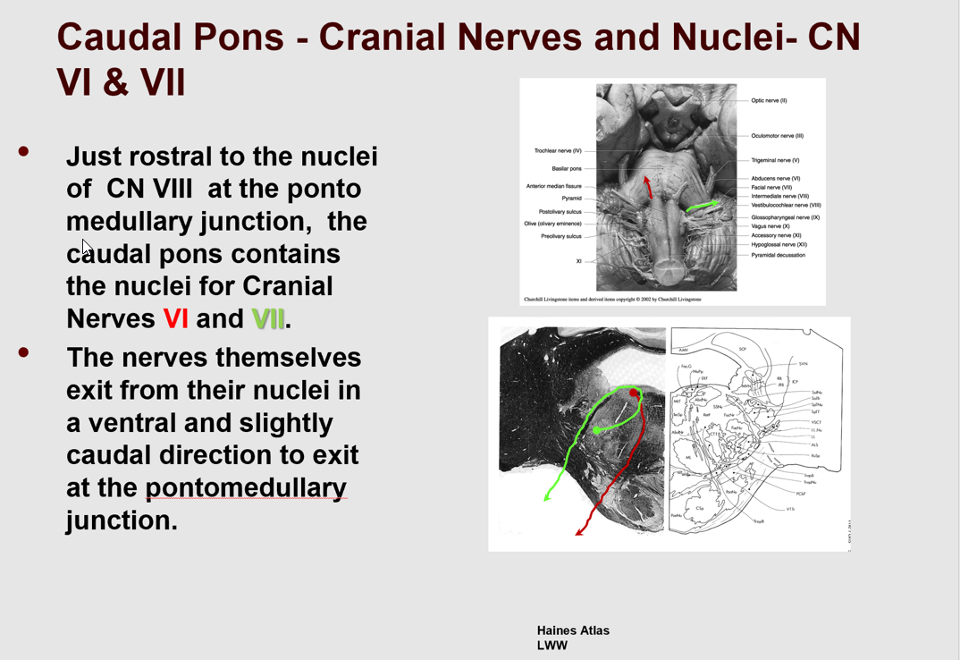
Describe CN VII:
Exits brainstem @?
Nerve Includes?
Describe the CNVII Motor Branch:
Internal nerve course
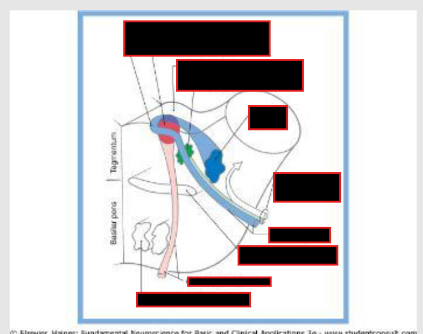
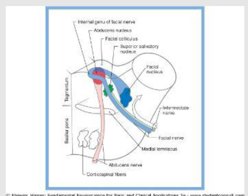
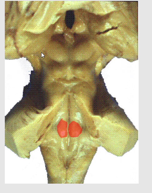
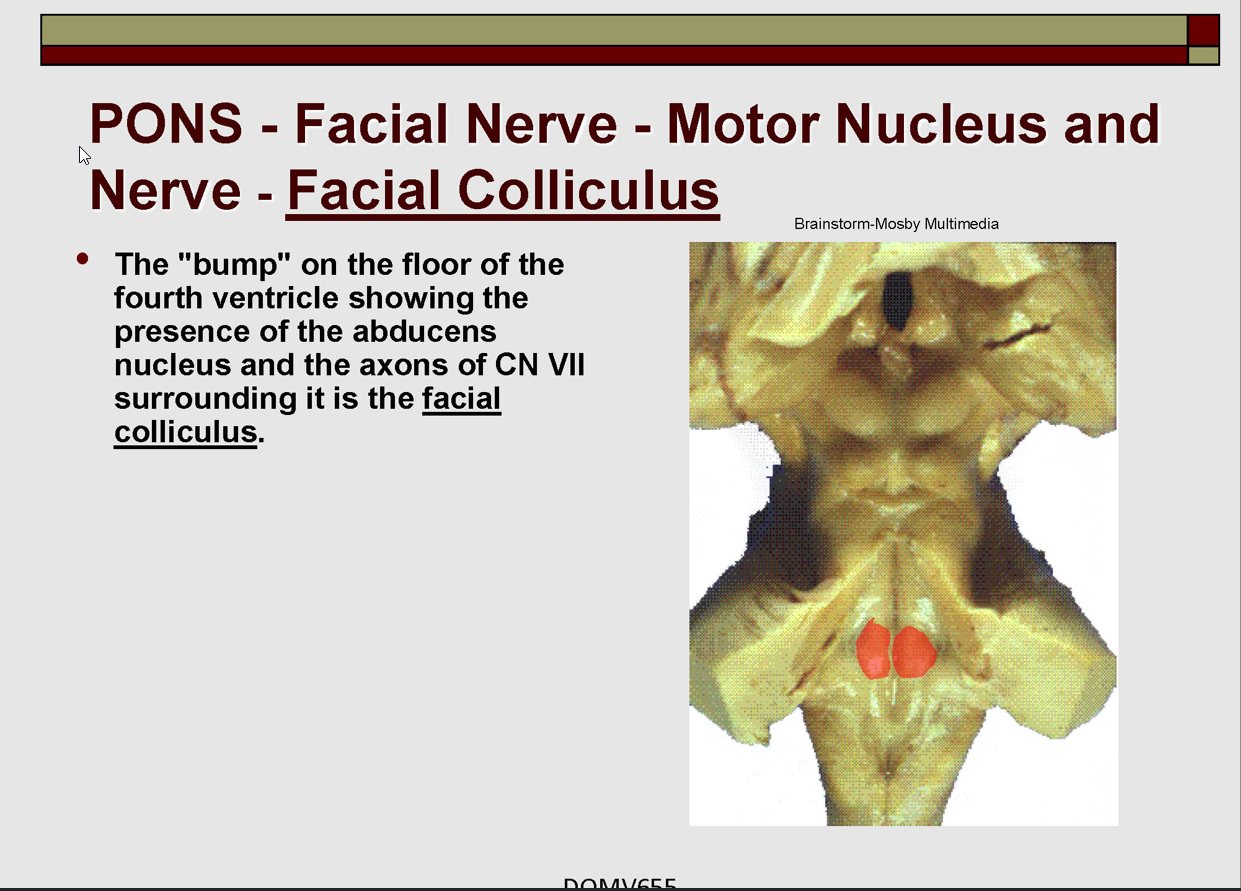
What is the Facial Colliculus:
Cortical Innervation?
Facial Nerve - CN VII
Exits brainstem:
anterior to VIII @ PMJ
nerve includes
motor root
smaller intermediate nerve lateral to it.
CNVII Motor Branch:
Internal nerve course
axons loop around (internal genu of facial nerve) the nucleus of CN VI Nucleus → exit ipsilaterally
Note: axons forming 'loop' = from the motor nucleus.
Facial Colliculus:
bump on floor of 4th ventricle
Shows presence of CNVI nucleus and axons of CN VII surrounding it
Cortical Innervation:
Bilateral: nucleus innervating muscles of the forehead
Contralateral: nucleus for muscles of the lower face
NOTE: This means that after a cortical or corticonuclear lesion function to the forehead may remain, but not to the lower face.
Describe PONS - CN VII - Intermedius Branch
Functions (3)
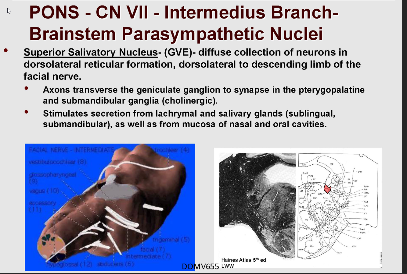
Describe the Superior Salivatory Nucleus- (GVE):
Location
Axon Pathway
Describe the Central autonomic connections to GVE:
Pathway
Function
Brainstem reflex circuits
Describe the pathway of Solitary Nucleus
Functions:
Parasympathetic functions (GVE):
lachrymal qlands
via pterygopalatine ganglion
mucous membranes of nose, hard and soft palates
salivary qlands-
via submandibular ganglion
submandibular, sublingual
Taste: (SVA)
anterior 2/3 of tongue
hard and soft palates
via geniculate ganglion.
GSA:
skin of ear wall of acoustic meatus and external tympanic membrane
Geniculate Ganglion → Nervus intermedius → Spinal Nuceus and tract of CNV
Superior Salivatory Nucleus- (GVE)-
Location:
in dorsolateral reticular formation
dorsolateral to descending limb of facial nerve
Axons:
transverse geniculate ganglion → pterygopalatine and submandibular ganglia (cholinergic).
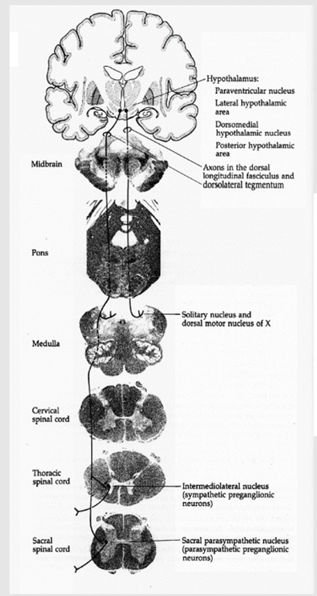
Central autonomic connections to GVE:
NOTE: this means Higher Brain systems → Intermedius Branch
Pathway:
limbic system and olfactory areas → hypothalamus → brainstem
via dorsal longitudinal fasciculus.
Functions
emotional responses
weeping
mouth watering to odors etc.
Brainstem reflex circuits
reflex lachrymation from V
gustatory stimulation of salivation.
Solitary Nucleus (rostral portion, gustatory nucleus, SVA)
Pathway:
Taste Sensory fibers on tongue/hard/soft palate → Chorda Tympani from geniculate ganglion → Brainstem as part of nervus intermedius → tractus solitarius → synapse in rostral portion of the nucleus → central teqmental tract to thalamus (VP) → Cortext (conscious perception of taste)
Solitary Nucleus
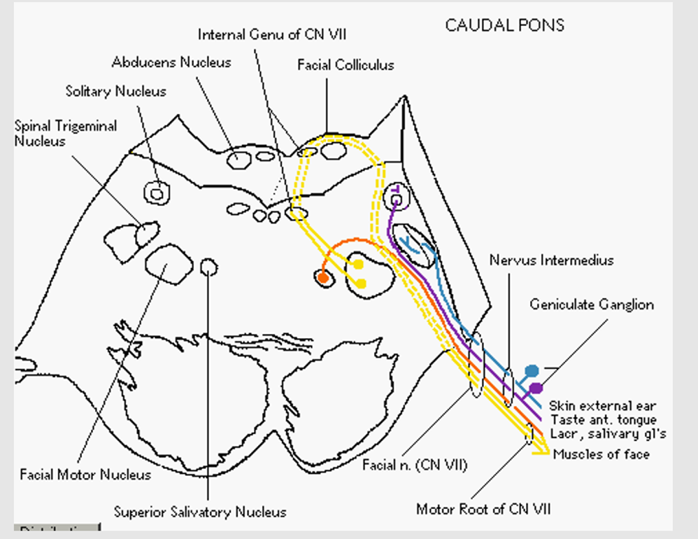
Abducens Nerve
Describe the Abducen Nerve
@ level?
Exits?
Function
Neuron Location?
Clinical Importance
Complex central connection w/? Via? involved in?
Abducens Nerve
Level of VI & VII
Exits in the inferior pontine sulcus
Function: GSE:
Ipsilateral Lateral Rectus
Neuron Location:
@ Abducens nerve
beneath axons of the facial nerve in the facial colliculus
Clinical Importance:
long intracranial course → one of the most frequently injured of cranial nerves
Complex central connections with oculomotor nuclei.
Via MLF (medial longitudinal fasciculus)
Involved in conjugate eye movements.
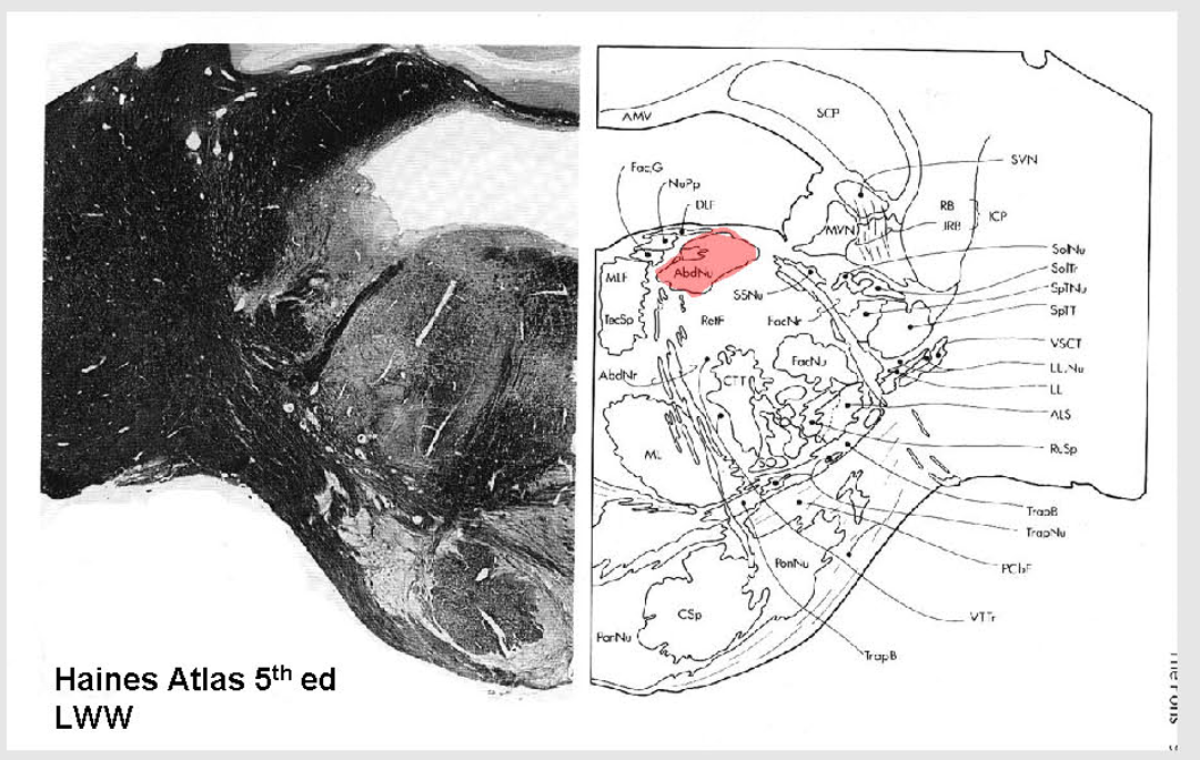
Abducens Nucleus
Describe the Reticular formation
Location
Function (3)
pontine reticular formation
Location: pontine tegmentum;
contains many nuclei that contribute to the reticular formation located over the entire brainstem.
Function:
forms complex connections btw CN nuclei for reflex and visceral functions.
rostral projections → thalamus
regulate consciousness,
caudal projections
regulate motor and sensory functions.
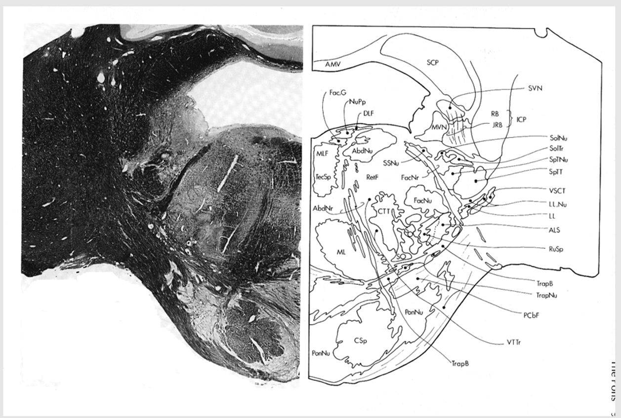
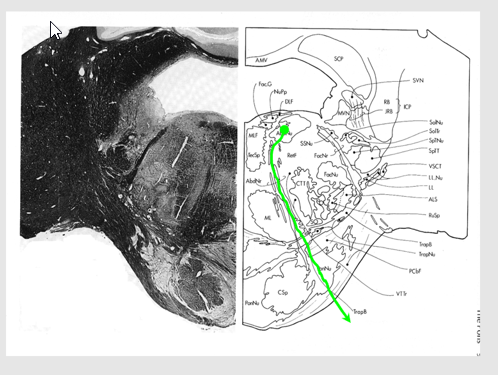
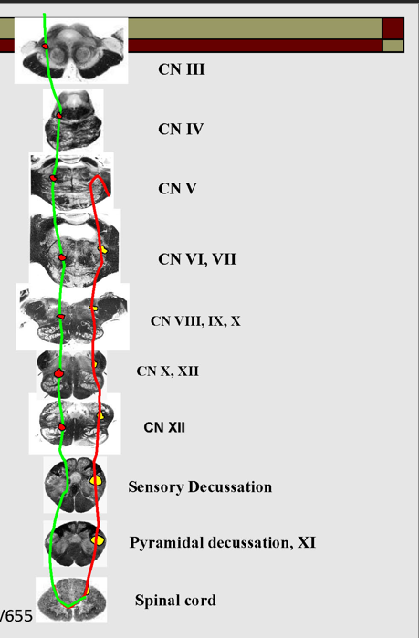
Describe the location of major tracts in the Caudal Pons
CST
Medial Lemniscus
ALS
Rubrospinal tract
Reticulospinals and vestibulospinals
spinal nucleus and tract of CN V
MLF and tectospinal (tectobulbospinal ) tracts
Others
location of major tracts in the Caudal Pons:
CST/corticonuclear (bulbar) run through the substance of the basilar pons
Medial Lemniscus:
still in the midline
begins to turn → somatotopy shifts → leg areas = more lateral
anterolateral system
still in lateral position w/in pontine tegmentum,
@ anterolateral edge w/ formation of middle cerebellar peduncle.
Rubrospinal Tract:
Dorsal to ALS
Reticulospinals and vestibulospinals
still being formed in this area, so they are not clear.
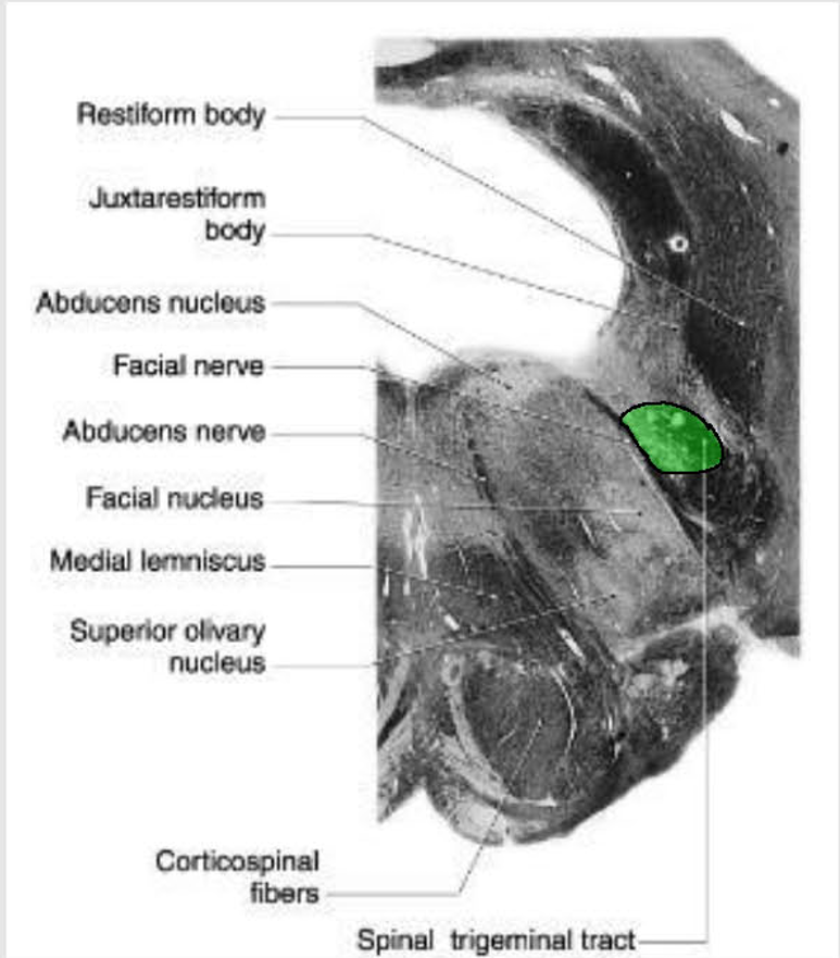
spinal nucleus and tract of CN V
still present as pars oralis
just dorsal to the exit of the facial nerve.
MLF and tectospinal (tectobulbospinal ) tracts
dorsal position near the midline.
will now carry motor information for eye movements.
Other Ascending/Descending → displaced into the pontine tegmentum
Draw out CST as it progresses up
Draw out the Medial Lemniscus as it progresses up
Draw out the ALS as it progresses up
Draw out the Spinal Nucleus tract of CNV as it progresses up
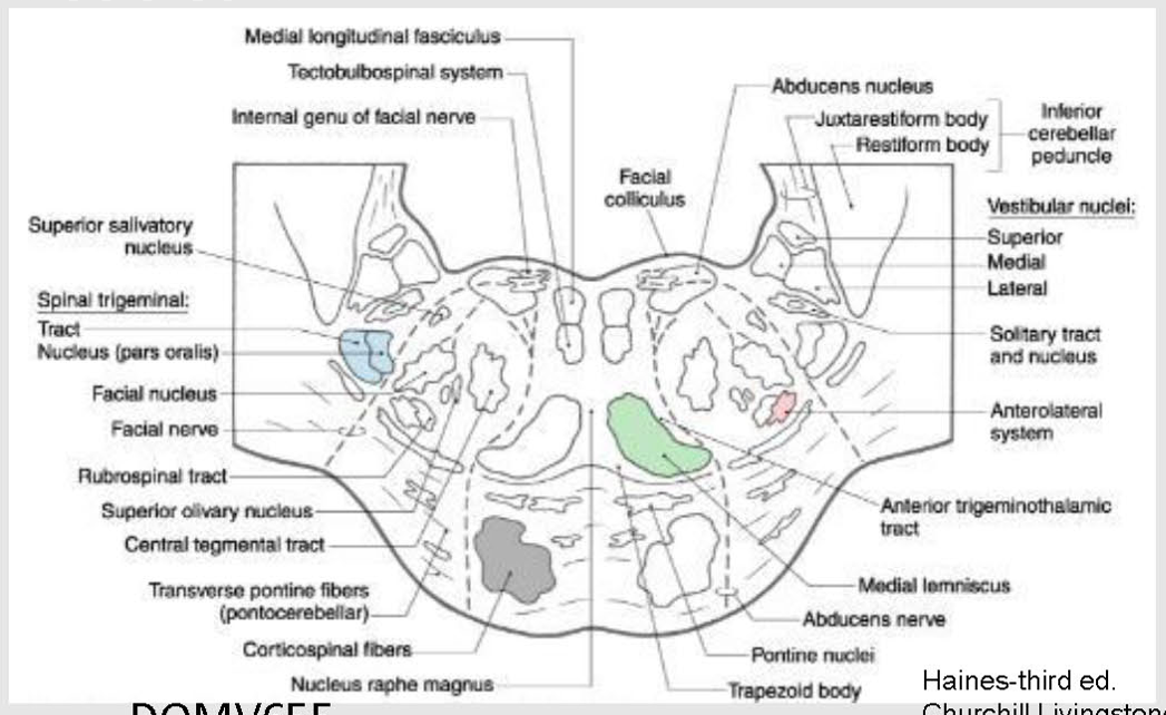
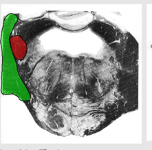
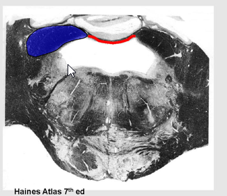
Describe the Inferior/Middle/Superior Cerebellar Peduncles
Locations?
Importance?
inferior cerebellar peduncle (restiform body, ICP)
enter cerebellum in dorsal and lateral position.
Includes:
vestibulocerebellar projections called juxtarestiform body
Middle Cerebellar peduncle:
between the pons and cerebellum
will wrap laterally around the inferior peduncle
superior cerebellar peduncles
in more rostral sections
Forms part of roof of 4th ventricle along with superior medullary velum,
CT membrane between them.
Draw out the Spinocerebellar tracts as it ascends
Inferior/middle cerebellar peduncle (middle = lateral)
Superior Cerebellar peduncle and superior medullary velum,
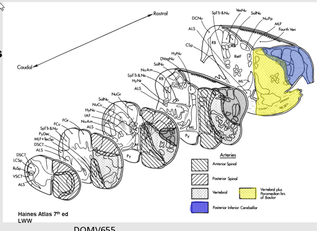
What are the Blood Supply to the Caudal Pons
Vertebral
Basilar
Posterior Inferior Cerebellar A
Anterior Inferior Cerebellar A
Vertebral + medial branches of basilar → Ventral and medial
PICA: Dorsolateral