Sensation and Perception Exam 3 (Touch, Taste, Vision, Vestibular)
 Call Kai
 Learn
Learn Practice Test
 Spaced Repetition
 Match
Match Flashcards
 Knowt Play
1/41

There's no tags or description
Looks like no tags are added yet.
Name | Mastery | Learn | Test | Matching | Spaced | Call with Kai |
|---|
No analytics yet
Send a link to your students to track their progress
42 Terms
Kinesthesis, Proprioception, and Somatosensation
Kinesthesis - perception of position and movement of our limbs in space (direction)
Proprioception - perception mediated by kinesthetic and vestibular receptors
Somatosensation - collective term for sensory signals from the body
Touch Mechanoreceptors
specialized structures that respond to mechanical stimulation
typically embedded on outer layer (epidermis) and underlying layer (dermis)
many types of touch receptors for different stimulation: vibration, deflection, fast/slow
Information from mechanoreceptors is transmitted to the brain via A-beta fibers
they signal “hey I got touched”
Kinesthetic Receptors and Muscle Spindles
Kinesthetic Receptors - sense of where limbs are and what kinds of movements are made
Ian Waterman - nerves connecting receptors to brain were destroyed, lacked kinesthetic senses had to relearn how to move
Muscle Spindle - located in muscle and senses tension
receptors in tendons → tension in muscles attached to tendons
receptors in joints → react when joint is bent to extreme angle
Thermoreceptors
signal information about changes in skin temperature
warmth fibers and cold fibers
body is constantly regulating internal temperature and respond when you make contact with objects warmer/cooler than your skin
Nociceptors + Pain
transmit information about noxious stimulation that could cause damage to the skin
transduce impending or ongoing damage to body’s tissue into a specialized neural signal = pain
Split into 2 groups
A-delta fibers - mid-sized, myelinated sensory nerve fibers that transmit pain and temperature signals
C fibers - narrow, unmyelinated sensory nerve fibers that transmit pain and temperature signals
You need to be able to sense pain → Miss C!
How do touch sensations travel?
passes through spinal cord first then
Spinothalamic Pathway - carries most information about skin temperature and pain (slower)
Dorsal column-medial lemniscal (DCML) pathway - carries signals from skin, muscles, tendons, and joints
In the brain signals arrive at the ventral posterior nucleus of the thalamus (VPN) and then routed to primary somatosensory cortex (S1)
How are touch sensations mapped onto the brain?
somatotopical representation - adjacent areas on skin connect to adjacent areas in brain
similar to retinotopy found in vision!
Homunculus - a map-like representation of regions of the body in the brain
Phantom Limb
sensation perceived from a physically amputated limb of the body
parts of brain listening to missing limbs not fully aware of altered connections
Analgesia and Endogenous Opiates
Analgesia - decreasing pain sensation during conscious experience (tylenol/advil)
body systems actively modulate pain perceptions and shut down pain in emergencies
Endogenous Opiates - chemicals released in body to block release or uptake of neurotransmitters that transmit pain
natural analgesia mechanism
externally produced substances like morphine, heroin, and codeine have similar effects
Gate Control Theory
system that transmits pain that incorporated modulating signals from the brain
Feedback circuit located in the substantia gelatinosa of dorsal horn of spinal cord
gate neurons block pain transmission → activated by extreme pressure, cold, or other stimulation
Rubbing/Holding a body part can activate gate neurons and help relieve pain
Cognitive Aspects of Pain
experience of pain includes nocieceptive sensation and emotional response
Cognitive and emotional aspects of pain are experienced in
Anterior Cingulate - perceived unpleasantness
Prefrontal Cortex - cognition and executive control
Pain Sensitization
Nociceptors provide signal to damage to body tissue
once damage occurs site can become more sensitive → Hyperalgesia
pain as a result of damage to nervous system → Neuropathic
no 1 pain medicine can alleviate all types of pain
Touch and Spatial Details
Two-Point Threshold - minimum distance which 2 stimuli are just perceptible as separate
varies across the body but is most accurate on fingertips, face, and toes
Fork prong demonstration
Smallest raised element that can be felt - 1 micrometer high = 75% accurate
Tactile Sensitivity and Acuity
Touch - differences sensed at 5ms
Vision - differences of 25ms
Audition - differences sensed at 0.01 ms
Haptic Perception
Knowledge of the world from sensory receptors in skin, muscles, tendons, joints (pressing snooze even when tired)
Geometric properties of objects are most important for visual recognition
Perception for action - less grip strength is needed after a while of holding
Acting for perception - metal bat will feel cold
Exploratory Procedure - stereotyped hand movement pattern used to contact objects to perceive their properties
Taste vs Flavor
Taste - sensations specifically from taste buds
Flavor - multimodal sensation associated with food or drink in the mouth
retronasal smell, somatosensation, trigeminal stimulation (spicy), astringency (pucker)
Taste Buds
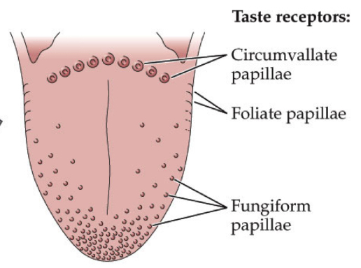
bumps on your tongue → papillae
Filiform Papillae - most bumps on your tongue, have nothing to do with taste
CONTAIN TASTE BUDS
Fungiform Papillae - bumps on edges of your tongue
Circumvallate Papillae - V of circles in the back
Foliate Papillae - folds on the back edges of your tongue
Taste Buds Structure + Function
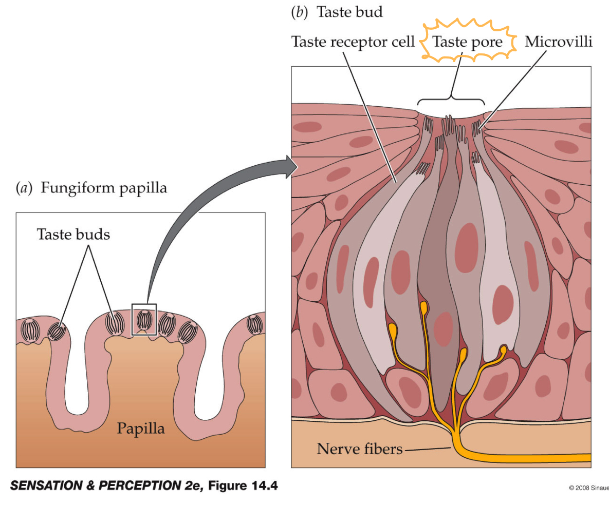
Each taste bud is a collection of taste receptor cells that make a neural response to tastants
Taste receptor cells extend microvilli into buds taste pore and contain taste receptors that the tastants bind to evoking a neural signal
Taste receptors determine what compounds each taste receptor cell responds to
papillae → taste bud → microvilli → taste receptor
Tastants
any stimulus that can be tasted
divided into 2 large categories
small charged particles that taste salty or sour: small ion channels in microvilli allow some types of charged particles to enter and not others
others are perceived via G protein-coupled receptors and taste sweet or bitter
Taste Receptors
determine what compounds each receptor cell responds to
salty receptors - ion channels that sodium can pass through depolarizing the cell and causing action potentials → salty
sour receptors - ion channels that let hydrogen ions pass, depolarizing the cell and causing action potentials → higher levels of acids so = sour
sweet and bitter receptors - special variety of G-protein coupled receptors that bind molecules with specific molecular shapes and release G-protein + action potentials
Umami
5th basic taste - essentially taste of protein
comes from receptors that detect monosodium glutamate (MSG)
almost nobody actually has a bad reaction to eating MSG
Umami receptors are found in the gut, suggesting the taste isn’t about tasting but knowing the nutrients
Taste Processing in the Central Nervous System
Pathway: taste buds → cranial nerves → medulla and gustatory thalamus → cortex
Insular Cortex - primary cortical processing area for taste, first receives taste information and gets olfactory information
Orbitofrontal Cortex - part of frontal lobe that lies above the bone containing the eyes
Receives projections from insular cortex
processes temperature, touch, smell, taste = integration area
Light + Vision
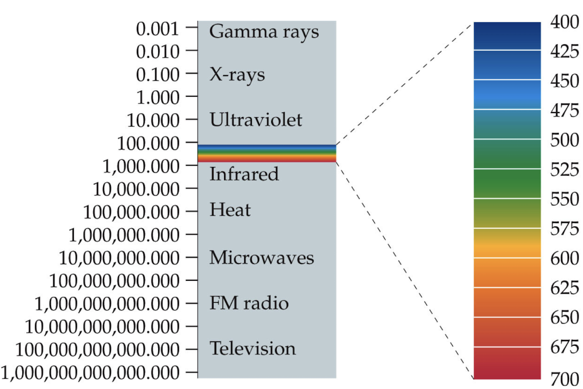
Light - wave, stream of photons, tiny particles that each consist of one quantum energy
Light can be absorbed (taken up/not transmitted), scattered (dispersed in an irregular fashion), reflected (redirected usually back to original point), transmitted (passed on through a surface), refracted (altered as it passes to another medium)
Human Eye
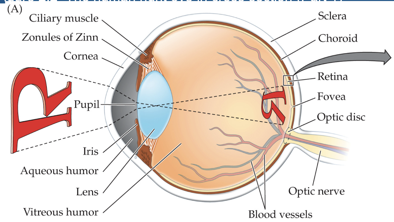
Cornea - transparent window into the eyeball
Aqueous Humor - watery fluid in anterior chamber
Crystalline lens - lens inside eye allows changing focus
Pupil - dark circular opening in the iris where light enters the eye
Vitreous Humor - transparent fluid that fills vitreous champer in the posterior of the eye
Retina - light sensitive membrane in back of the eye and contains rods+cones, send info to brain through the optic nerve
Accomodation
the lens in the eye refracts light to focus the image on the retina
Accommodation - process where lens changes shape altering refractive power and focusing on different focal planes in the world
Using ophthalmoscope doctors can view back of patients eye → fundus
Eye Problems
Presbyopia - unable to accommodate lens sufficiently to focus on nearby objects, caused by sclerosis (hardening of lens) and loss of elasticity
Cataracts - opacities in the lens that develop through age/illness
Emmetropia - no refractive error, image is properly focused on the retina
Problems with Refraction
Myopia - light entering eye is focused in front of retina and distant objects can’t be seen sharply (nearsighted)
Hyperopia - light entering the eye is focused behind the retina (farsighted)
Astigmatism - unequal curving of one or more refractive surfaces of the eye, usually cornea
Blind Spot + Depth Cues
Blind Spot - optic disc = place where the nerve leaves the eye
your brain fills in the space you can’t actually see so you don’t perceive a gap in your vision
Pictoral Depth Cues - cue to distance/depth used by artists to depict 3D depth in 2D pictures
Anamorphosis - use of rules of linear perspective to create a 2D image so distorted it looks correct when viewed from a specific angle or mirror
Photoreceptors
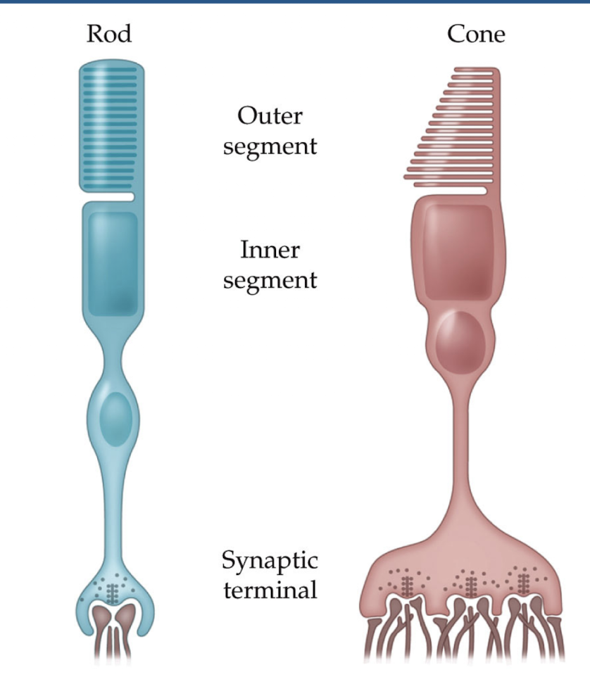
Photoreceptors - cells in the retina that initially transduce light energy into neural energy
Rods - photoreceptors specialized for night vision
respond well in low light, no color processing, gather light over large areas
Cones - photoreceptors that are specialized for daylight vision and fine visual acuity and color
respond best with lots of light, gather light over small areas
very poor color vision and spatial resolution in your periphery
Visual Transduction
Light activates rhodopsin molecules
Each rhodopsin molecule activates hundreds of transducin molecules
Each transducin molecule activates hundreds of phosphodiesterase molecules
Each phosphodiesterase hydrolyzes (inactivates) hundreds of cGMP molecules
drop in cGMP causes hyperpolarization of photoreceptor
hyperpolarization reduces transmitter release → neural signal
light → rhodopsin → transducin → phosphodiestrase → cGMP → hyperpolarization of photoreceptors → neural signal
Photoreceptor Dark Current
when there is no light cells are constantly depolarized by Na+ and Ca2+ currents in the outer segment
constantly releasing neurotransmitter glutamate
Light causes reduction in dark current → decrease in transmitter release
decrease in neural signals = light on that spot of the retina
Specialization and Parallel Processing
retinal signals are distributed to many parts of the circuit which all analyze it at the same time → Parallel Processing
Each part of the circuit is specialized to certain kinds of processing
detection of motion, faces, color, absence of light, fine spatial detail
Path of image processing
Eye → photoreceptors, bipolar cells, retinal ganglion cells
Lateral geniculate nucleus in the thalamus
Primary visual cortex
Receptive Fields
Receptive Field - region in space which stimuli activate neurons
critical conept across all sensory systems!
Photoreceptors have simple, round receptive fields
circular area in space where light will cause them to respond (reduce neurotransmitter release)
this sets the limit on how small of a stimulus we can discriminate
Ex. stripes or grey block? (both light and dark parts fall into receptive field of cone)
size of photoreceptor = limit to what you can see
Retinal Ganglion Cells
Retinal Ganglion Cells → Center Surround Receptive Field
ON-center ganglion cells - excited by light falling on the center and inhibited by light falling in the surround
OFF-center ganglion cells - inhibited by light falling on the center and excited by light falling on the surround
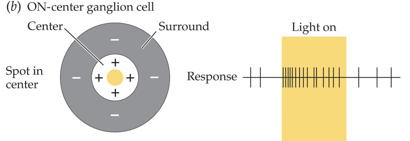
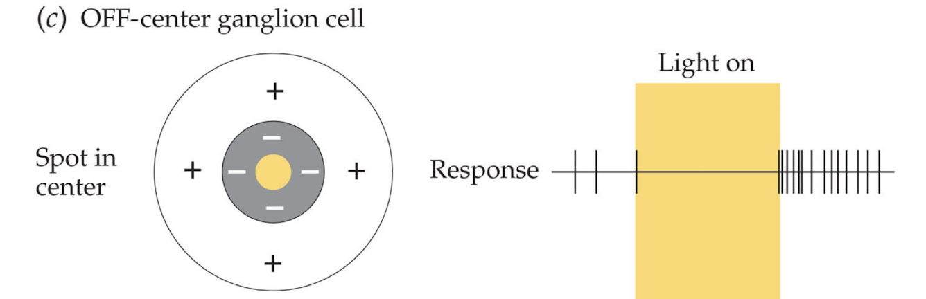
Center-surround receptive fields are tuned to respond to sine wave patterns with specific frequencies → specific spatial frequency + phase + place in visual world
Primary Visual/Striate Cortex
gets input from lateral geniculate nucleus carrying basic info from the eyes
cells here respond best to bars of light/dark rather than spots
Topographical Mapping - points in physical space are represented in similar points in the brain (picture mapped onto brain)
Cortical Magnification - dramatic scaling of information from different parts of visual field
amount of cortex devoted to processing the fovea is much more than that devoted to processing periphery
Orientation Tuning - neurons respond more to bars of certain orientations, response rate falls off with angular difference of bars
Hubel and Wiesel
cortical neuron responds to oriented bars of light might receive input from several retinal ganglion cells
string several retinal ganglion cells together → oriented bar
Cell that is tuned to any orientation you want could be created in cortex by connecting it up with the appropriate retinal ganglion cells
Object Recognition
Mid-Level Vision - process which edges are identified and linked together to form discrete objects
figure-ground assignment - brain decides what is background vs object
strongly influenced by expectations about the world
brain rejects accidental viewpoints → combinations of visual inputs that are very unlikely
Object Recognition - process which brain connects visual stimuli with understanding of how objects work
naive template matching → doesnt work because same object can look varyingly different (cowiest cow)
Depth Perception
based on our prior understanding of the world → ex overlapping pennies = 2 pennies
visual systems use depth cues when interpreting size of objects → most likely explanation based on prior information
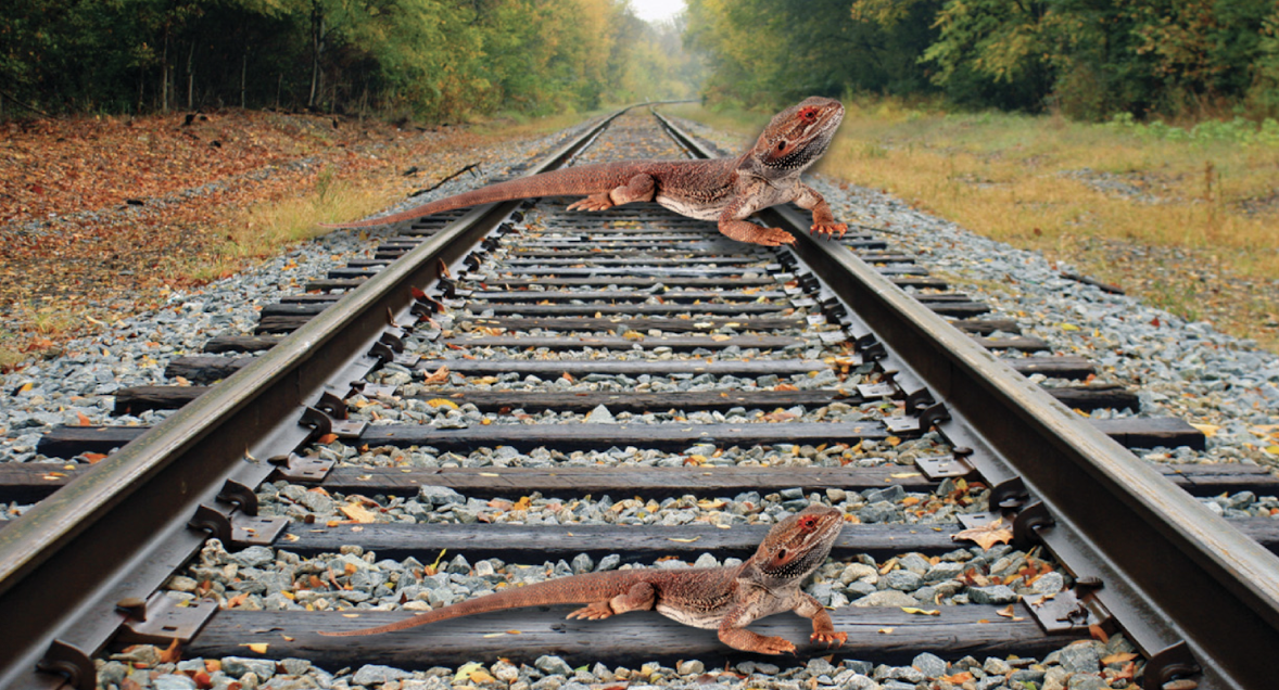
Calculating Depth Perception
Binocular Disparity - differences between the two retinal images of the same scene
Ex. finger in front different eye different position
Stereopsis - perception of 3 dimensionality of the world based on binocular disparity (not available with monocular vision)
Ex. putting on pen cap with one eye closed
Motion Parallax - images closer to observer seem to move faster than images farther away → brain uses this information to calculate distances of objects
Color Vision + Problem of Univariance
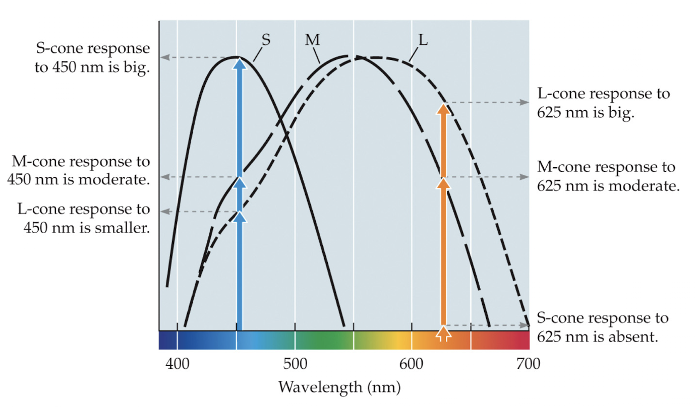
Problem of Univariance - infinite set of different wavelength intensity combinations can elicit exact same response from photoreceptor
1 photoreceptor can’t make color discriminations based on wavelength → why you can’t see color with just rods
Cone photoreceptors come in 3 varieties
S-cones - preferentially sensitive to short wavelengths (blue)
M-cones - preferentially sensitive to middle wavelengths (green)
L-cones - preferentially sensitive to long wavelengths (red)
Metamers show color is not a physical property but psychophysical instead
Trichromacy + Metamers
Trichromacy - color of any light is defined in our visual system by relationships of 3 numbers, outputs of 3 recepter types/cones
Metamers - different mixtures of wavelengths that look identical, any pair of stimuli perceived as identical in spit of physical differences
show color is a mental construct not just wavelengths
Middle Vision (Gestalt)
Gestalt Grouping Rules - describe when elements in an image will appear to group together
Similarity + Proximity
Good Continuation - 2 elements group together if they lie on the same contour
Texture Segmentation - carving image into regions of common texture properties
Parallelism + Symmetry
Common Region + Connectedness
Color Perception
the 7 colors of the rainbow are stupid and made up… what even is indigo