Quiz 1 LOs: Diagnosis of Oral Lesions
1/42
There's no tags or description
Looks like no tags are added yet.
Name | Mastery | Learn | Test | Matching | Spaced | Call with Kai |
|---|
No analytics yet
Send a link to your students to track their progress
43 Terms
LO1: List the diagnostic categories that contribute to the diagnostic process
Medical / Dental History
Intraoral Exam / Extraoral Exam
Radiographs
Confirmatory tests: biopsy/referral to specialists --> Laboratory/Microscopic/Surgical
Therapeutic
Differential findings
What do we include in a description of oral lesions?
clinical appearance, consistency, color, size, texture
What are the terms we use to describe the appearance soft tissue lesions?
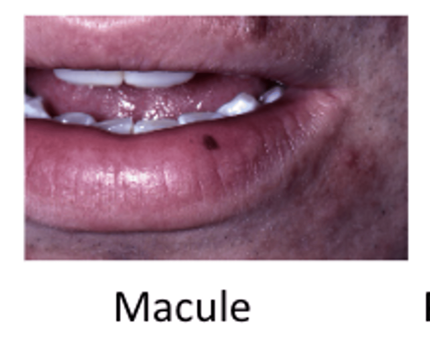
Macule
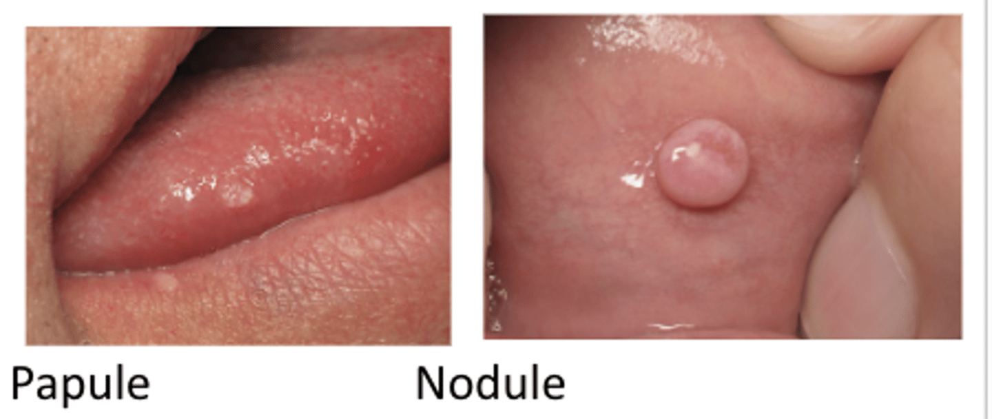
Papule/nodule
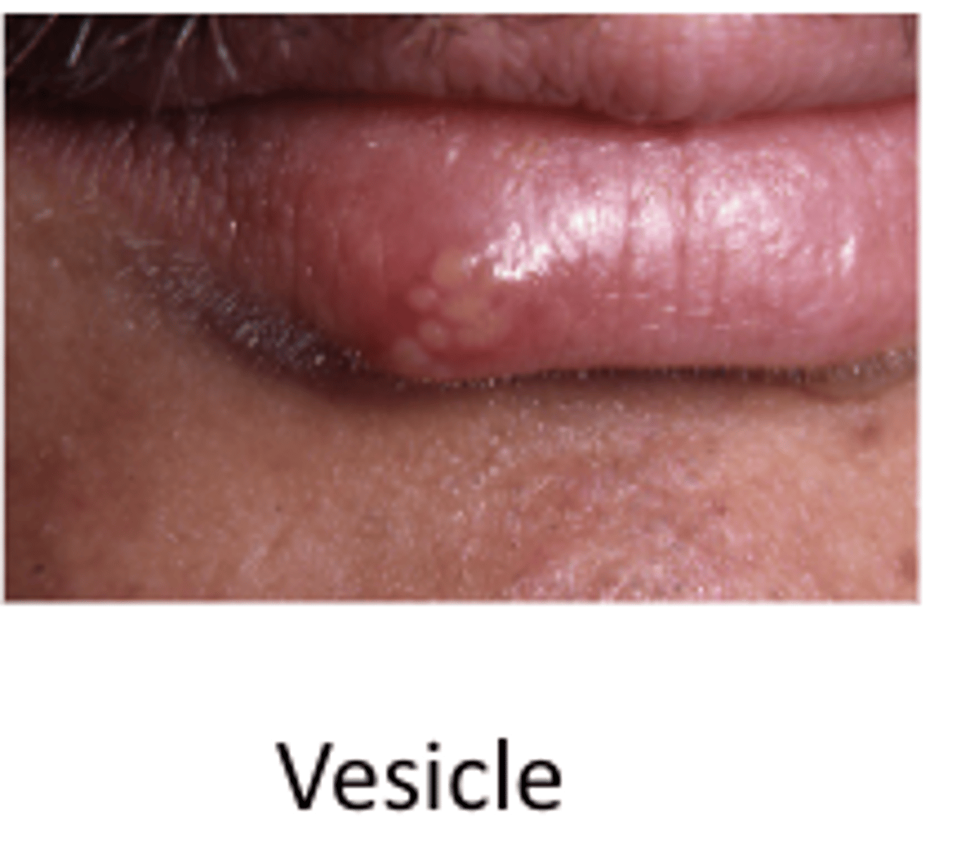
Vesicle/bulla
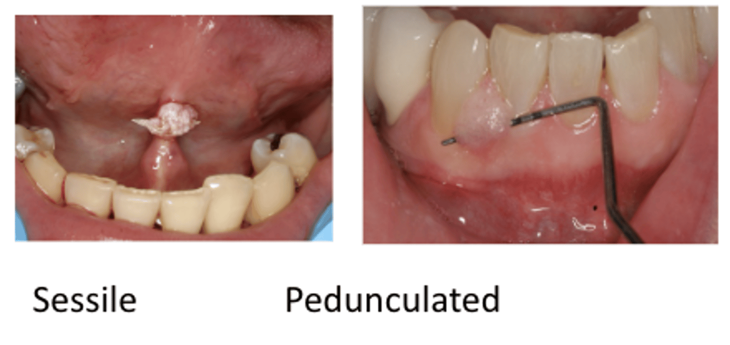
Pedunculated/sessile
Macule
flat lesion, pigmented
Papule/nodule
raised lesion
nodule > 1 cm
papule < 1 cm
Vesicle/bulla
fluid filled raised area
bulla > 5 mm
vesicle < 1 cm
Pedunculated/sessile
sessile has a raised surface
pedunculate needs to the probe to raise it (mushroom shape)
What are the terms we used to describe soft tissue consistency?
soft
firm
fluid-filled
What are the terms we used to describe the color of the lesion?
Normal/white/red/pigmented (black/blue)
Leukoplakia - White patch
Erythroplakia – Red patch
Erythema – red surface
What units do we use for size of the lesion?
cm or mm
What are the terms we used to describe the surface texture of the lesion?
Smooth
Papillary/verrucous --> irregular
Ulcerated --> grey center
How do you describe a jaw lesion?
location, growth pattern, symptoms, others
What terms do you use to describe location of a jaw lesion?
Maxilla/Mandible
Anterior/Posterior
What terms do you use to describe growth pattern of a jaw lesion?
Swelling (expansile/non-expansile)
Slow/fast
growing
Breaking through bone
Ulceration/inflammation
What terms do you use to describe symptoms of a jaw lesion?
pain, paresthesia, exudate
What other terms do you use to describe a jaw lesion?
systemic signs and symptoms --> fever, fatigue, pigmentation, malignancy, endocrine dysfunction etc
LO2: What is periapical cemento-osseous dysplasia? (cementoma)
form of COD that is confined to the bone around the roots of the md anterior teeth
LO2: What is the relationship between race, sex, and age and periapical cemento-osseous dysplasia? (cementoma)
black women in their 30s
LO2: What is the radiographic appearance of periapical cemento-osseous dysplasia? (cementoma)
a radiolucent area at the apex of a tooth on a radiograph
LO3/4: Define leukoplakia
white lesion that cannot be rubbed off and cannot be diagnosed through clinical characteristics alone
LO3: Define erythroplakia
red lesion that cannot be diagnosed on the basis of clinical features alone.
LO4: Describe tori
excessive growth of normal bone
common in ADULTS
What race and sex is torus palatinus most common in?
native american females
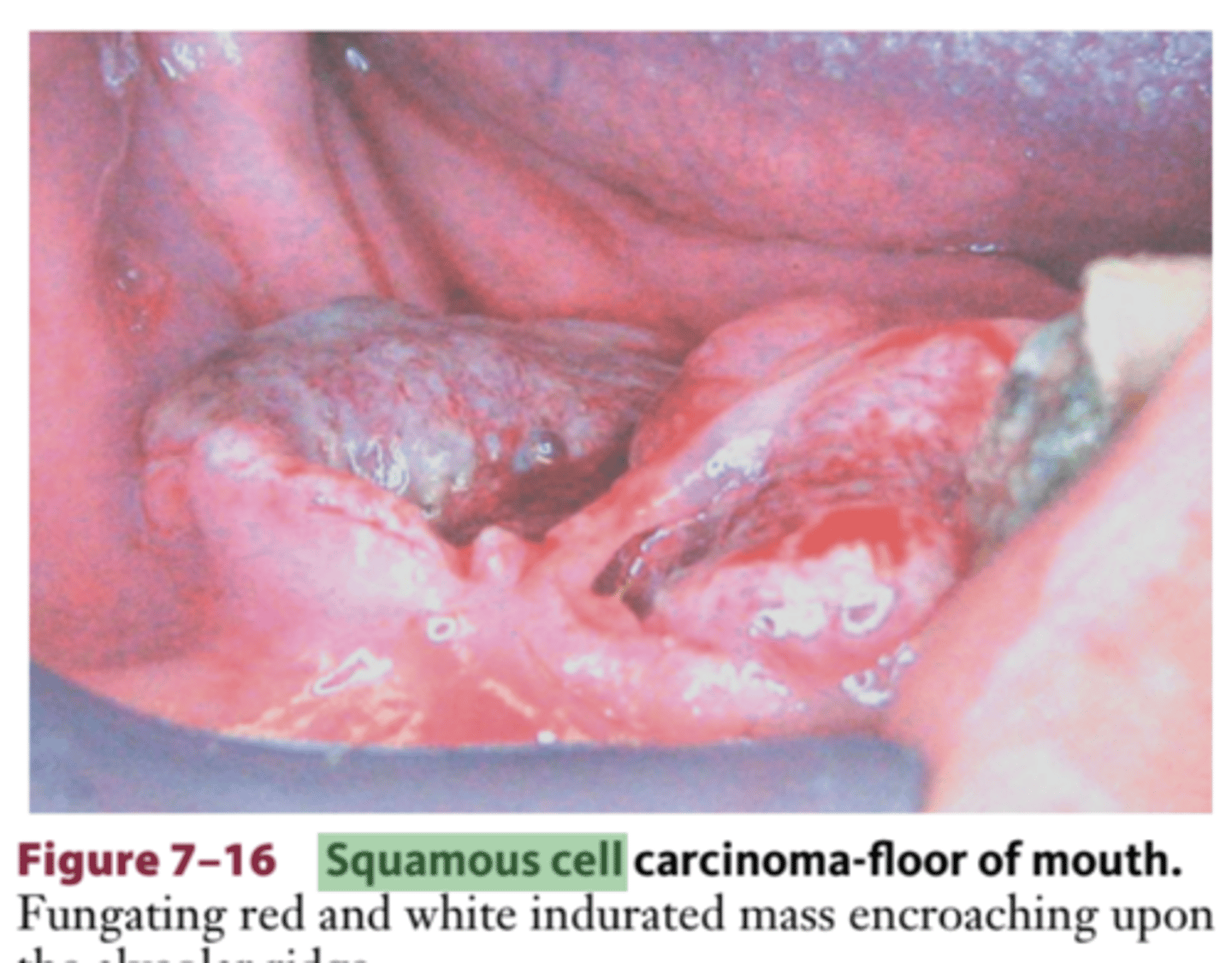
LO4: Describe squamous cell carcinoma
malignant transformation
of the squamous epithelium lining of the oral cavity
carcinomas: malignant tumors arising from cells that originated
from embryonic ectoderm
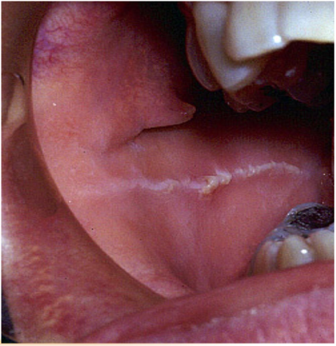
LO4: Describe linea alba
appears as a white line along the plane of occlusion
unilateral or bilateral
tissue becomes keratinized
LO4: Describe erythema migrans
usually asymptomatic, etiology unknown
abundance of both acute and chronic inflammatory cells
LESIONS ON MUCOSAL SURFACES --> red, irritated
lesions develop and then spontaneously regress, then appear again later
LO4: Describe nutritional deficiencies
varied --> can include glossitis, pallor, etc.
LO4: Describe a Vitamin D deficiency
result in bone loss and increased inflammation
LO4: Describe a protein deficiency
cell-mediated immunity, complement system, phagocyte activity and production of cytokines
LO4: Describe an antioxidant and Omega-3 defiency
increases risk for perio
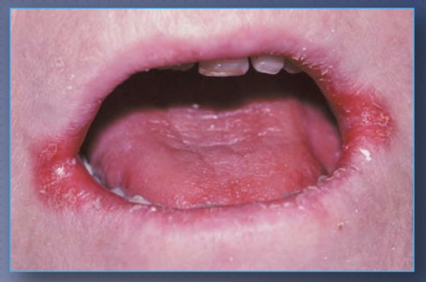
LO4: Describe angular cheilitis
describe redness and cracking at the corners of the mouth
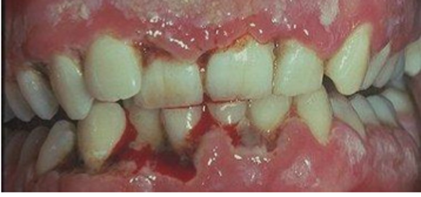
LO4: Describe necrotizing ulcerative gingivitis
Punched out papilla
Profuse bleeding
Fever
Fatigue
Swollen lymph nodes
Rapid onset
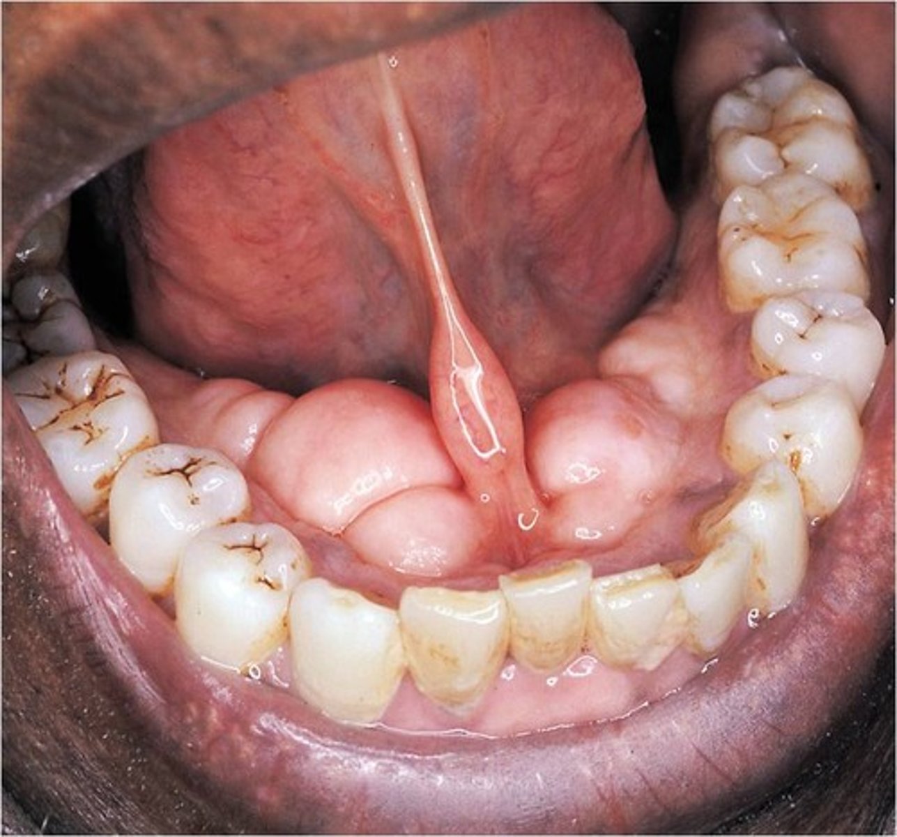
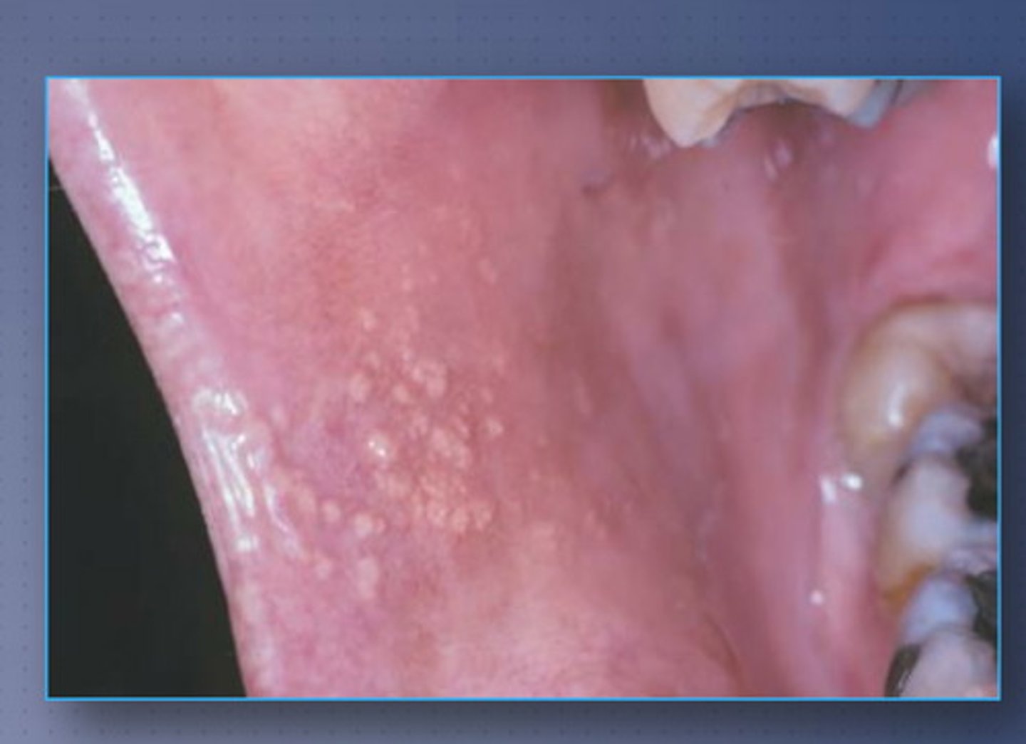
LO4: Describe fordyce granules
oil glands of skin that appear as yellow nodules along buccal and labial mucosa
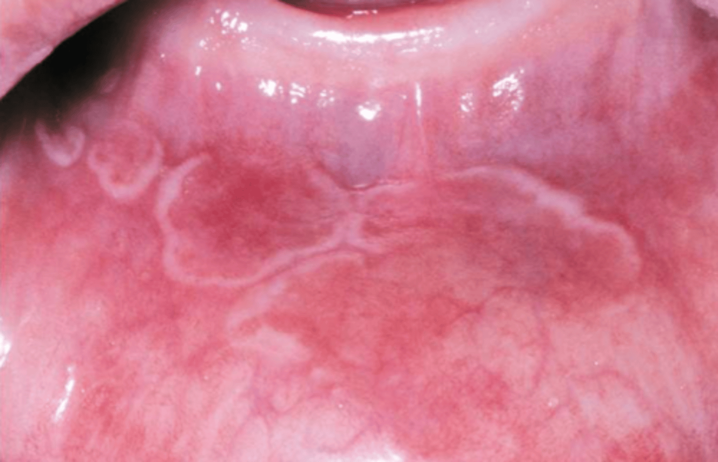
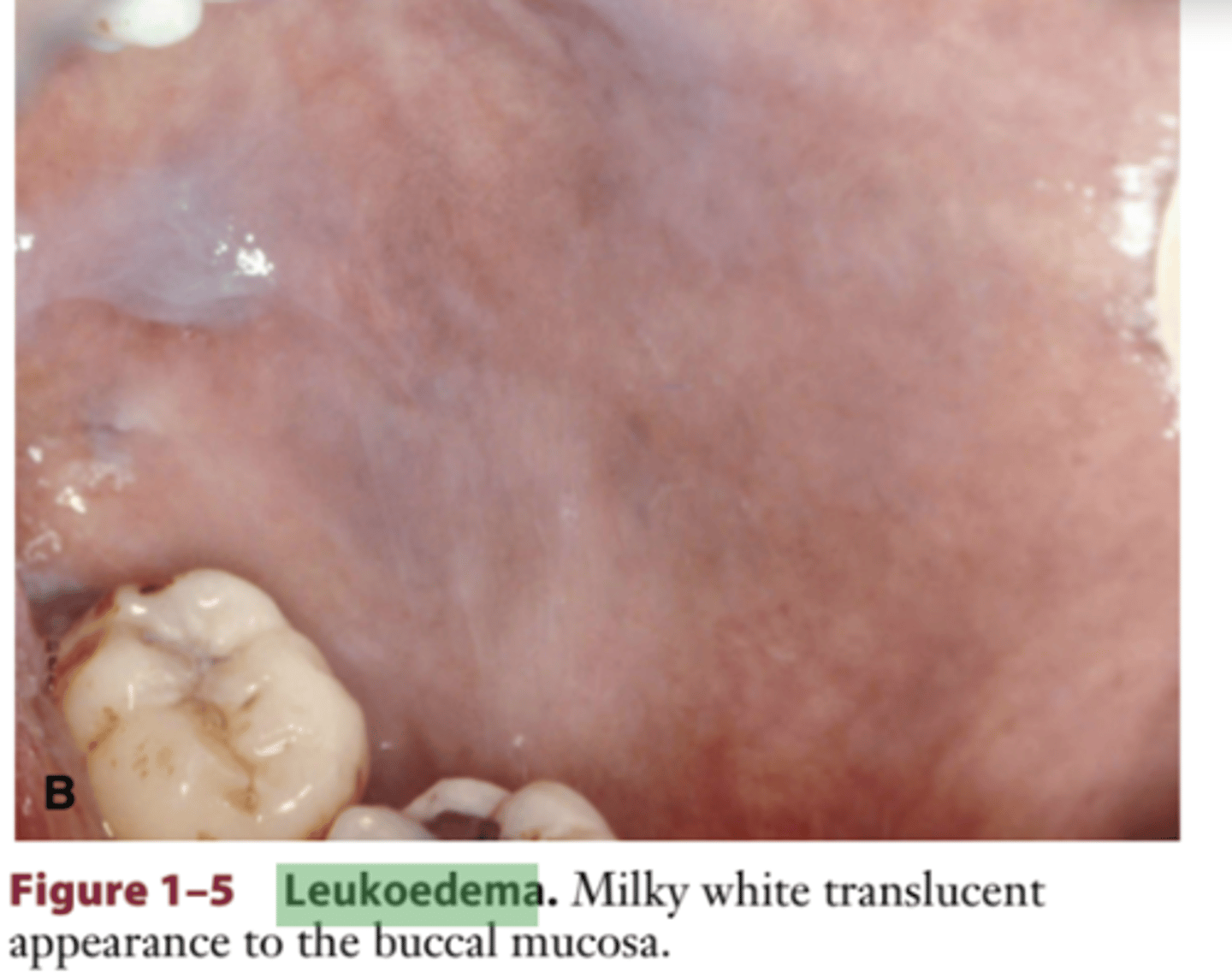
LO4: Describe leukoedema
generalized translucent or
opalescent covering, located over the buccal mu-
cosa
lesion initially appears
translucent white or opalescent and then disappears when the tissue is stretched
most common in BLACK individuals
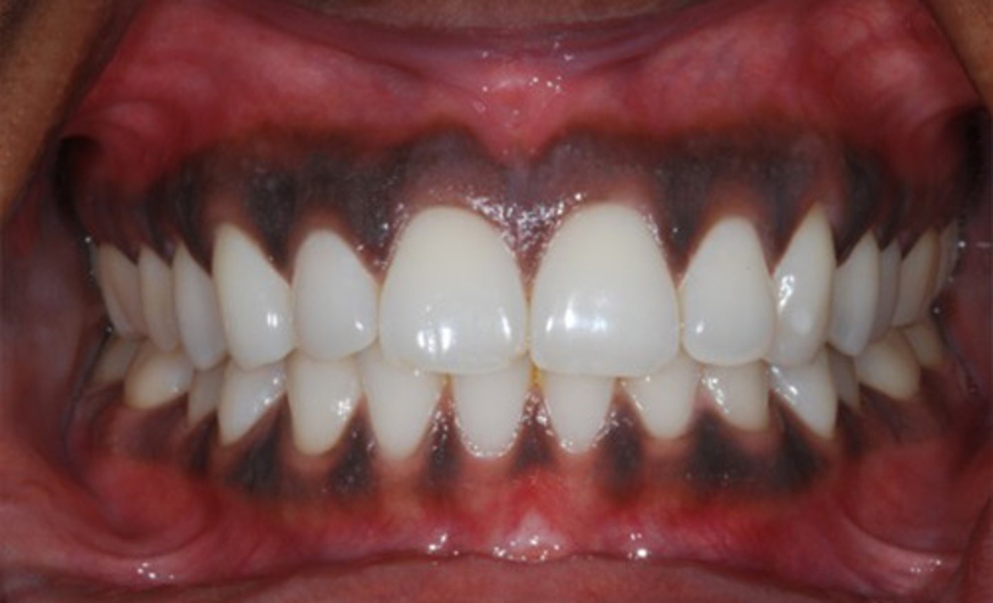
LO4: Describe melanin pigmentation
diffuse or localized brown pigmentation of the mucosa
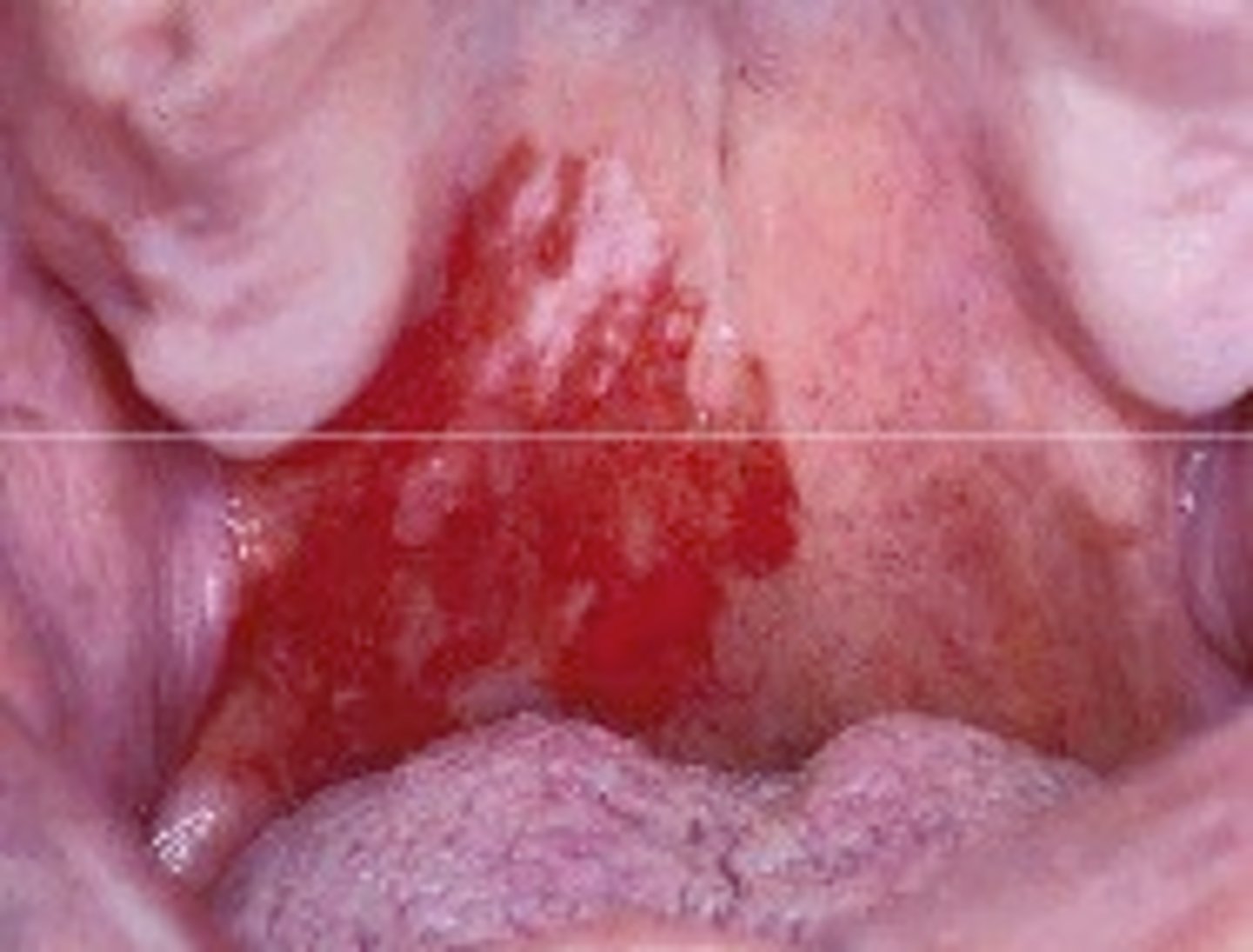
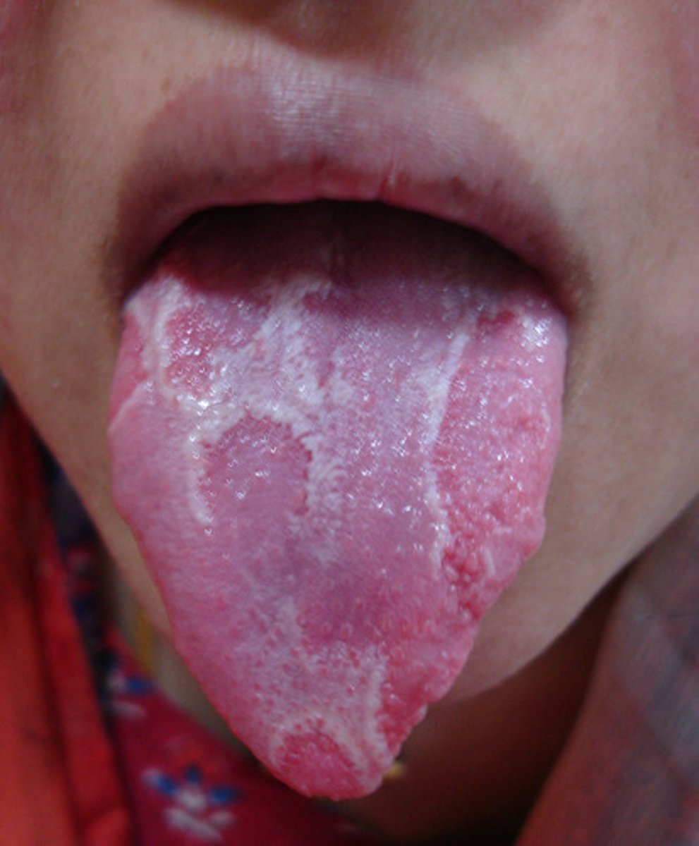
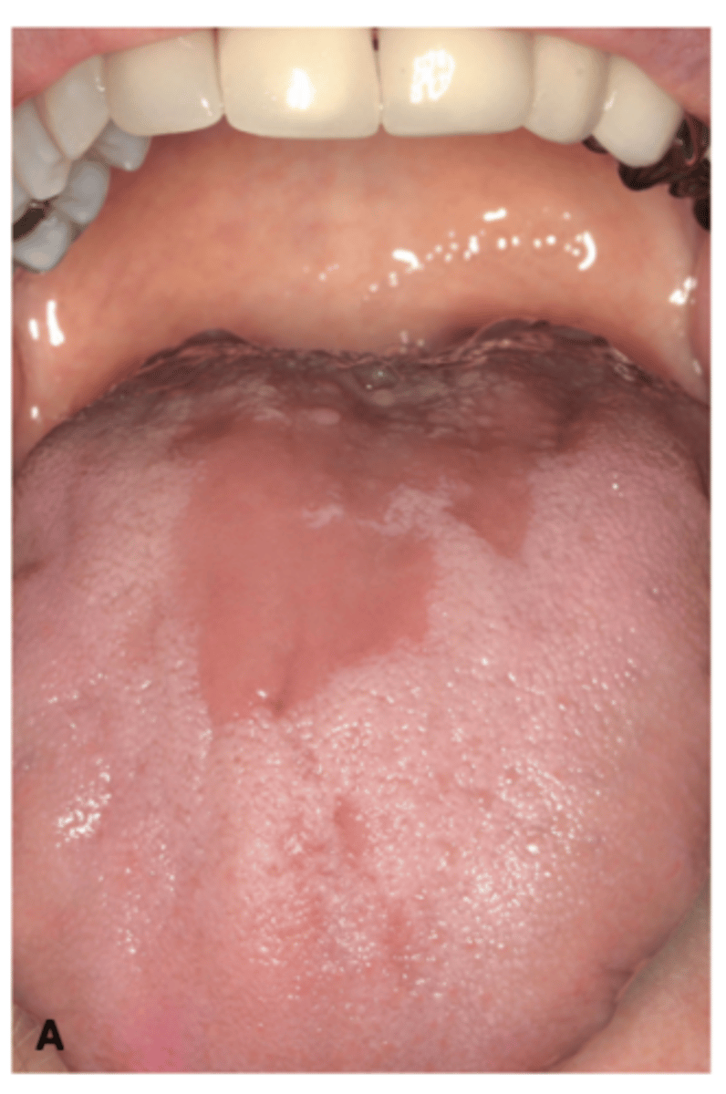
LO4: Describe erythema migrans --> geographic tongue
usually asymptomatic, etiology unknown
abundance of both acute and chronic inflammatory cells
LESIONS ON TONGUE--> red, irritated
asymmetrical, bordered areas of ery-
thematous mucosal atrophy, surrounded by white or
yellowish circinate (circular or ring-shaped), slightly
elevated borders
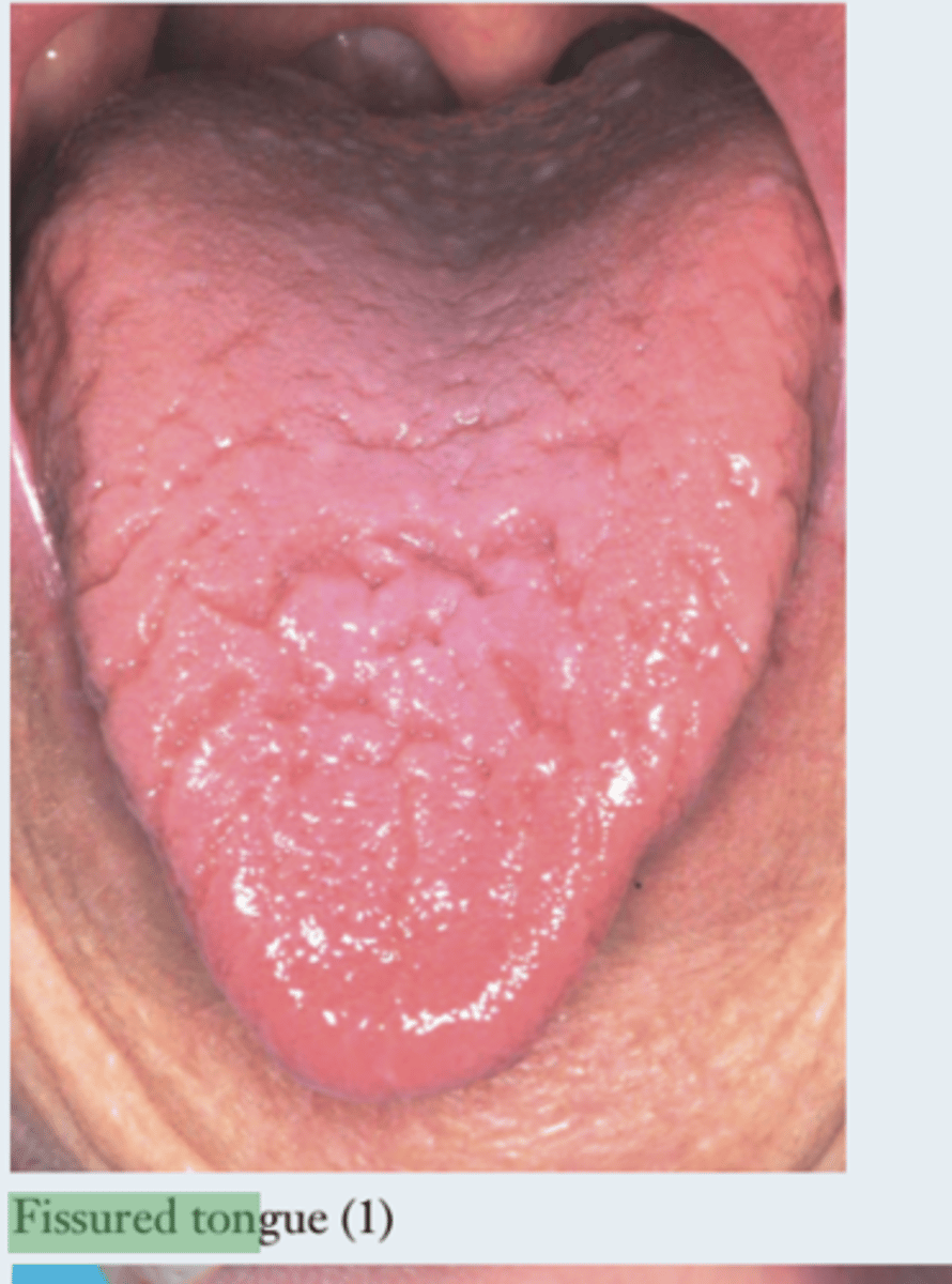
LO4: Describe fissured tongue
deep grooves present on the dorsum on the tongue
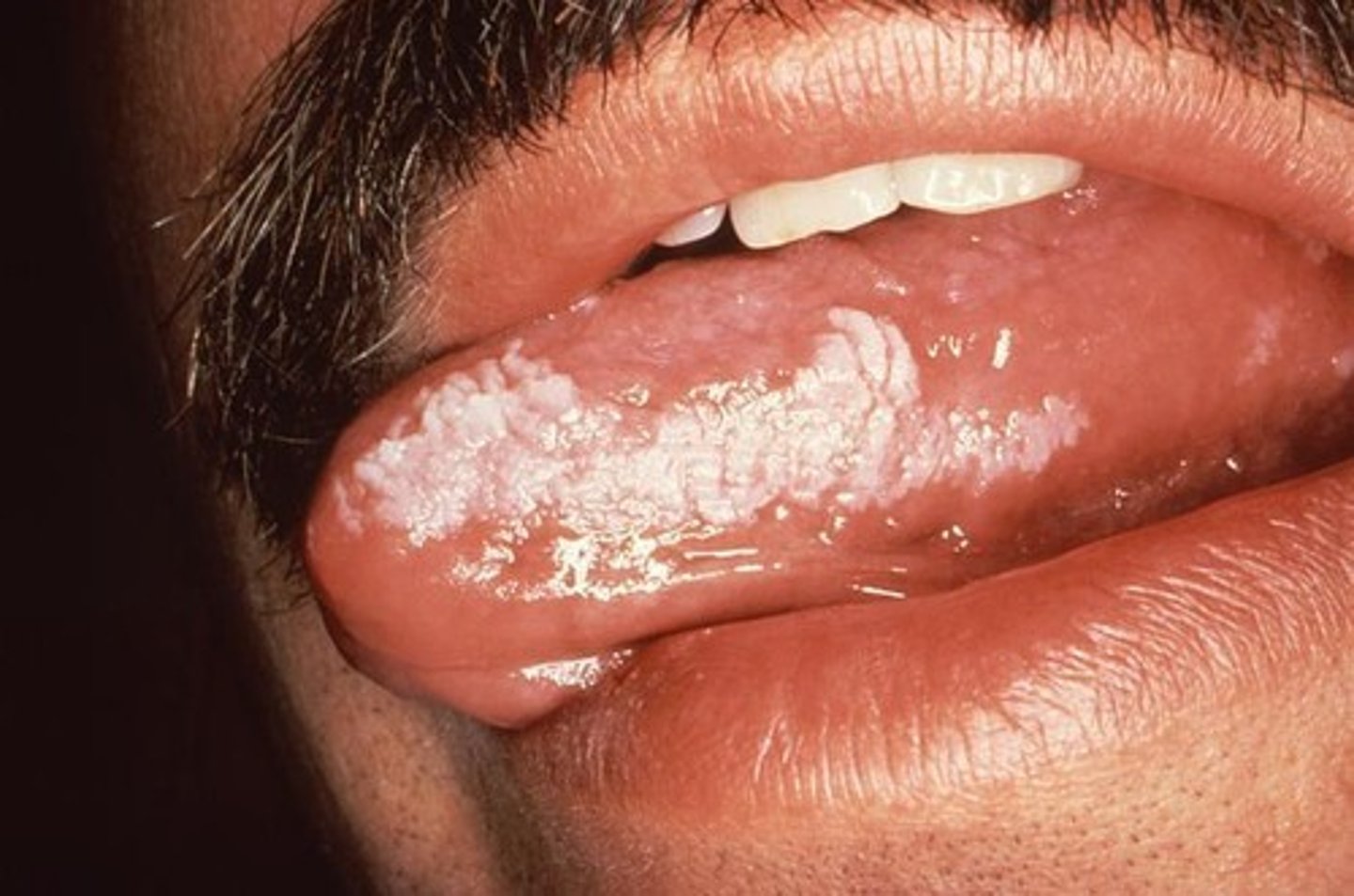
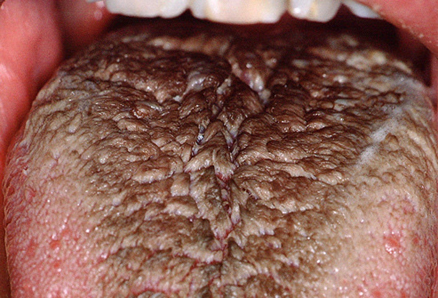
LO4: Describe hairy tongue
elongated filiform papillae that
produce a hair-like appearance
can pick up
yellow or brown stain and present an aesthetic problem for the patient
LO4: Describe median rhomboid glossitis (central papillary atrophy)
localized loss of the filiform papillae creating a smooth, red, rhomboid-shape at the midline on the dorsal surface of tongue
more common in ADULTS
LO5: Define variant of normal
Anatomical structures or conditions that deviate from the common presentation BUT are considered within the range of normal variation and are not pathological
LO5: What are some examples of the tongue that are variants of normal?
fordyce granules, tori, melanin pigmentation, linea alba, leukoedema
LO6: Define lingual thyroid
normal thyroid tissue
remains on the dorsum of the tongue --> it is ectopic, meaning out of place
most commonly a mucosal-colored round nodule, reaching up to 4 cm in diameter
affects WOMEN more than men
LO6: What are the 3 clinical symptoms associated with lingual thyroid?
1. Dysphasia--> language disorder that affects how you speak and understand language
2. Dysphonia --> difficulty speaking
3. Dyspnea --> shortness of breath