A&P1 - Exam 2 - Ch. 4 + 5
1/82
There's no tags or description
Looks like no tags are added yet.
Name | Mastery | Learn | Test | Matching | Spaced | Call with Kai |
|---|
No analytics yet
Send a link to your students to track their progress
83 Terms
List the general tissue categories (+know what their general function is)
Epithelial - covers exposed surfaces, lines internal passageways, and forms glands
Connective - fills internal spaces, supports other tissues, transports material, and stores energy
Muscle - specializes for contraction
Nervous - carries electrical signals from one part of the body to another
What are the apical and basal surfaces?
These are important surface layers of epithelial cells, which creates the strong polarity of specialized epithelial cells.
The apical surface is the top (most superficial) layer of the epithelial cell, exposed to the external or internal environment.
The Basal surface is the bottom (most deep) layer of the epithelial cell, attached to other ET cells beneath it, or other tissue.
The specialized epithelial cell is often divided into two functional groups, meaning the cell _________ (or having what effect?)
the cell has strong polarity
What are the characteristics of the apical surface? What general tissue type is it a part of?
The apical surface is the most superficial layer of an epithelial cell. It is exposed to the external or internal world. It contains microvilli and/or cilia.
The microvilli allow for an increased surface area (for absorption or secretion).
The cilia move fluids and other materials over the surface.
Microvilli and cilia are a characteristic of which epithelial cell layer/surface?
Apical surface
What are the characteristics of the basal surface? What general tissue type is it a part of?
The basal surface is the surface that is on the bottom of an epithelial cell and serves as an attachment to other epithelial cells below. Or if it is the bottommost ET cell, it attaches to other tissues below.
Name the general characteristics of epithelial tissue.
Polarity: Difference between exposed (apical) surface and attached (basal) surface
Cellularity: epithelial cells are tightly bound by cell junctions
Attachment: The base of the epithelium is bound to a noncellular basement membrane
Avascularity: no blood vessels
Regeneration: continuously replaced by division of stem cells
Epithelial tissue is tightly bound by ________
Cell junctions
The base of epithelium (basal surface) is bound to what two things?
the noncellular basement membrane; as well as attached to tissue below
Epithelial tissue is (vascular/avascular)
avascular
Connective tissue is (vascular/avascular)
Mostly vascular — some tissue such as cartilage is entirely avascular
Muscle tissue is (vascular/avascular)
vascular
Nervous tissue is (vascular/avascular)
vascular (?)
What are the main specializations of epithelial tissue? (name 3)
1) move fluids over epithelium for protection and lubrication
2) move fluids through epithelium to control permeability
3) produce secretions for protection and chemical messaging
all very fluid and secretion based!
What maintains the integrity of the epithelia?
1) Intercellular connections
2) attachment to basement membrane
3) epithelial maintenance and repair
Transmembrane proteins that attach opposing plasma membranes and plasma membranes to the extracellular matrix.
____________
Cell adhesion molecules (CAMs)
Cell adhesion molecules (CAMs)
Transmembrane proteins that attach opposing plasma membranes and plasma membranes to the extracellular matrix.
List the types of cell junctions
gap junctions
tight junctions
desmosomes
Gap junctions (function/characteristics)
1 of 3 main types of cell junctions
Hold cells together by interlocking transmembrane proteins (connexons), which allow passage of small molecules and ions
Tight junctions (function/characteristics)
1 of 3 types of cell junctions
They interlock membrane proteins that bind plasma membranes
They have a lumen which is a passageway surrounded by cells.
They work to prevent passage of water and solutes between cells from lumen to underlined cells and tissues
Gap junctions and tight junctions both interlock proteins. Name the specific protein each interlocks and what the outcome of this is.
Gap junctions: transmembrane proteins (or connexons) — this will allow passage of small molecules and ions
Tight junctions: interlock membrane proteins that bind plasma membranes together — this works to prevent passage of water and solutes between cells from lumen to underlined cells and tissues
Which of the cell junctions binds plasma membranes together? How?
Gap junctions
Tight junctions
Desmosomes
Both tight junctions and desmosomes bind plasma membranes together
Tight junctions use membrane proteins to do so while desmosomes use CAMs (cell adhesion molecules) and proteoglycans
Desmosomes (function/characteristics)
CAMs and proteoglycans link opposing plasma membranes
There is a dense area of each desmosome that is connected to cytoskeleton
What are the two types of desmosomes? Differentiate between the 2.
Spot desmosomes - small discs that stabilize cell shape
Hemidesmosomes - look like half of a spot desmosome; they anchor cells to the basement membrane
What type of cell junction anchors cells to the basement membrane?
Desmosomes; specifically - hemidesmosomes
What are the three classifications of epithelia based on shape?
Squamous; cuboidal; columnar
What is the classification of epithelia that have 1 layer? Multiple layers?
Simple epithelium
Stratified epithelium
Describe the following classifications of epithelia:
squamous
cuboidal
columnar
Squamous — thin and flat, irregularly shaped
cuboidal — boxy with central nucleus
columnar — tall and slender rectangles
Function and main locations of simple squamous epithelia
Function: Absorption and diffusion (in the lining of lung alveoli)
It also makes up the mesothelium (body cavity lining) and endothelium (heart and blood vessel lining)
Function and main locations of stratified squamous epithelia
Function: protects against mechanical stresses/abrasion in the top layer of skin, lining of mouth, and anus
It also has added strength and water resistance from added keratin (we can also say this epithelium is keratinized)
Stratified squamous epithelia has added strength and water resistance due to what
Stratified squamous epithelia is keratinized (has keratin)
Function and main locations of simple cuboidal epithelia
Function: secretion and absorption
Location: forms glands, lines kidney tubules
Function and main locations of stratified cuboidal epithelia
Function/location: forms ducts of sweat and mammary glands
fun fact, they’re relatively rare!
Function and main locations of transition epithelium
cells appear cuboidal when not stretched and squamous when stretched
they tolerate stretching repeatedly without damage
forms lining of urinary bladder
What specific tissue type can undergo repeated stretching without damage?
transitional epithelium (think: it transitions through different forms through stretching!)
Function and main locations of simple columnar epithelium
Function: absorption and secretion
Location: stomach, small intestine, large intestine
The function of simple epithelia, despite shape classification, tends to be …
Absorption and secretion
Function and main locations of stratified columnar epithelium
Relatively rare
Function: protection
Location: pharynx, anus, and urethra
Characteristics, function, and main locations of pseudostratified columnar epithelium
appears layered but is actually only one layer
cells typically have cilia
Function: protection, secretion, moving mucus (with the cilia!)
Location: lining of nasal cavity, trachea, and bronchi
What is glandular epithelia
glands are collections of epithelial cells that produce secretions
What are the two types of glandular epithelia? What are their key characteristics?
Endocrine: release hormones into the bloodstream: do NOT have ducts
Exocrine: produce exocrine excretions onto epithelial surfaces; DO have ducts
Endocrine glands (do/do not) have ducts while exocrine (do/do not) have ducts
endocrine: NO ducts
exocrine: YES ducts!
What structure do exocrine glands discharge by? Where is their target?
They use ducts to discharge exocrine secretions onto epithelial surfaces
What is the classification of goblet cells? What is their function?
They are a unicellular exocrine gland
Location: epithelia of intestines
Function: secrete mucin which, when mixed with water, produces mucus
Unicellular vs multicellular exocrine glands
Unicellular exocrine glands are composed of a single cell that excretes product onto a surface
Multicellular on the other hand, is composed of many cells the come together to form complex secretory units and ducts
Merocrine secretion, apocrine secretion, and holocrine secretion all fall under what category?
Exocrine secretion
Merocrine secretion (how it functions + example of a gland)
exocrine secretion;
product is released by secretory vesicles (exocytosis)
merocrine sweat glands
Apocrine secretion (how it functions + example of a gland)
Product is released by shedding cytoplasm
e.g. mammary glands
Holocrine secretion (how it functions + example of a gland)
Product released by cells bursting and dying (then replaced by stem cells)
e.g. sebaceous glands
Sebaceous glands have what type of secretion?
Holocrine (product released by cells bursting and dying); exocrine
Mammary glands have what type of secretion?
Apocrine (product release via shedding cytoplasm); exocrine
Merocrine sweat glands have what type of secretion?
merocrine secretion (product released via secretory vesicles
Which category of exocrine secretion works by releasing product by secretory vesicles? Give an example
Merocrine; merocrine sweat glands
Which category of exocrine secretion works by shedding cytoplasm? Give an example.
Apocrine; mammary glands
Which category of exocrine secretion works by bursting and dying to release product, then being replaced by stem cells? Give an example.
Holocrine secretion; sebaceous glands
Which type of gland secretes watery secretions that contain enzymes?
Serous glands
Which type of gland secretes mucins that form mucus?
Mucous glands
Which type of gland secretes a mixed population of serous and mucous gland cells?
Mixed exocrine glands
What is the the name of diagnostic tests that are done by studying shed epithelial cells? What is a common example?
exfoliative cytology
pap smear
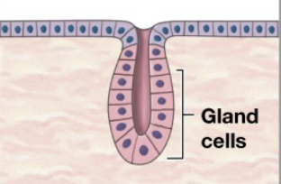
What is this?
Simple tubular gland
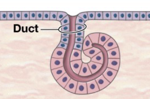
What is this?
Simple coiled tubular gland
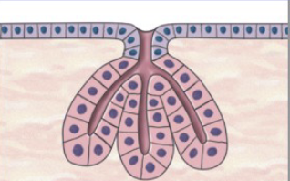
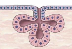
What is this?
Simple branched tubular gland
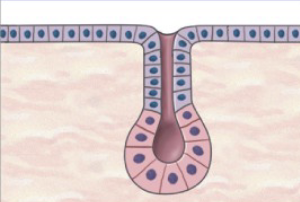
What is this?
Simple alveolar (acinar) gland
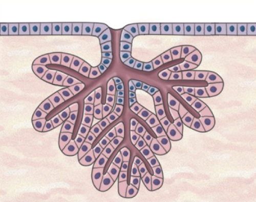
What is this?
Simple branched alveolar gland
What is this?
Compound tubular gland
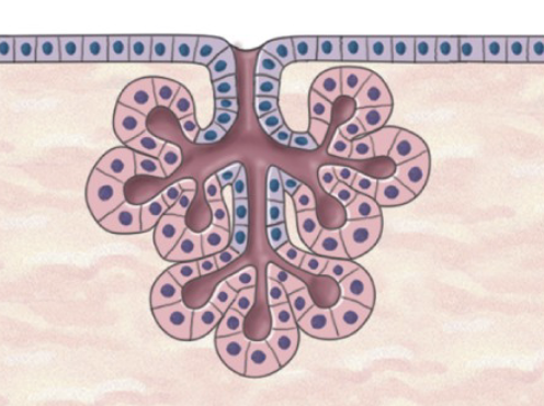
What is this?
Compound alveolar (complex acinar) gland
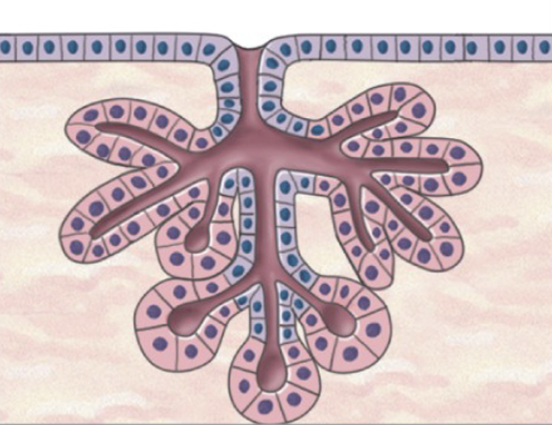
What is this?
Compound tubulo-alveolar gland
What tissues are considered connective tissue proper?
Dense and loose connective tissues; they must PHYSICALLY connect tissue
types:
Loose: areolar tissue, adipose tissue, and reticular tissue
Dense: dense regular, dense irregular, and elastic tissue
What are the three fibers of connective tissue proper?
Collagen, reticular, and elastic
What the cells build protein fiber cells? expand.
Fibroblasts;
they are the most abundant fixed cells of connective tissue
they secrete the component of ground substance and protein fibers
hint: suffix “-blast” refers to builder cells in this context
What is the viscous material in areolar CT and that makes the stroma of organs?
Ground substance
What are the 3 classes of connective tissue?
Connective tissue proper (viscous matrix rich in protein fibers)
Fluid connective tissue (watery matrix w dissolved proteins)
Supporting connective tissue (matrix w densely packed fibers)
What is released by mast cells? What is their function?
Histamine and Heparin
Histamine: stimulates inflammation after injury or infection
Heparin: anticoagulant (prevent clotting)
Name the types of macrophages. What tissues they are found in? What is their function?
Types: Fixed macrophages and free macrophages
Location: scattered through CT (present in almost every tissue)
Function: Large phagocytes engulf damaged cells and pathogens and release chemicals that activate immune defense.
Name the types of microphages. What tissues they are found in? What is their function?
Types: Neutrophils and eosinophils.
Location: Normally circulate in the blood; leave the bloodstream to enter peripheral tissues at sites of injury or infection.
Function: Small phagocytes that remove cellular debris and attack microorganisms. Neutrophils act quickly; eosinophils target material coated with antibodies.
Name the types of mast cells. What tissues they are found in? What is their function?