Lecture 6
1/11
There's no tags or description
Looks like no tags are added yet.
Name | Mastery | Learn | Test | Matching | Spaced | Call with Kai |
|---|
No analytics yet
Send a link to your students to track their progress
12 Terms
How can we identify the unknown proteins?
Western Blotting
Transfer the proteins onto a nitrocellulose membrane
Use antibodies to identify specific proteins
How to make antibodies
Purify your protein
Inject into an animal (rabbit or goat)
Bleed animal after month or so
Purify antibody from the blood
Staining of the Blots
Protein stain
No different from the stained PAGE Gel
Use antibodies to identify specific proteins
But wont the antibodies stick non-specifically to the nitrocellulose?
Incubate blot with Bovine Serum Albumin
Now no other proteins can stick to the blot
Stain” Blot with the Primary Antibody
incubate with rabbit anti-mouse “C” antibody
Use pre-Stained Standard Proteins
Specific antibody binds only to C
antibodies are invisible, What are our options
Directly Label the antibody
Fluorescent compound
Radioactive compound
Bind an enzyme to the antibody)
Use a labeled Secondary antibody
Enzyme Conjugated Goat anti-rabbit Immunoglobulin antibody
• Immunoglobulin = generic name for the antibody protein
• Antibody = used for specific antibodies
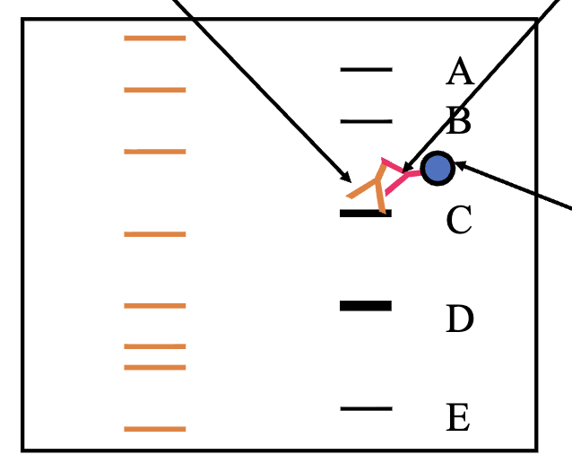
Labeled Secondary Antibody binds to the Primary Antibody
Primary Antibody (rabbit anti-mouse C protein Binds to the specific C protein)
Secondary Antibody (enzyme labeled goat anti-rabbit Ig Binds to the Primary Antibody)
Enzyme linked to the Secondary Antibody
Secondary Antibodies
must be from a different species than the primary antibody
Primary Ab is Goat anti-???
• Secondary must be from some animal other than a goat!
• Rabbit anti-goat Ig
• Mouse anti-goat IgIf primary antibody = mouse anti-phospho-tyrosine
• Secondary antibody must be goat, rabbit, donkey, etc. (anti-mouse Immunoglobulin)
What Enzyme to use to label the secondary antibody?
Inexpensive
Easy to make
Has substrates that form insoluble, colored precipitates
• Horseradish Peroxidase (HRP)
• Alkaline Phosphatase (AP) – We will use this enzymeSubstrate?
• BCIP (5-bromo-4chloro-3-indolyl phosphate)
• Acridan Based substrate (CDP-Star in this case)
Degradation of the Substrate
Alkaline Phosphatase (AP) hydrolyzes CDP-Star to remove a phosphate
The intermediate dioxetane phenolate anion is formed and once it decomposes light is emitted at 475 nm
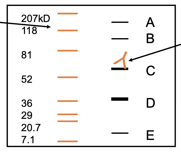
Result: Only the “C” Band stains
We can visualize the “C” protein band
Estimate the size of the “C” protein as about 55 kDa
This “C” protein is NOT carbonic anhydrase!
Staining of the blots
EGF binding to the EGF-receptor
• Activates the EGF-R which is a Kinase
• EGF-R phosphorylates itself and other proteins at tyrosine residues
• This sets off a kinase tyrosine phosphorylation cascade signaling pathway
Stain for Tyrosine phosphorylated proteins
EGF stimulation should result in the tyrosine phosphorylation of several proteins
• EGFR activates other kinases
• These kinases activate other kinases
• Kinase cascadeShows that the pathway has been activated
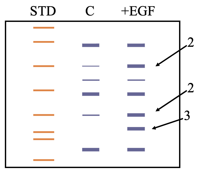
What We Expect to See:
Controls compared to EGF Treated:
1. More bands
2. Some heavier stained
3. Some bands in the EGF treated but not in the control