CH 3 end of chapter questions. Knowledge Comprehension and Multiple Choice
1/23
There's no tags or description
Looks like no tags are added yet.
Name | Mastery | Learn | Test | Matching | Spaced | Call with Kai |
|---|
No analytics yet
Send a link to your students to track their progress
24 Terms
Fill in the following blanks.
a. 1 µ = ____mm
1 µm = 10⁻⁶ m
1 nm = 10⁻⁹ m
1 µm = 1000 nm or 10^3 nm
b. 1 ___ = 10^-9 m
1 nm = 10⁻⁹ m
c. 1 µm = ___nm
1 µm = 1000 nm or 10^3 nm
Which type of microscope would be best to use to observe each of the following?
- a stained bacterial smear
- unstained bacterial cells: the cells are small, and no detail is needed
- unstained live tissue when it is desirable to see some intracellular detail
- a sample that emits light when illuminated with ultraviolet light
- intracellular detail of a cell that is 1 µm long
- unstained live cells in which intracellular structures are shown in color
- Compound light microscope
- Darkfield microscope
- Phase-contrast microscope
- Fluorescence microscope
- Electron microscope
- Differential interference contrast microscope
Calculate the total magnification of the nucleus of a cell being observed through a compound light microscope with a 10× ocular lens and an oil immersion lens
Ocular Lens Magnification × Oil Immersion Lens Magnification = Total Magnification of Specimen
10× × 100× = 1000×
Why is a mordant used in the Gram stain? In the flagella stain?
Mordant is used in a Gram stain to combines with the basic dye and form a complex that will not wash out of gram-positive cells.
Why is a mordant used in the flagella stain?
Mordant is in a flagella stain, to accumulate on the flagella so that they can be seen with a light microscope.
What is the purpose of a counterstain in the acid-fast stain?
A counterstain stains the colorless non-acid-fast cells so that they are easily seen through a microscope.
What is the purpose of a decolorizer in the Gram stain?
In the Gram stain, the decolorizer removes the color from gram-negative cells.
What is the purpose of a decolorizer in the acidfast stain?
In the acid-fast stain, the decolorizer removes the color from non- acid-fast cells
The maximum magnification of a compound microscope is (a) ; that of an electron microscope, (b) . The maximum resolution of a compound microscope is (c) ; that of an electron microscope, (d) . One advantage of a scanning electron microscope over a transmission electron microscope is (e) .
The maximum magnification of a compound microscope is 1500×; that of an electron microscope, 10,000,000×. The maximum resolution of a compound microscope is 0.2 µm; that of an electron microscope, 10 picometers. One advantage of a scanning electron microscope over a transmission electron microscope is seeing 3D detail.
DRAW IT A sputum sample from Calle, a 30-year-old Asian elephant, was smeared onto a slide and air dried. The smear was fixed, covered with carbolfuchsin, and heated for 5 minutes. After washing with water, acid-alcohol was placed on the smear for 30 seconds. Finally, the smear was stained with methylene blue for 30 seconds, washed with water, and dried. On examination at 1000×, the zoo veterinarian saw red rods on the slide. What microbe do these results suggest?
An acid-fast bacterium (Mycobacterium)
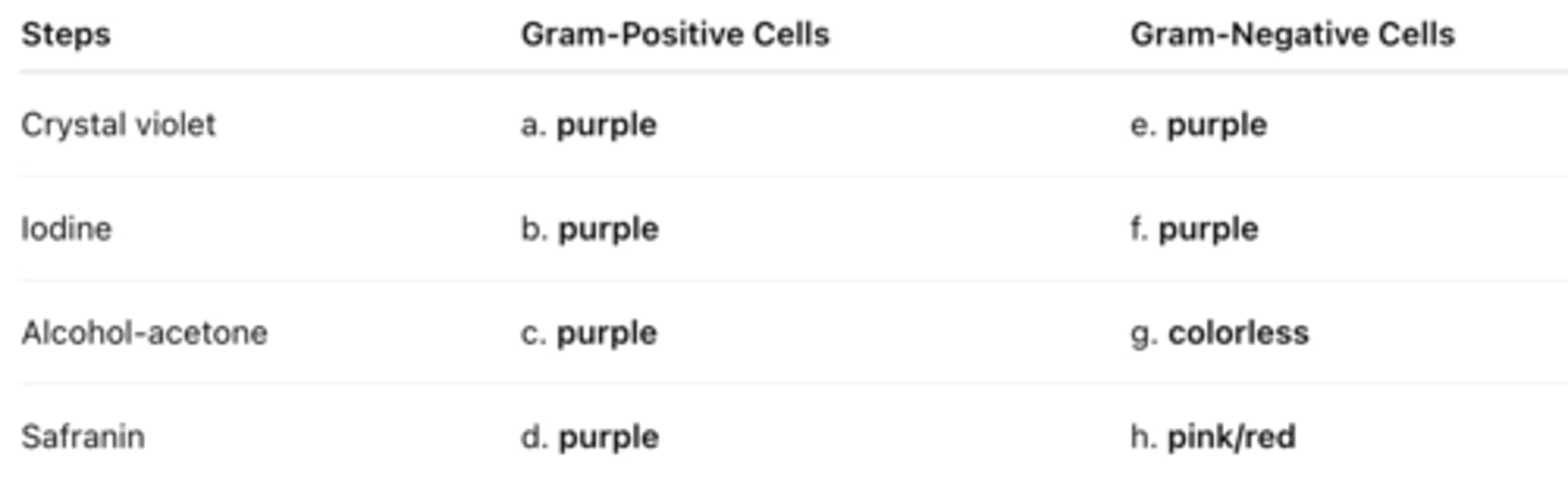
9. Fill in the following table regarding the Gram stain:
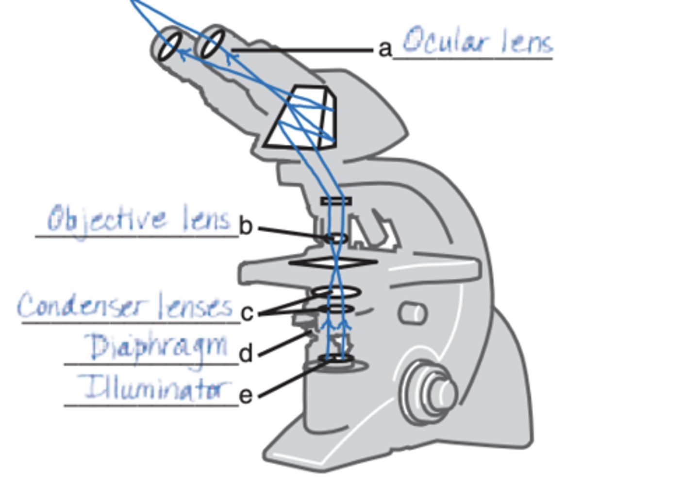
DRAW IT Label the parts of the compound light microscope in the following figure, and then draw the path of light from the illuminator to your eye.
Assume you stain Bacillus by applying malachite green with heat and then counterstain with safranin. Through the microscope, the green structures are
- cell walls.
- capsules.
- endospores.
- flagella.
- impossible to identify.
endospores.
Three-dimensional images of live cells can be produced with
- darkfield microscopy.
- fluorescence microscopy.
- transmission electron microscopy.
- confocal microscopy.
-phase-contrast microscopy.
confocal microscopy.
Carbolfuchsin can be used as a simple stain and a negative stain. As a simple stain, the pH is
-2.
- higher than the negative stain.
- lower than the negative stain.
- the same as the negative stain.
higher than the negative stain
Looking at the cell of a photosynthetic microorganism, you observe the chloroplasts are green in brightfield microscopy and red in fluorescence microscopy. You conclude:
- chlorophyll is fluorescent.
- the magnification has distorted the image.
- you're not looking at the same structure in both microscopes.
- the stain masked the green color.
-none of the above
chlorophyll is fluorescent.
Which of the following is not a functionally analogous pair of stains?
-nigrosin and malachite green
-crystal violet and carbolfuchsin
-safranin and methylene blue
-ethanol-acetone and acid-alcohol
-All of the above pairs are functionally analogous
-nigrosin and malachite green
Which of the following pairs is mismatched?
- capsule—negative stain
- cell arrangement—simple stain
- cell size—negative stain
- Gram stain—bacterial identification
- none of the above
none of the above
Assume you stain Clostridium by applying a basic stain, carbolfuchsin, with heat, decolorizing with acid-alcohol, and counterstaining with an acidic stain, nigrosin. Through the microscope, the endospores are 1 , and the cells are stained 2 .
- 1—red; 2—black
- 1—black; 2—colorless
- 1—colorless; 2—black
- 1—red; 2—colorless
- 1—black; 2—red
1—red; 2—colorless
Assume that you are viewing a Gram-stained field of red cocci and blue rods through the microscope. You can safely conclude that you have
- made a mistake in staining.
- two different species.
- old bacterial cells.
- young bacterial cells.
- none of the above
two different species.
In 1996, scientists described a new tapeworm parasite that had killed at least one person. The initial examination of the patient's abdominal mass was most likely made using
- brightfield microscopy.
- darkfield microscopy.
- electron microscopy.
- phase-contrast microscopy.
- fluorescence microscopy.
brightfield microscopy.
Which of the following is not a modification of a compound light microscope?
- brightfield microscopy
- darkfield microscopy
- electron microscopy
- phase-contrast microscopy
- fluorescence microscopy
electron microscopy