Approach to skin nodules and nodular dermatitis
1/42
There's no tags or description
Looks like no tags are added yet.
Name | Mastery | Learn | Test | Matching | Spaced | Call with Kai |
|---|
No analytics yet
Send a link to your students to track their progress
43 Terms
Define nodule
A circumscribed solid elevation greater than 1cm in diameter and usually extends into deeper layers of skin
What do nodules usually result from?
Inflammatory cells
Neoplastic cells
Deposition of fibrin or crystals (e.g.- calcinosis cutis)
What are some ddx of nodules?
How can you describe the clinical presentation of a nodule?
Location
Number
Size
Behaviour (acute vs gradual)
Aspect/clinical features
Warm and/or painful
Hard, soft, elastic, fluctuant, movable, fixed
Alopecic, smooth/rough surface
Ulcerated (possible presence of draining tracts)
Hyper/hypopigmented
How can you diagnose the cause of an nodule?
Cytology
FNA
Apposition (if ulcerated/dischargin)
Will either be diagnostic or direct you to further testing (culture, special stains, immunocytochemistry)
What steps may you take after cytological testing for a nodule?
Histology
Excision of whole nodule
Punch/wedge biopsy
Depends on clinical presentation
(aseptic collection of sample for culture)
What further diagnostic tests can be done on nodules?
Immunohistochemistry
Special stains
PCR
Biochem, urinalysis (e.g.- calcinosis cutis)
Serology (e.g. Leishmania, Toxoplasma, Neospora, Cryptococcus)
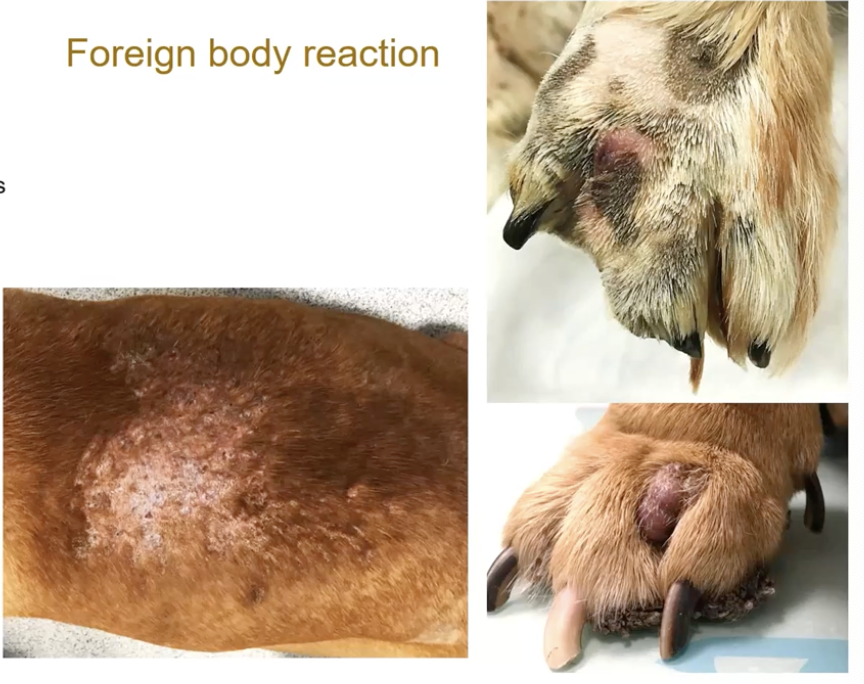
What are some common causes of foreign body reaction leading to nodules?
Plant material
Grass awns
Embedded insect mouth parts
Suture material
Porcupine quills
Endogenous: e.g.-
hair, sebum, keratin
Calcium salt
Tyrosine salt
What commonly causes an infectious nodule?
Abscesses
Penetrating wounds, bites, foreign bodies
Proliferation of bacteria involved
Dog: Staphylococcus
Cat: Polymicrobial
Pasteurella, Staph, Strep
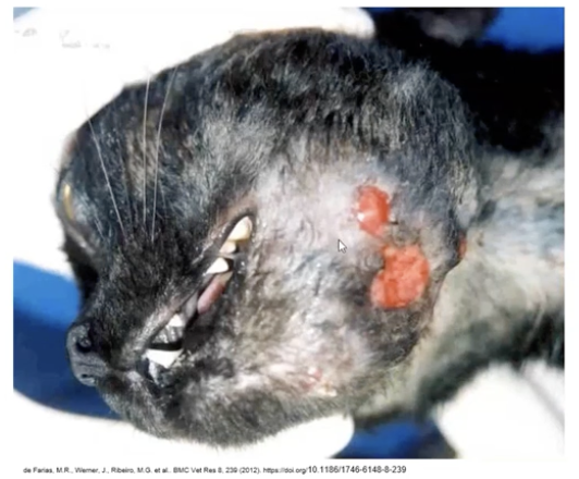
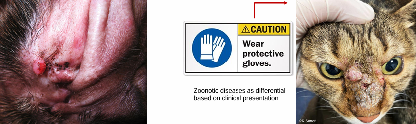
When you have nodules on the face in cats what should you consider?
Rodent bites so Mycobacteria and Poxvirus (more ulcerative than nodular)
What filamentous bacteria can cause nodules?
Actinomyces
Nocardia
Actinobacillus
What are the clinical signs of a nodule caused by filamentous bacteria?
Nodules and abcesses with ulcers, draining tracts and cellulitis
Anywhere in body usually from bite wounds or penetrating foreign bodies
Serosanguineous exudate
Possible systemic signs
How are nodules caused by filamentous bacteria diagnosed?
Cytology (gram, ZN stain)
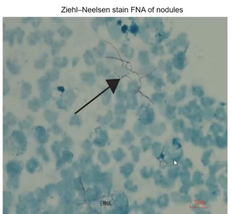
Histology
Culture
Molecular technique (PCR, gene sequencing, MALDI-TOF)
How are nodules caused by filamentous bacteria treated?
Surgical drainage and antimicrobial therapy
Long courses
What are the two main presentations of a nodule caused by mycobacteria (saprophytic)?
Dog: Canine leproid granuloma (Short coated breeds, boxers ++)
Cat: Feline leprosy syndrome (++Outdoor male cats)
What are the clinical signs of a mycobacteria nodule?
Single or multiple, firm, well-circumscribed nodule in skin or subcutis
Peripheral lymphadenomegaly
CLG usually self-limiting
FLS progressive and occasionally aggressive clinical course
(extremely fastidious and generally uncultivable)
How is a mycobacteria nodule diagnosed?
Cytology
Histology
Culture- generally uncultivable, needs specialist mycobacterial culture and subsequent genotyping at lab
PCR
How are CLG and FLS treated?
CLG- spontaenous regression in 1-3 months, can persist in immunocompromised
FLS- progressive, empirical combination of 2/3 antibiotics, surgical excision but possible recurrence
Long courses in some cases life long
What atypical/non tuberculous mycobacteria can cause nodules?
M. avium complex
M. fortuitum
M. thermoresistible
(Penetrating wounds in cats> dogs)
What are the clinical signs of nodules caused by atypical/ non tuberculous mycobacteria?
Granulomatous panniculitis: single/ multiple nodules, plaques, macules and diffuse swelling- multiple punctate ulcers and draining tracts
Pyogranulomatous lobular pneumonia
Disseminated systemic disease
How are atypical/ non tuberculous mycobacteria treated?
Empirical antibiotic treatment +/- surgical intervention
What is the main source of M tuberculosis mycobacteria causing nodules?
Mainly from infected wild rodents
Cats>dogs
(M.microti, M.bovis, M.tuberculosis)
What are the clinical signs of a nodule caused by M. tuberculosis?
Male outdoor cats- face, extremities, tail base, perineum
Firm nodules, ulcerations, non-healing wounds with draining tracts-> thick yellow to green fluid and systemic signs
Localised or generalised lymphadenopathy
How is a nodule caused by M. tuberculosis diagnosed?
Cytology
Histology
Culture- specialist and subsequent genotyping at lab
PCR
How is a nodule caused by M. tuberculosis treated?
Notifiable disease- owner might need to be screened at TB clinic
Euthanasia/ empirical multidrug regimens
What are the types of nodules caused by dermatophytes? What aetiological agents cause them?
Dermatophytic pseudomycetoma (Cats- persian and DLH+)
M. canis
Nodular dermatophytosis (Dogs)
M gypseum, T. mentagrophytes, M. canis
How does Dermatophytic pseudomycetoma present?
Deep dermal and or subcut infection
Painless, single or multifocal ulcerated dermal nodules, yellow granular discharge
No history of skin trauma
Neck, dorsum, tail, flanks or limbs
How is Dermatophytic pseudomycetoma diagnosed and treated?
D: cytology, histopath, culture from exudate or FNA
T: systemic antifungals (itraconazole, ketoconazole, terbinafine) +/- surgery
How does Nodular dermatophytosis present?
Single lesion commonly
Or multiple erythematous, alopecic, exudative nodules
On head, neck, limbs
How is Nodular dermatophytosis diagnosed and treated?
D: cytology, histopath, culture from exudate or fresh tissue
T: systemic antifungal (itroconazole, ketoconazole)
How do fungal subcutaneous nodules present?
Traumatic implant of saprophytic organisms on soil and vegetation
Significant tissue destruction and inflammation
Chronic and localised to disseminated in immunocompromised
How are subcutaneous fungal nodules diagnosed and trated?
D: cytology, histopath, fungal culture, PCR, Serology (important for cryptococcus)
T: Ideally based on culture, azoles main systemic antifungal, amphotericin B, terbinafine
What are some differnt types of fungal subcutaneous nodules?
Mycetomas- pyogranulomatous nodules that contain tissue grains or granules colonies of organisms and necrotic debris
Pigmented fungi
Unpigmented fungi composed of dense
Chromomycosis- subcutaneous and systemic disease associated with pigmented fungal elements
pigmented hyphal elements but NOT grains in tissues
Hyalohyphomycosis
Unpigmented
Cryptococcosis
In the environment (soil, trees, bird droppings)
Most common systemic
Sporotrichosis mycosis in cats
Emerging zoonotic disease – mainly in Latin America (tropical/subtropical climates) but spreading
Hunting dogs, outdoor cats ++
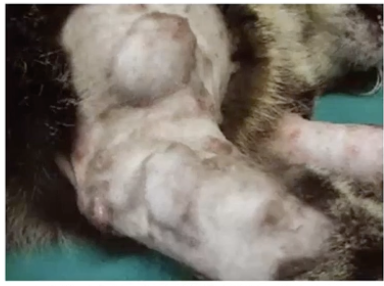
What protozoa can cause nodular skin lesion?
Toxoplasma gondii
Neospora caninum
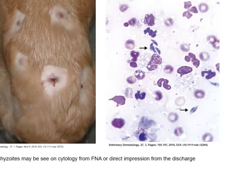
Leishmania spp
All rare
How can nodules due to protozoa be diagnosed?
Cytology
Histopath + IHC + PCR +/- DNA sequencing (distinction Toxo/Neospora can be challenging)
Serology
What parasite can cause nodular skin lesions?
Dirofilaria repens
Transmitted by mosquitoes
How can nodules due to parasites be diagnosed?
FNA can show microfilariae
Ultrasound evaluation
Histopathology
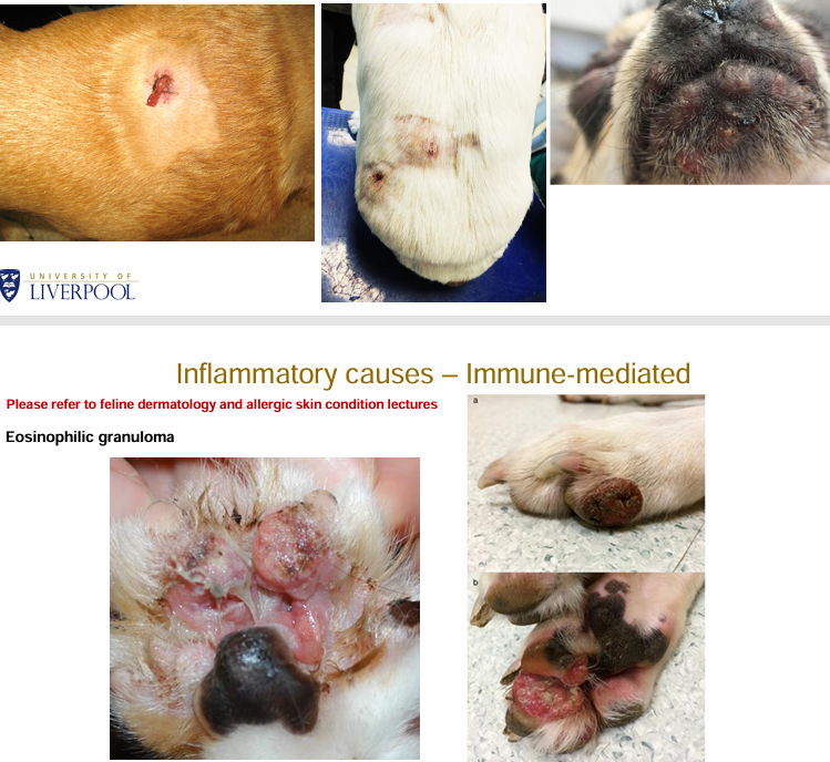
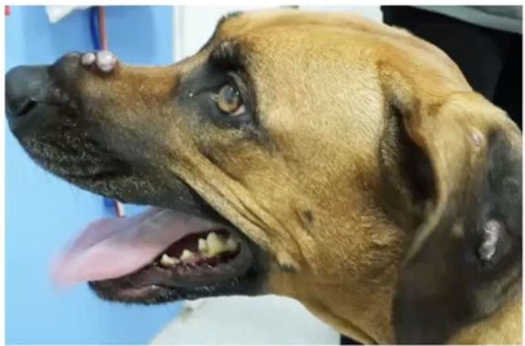
What immune mediated conditions can cause nodules?
(Juvenile) Sterile granulomatous dermatitis and lympadenitis
Sterile pyogranulomatous dermatitis and panniculitis
Eosinophilic granuloma
What are the two main types of histiocytic proliferative disorders?
Neoplastic
Histiocytomas (solitary lesions)
Canine cutaneous langerhans cell histiocytosis (multiple histiocytomas)
Histiocytic sarcoma
Feline progressive histiocytosis
Reactive
Cutaneous histiocytosis (inflammatory lymphohistiocytic proliferative disorder that primarily involves skin and subcutis)
Systemic histiocytosis (generalized histiocytic proliferative disease)
How does cutaneous histiocytosis present grossly?
multiple cutaneous and subcutaneous nodules up to 4 cm diameter – non-painful, non-pruritic
skin ulceration common
may disappear spontaneously or regress and appear at new sites simultaneously
face, nose, neck, trunk, extremities (including foot pads), perineum, and scrotum
How does systemic histiocytosis present grossly?
involves skin, ocular and nasal mucosae, and peripheral lymph nodes
lung, liver, bone marrow, spleen, peripheral and visceral lymph nodes, kidneys, testes, orbital tissues, nasal mucosa
How are reactive histiocytic proliferative disorders diagnosed?
histopath + IHC
How are reactive histiocytic proliferative disorders treated?
Glucocorticoids, ciclosporin