Lecture 3 - microscopy cultivation and pathogens
 Call Kai
 Learn
Learn Practice Test
 Spaced Repetition
 Match
Match Flashcards
 Knowt Play
1/22

There's no tags or description
Looks like no tags are added yet.
Name | Mastery | Learn | Test | Matching | Spaced | Call with Kai |
|---|
No analytics yet
Send a link to your students to track their progress
23 Terms
Define magnification, resolution.
ability to enlarge an image
the ability to distinguish two adjacent objects as distinct and seperate.
Limit of resolution for light microscope is about 0.2 μ m.
magnification of 1000x needed for 0.2nm diameter resolution
Different types of several types.
bright field, phase contrast, differential inferences contrast, dark field, fluroescence
What is the general structure of a microscope?
Compound light microscope uses visibel light to illuminate cells
The condenser focuses the light
Objective lense magnifies (10-100x)
Ocular lense magnifies (10-30x)
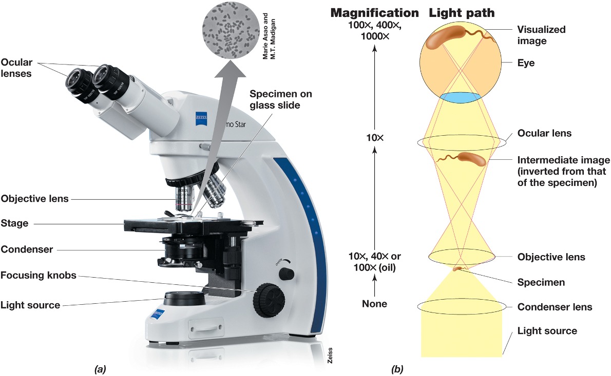
How do we calculate total magnification?
total magnification = objective magnification x ocular magnification
Explain what bright field scope is?
When specimens are visualized because of differences in contrast (absorbing or scattering light differently) between specimen and surrounding.
Pigmented microbes add contrast
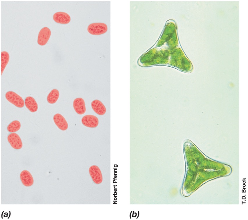
What is staining and how is it done?
staining increases the contrast for bright field microscopy and uses dyes that are organic compounds that binds to speciifc cellular materials.
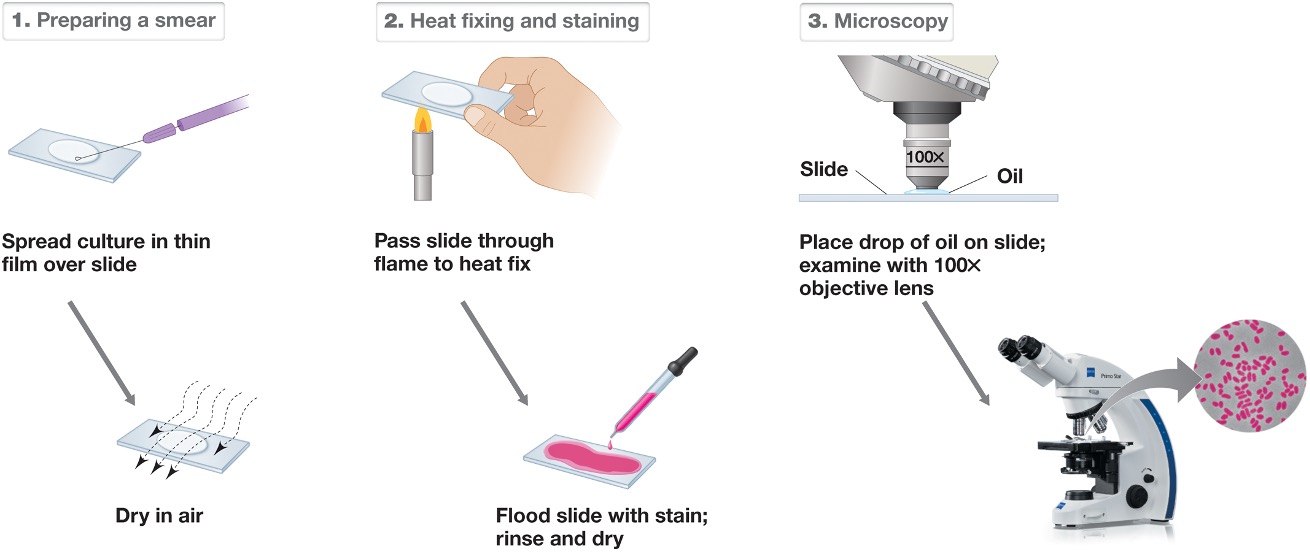
What are basic dyes?
positively charged, bind strongly to negatively charged cell components
examples are methylene blue
simple stains use dried cells
What is a differential stain? And what is an example?
Render different kinds of cells in different colors.
Gram stain is an example which causes differences because of cell wall structure
Gram positive bacteria appear purple and gram negative appear pink.
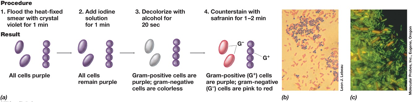
Explain the mechanism behind the Gram staining.
•Gram-positive bacteria have a thick peptidoglycan layer that retains the crystal violet-iodine complex, staining them purple.
•Gram-negative bacteria have a thin peptidoglycan layer and an outer membrane, which does not retain the crystal violet and instead takes up the counterstain (safranin).
What is phase contrast microscopy?
improves image contrast of unstained live cells
the phase ring amplifies differences in the refractive index af the cell
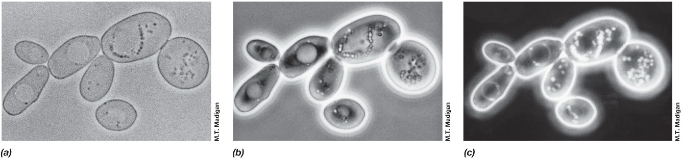
What is dark field microscopy?
Light reaches the specimen from the sides.
Only light scattered by specimen reaches the lens.
Image appears light on a dark background.
Better resolution than light microscopy.
Excellent for observing motility (flagella).
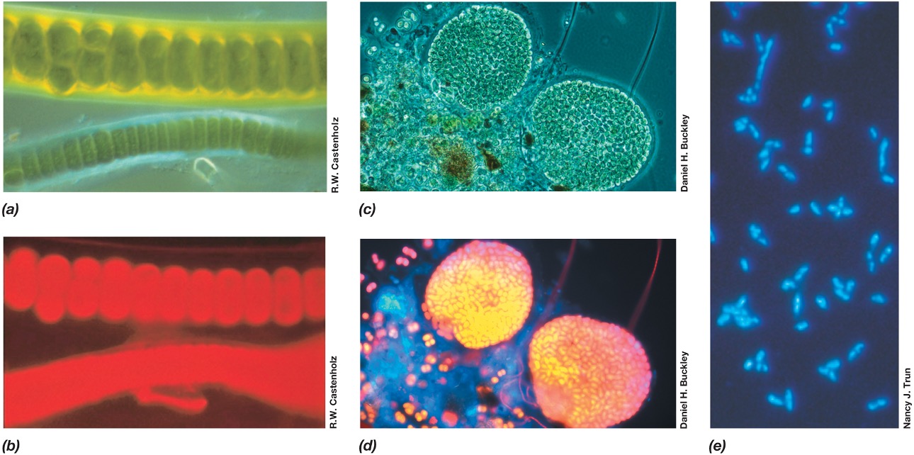
What is fluroescence microscopy?
Used to visualize specimens that fluoresce (emit light)
Cells appear to glow on black background due to filters.
Fluoresce naturally (autofluorescence) or after they have been stained with a fluorescent dye such as DAPI.
Widely used in clinical diagnostic microbiology and microbial ecology for enumerating bacteria.
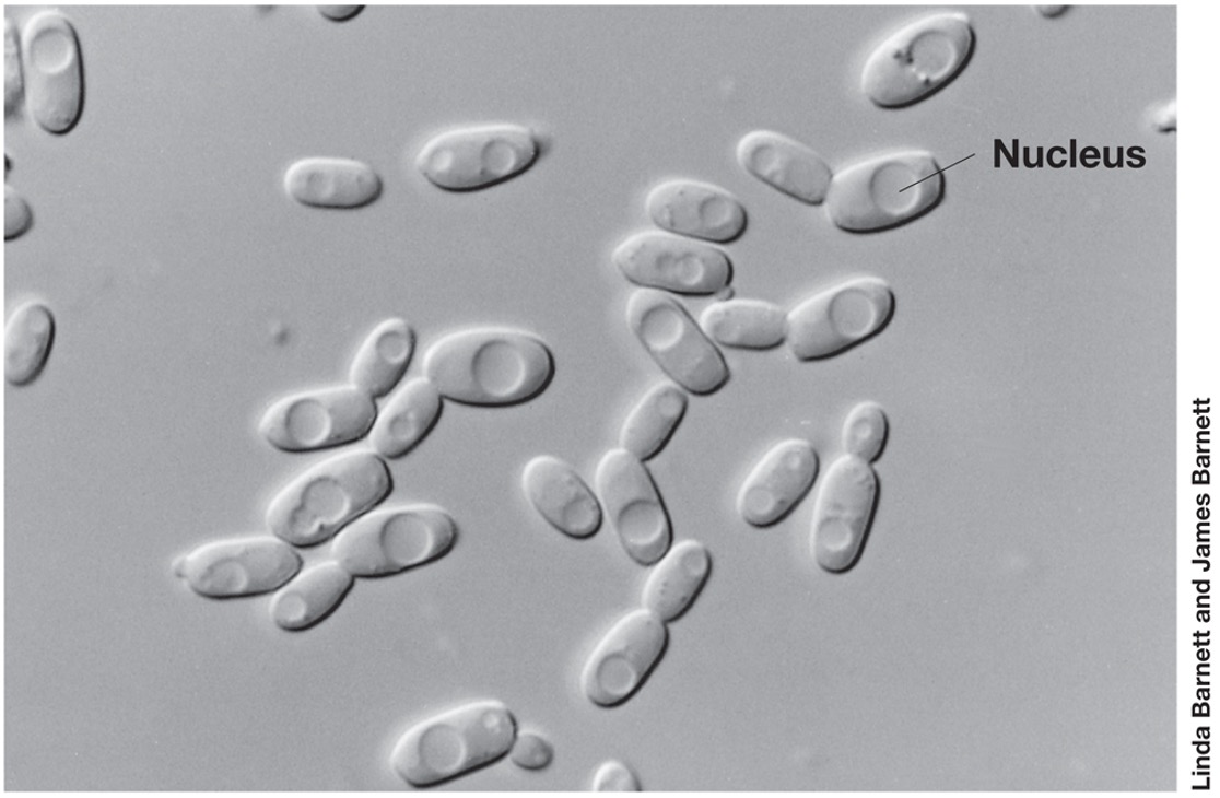
What is Differential interference contrast (DIC) microscopy?
uses a polarizer to create two distinct beams of polarized light (light in single plane)
gives structures such as nuclei and bacterial inclusions a three-dimensional appearance
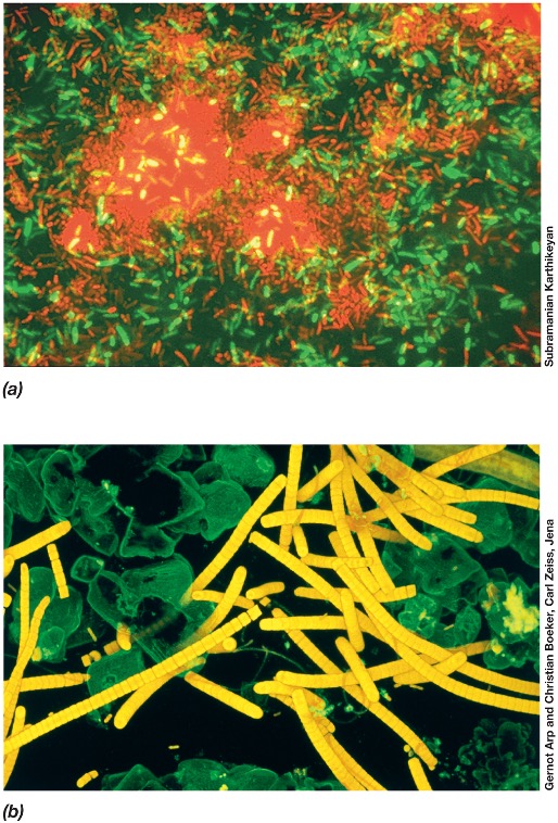
What is Confocal scanning laser microscopy (CSLM)??
using computerised fluorescent microscope coupled with laser source to make a 3D image
focus on single layers of specimen
3D image can be made
What is electron microscopy?
use of electrons instead of visible light to image cells and structures
electromagnets functioning as lenses
operation in vaccum
types: transmission/scanning electron microscope
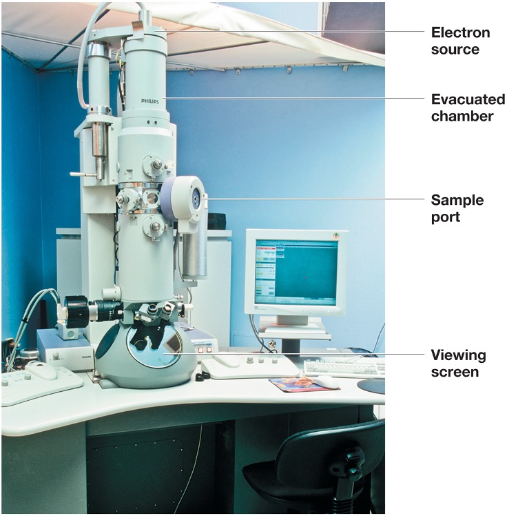
What is transmission electron microscopy?
Much greater resolving power (0.2 n m) than light microscope.
Enables visualization of structures at the molecular level.
Specimen must be very thin (20–60 n m) and stained with high atomic weight substances that scatter electrons well and improve contrast.
Negative staining allows direct observation of intact cells/components. (Figure 1.29b).
Electron cryotomography (cryo E T): T E M used to obtain three-dimensional images.
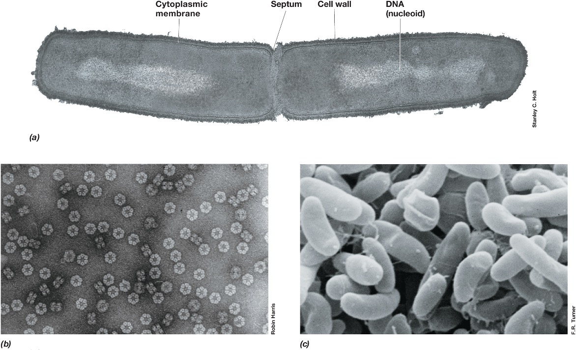
What is scanning electron microscope?
Specimen is coated with a thin film of heavy metal (e.g., gold).
An electron beam scans the object.
Scattered electrons are collected and projected to produce an image. (Figure 1.29c)
Even very large specimens can be observed.
magnification range of 15–100,000× only surface visualized
What is asepctic technique?
collection of practices that allow preparation and maintenance of sterile (no living organisms) media and solutions
What did louis pasteur discover?
discovered that living organisms discriminate between optical isomers
alcoholic fermentation was biological
diaproved theory of spontaneous generation
What did robert koch discover?
link between microbes and infectious diseases
identifies agents of TB etc.
developed solid media for obtaining pure cultures of microbes
etc.
Why did bacteria become excellent models of life?
Because they were easy to grow under controlled conditions.
How was the first tree of life made?
Ribosomal RNA present in all cells made it possible to build the first tree of life.
Woese discovered Archaea using this rRNA
Discovered that evolutionary relationships can be revealed by rRNA analysis
What is a phylogenetic tree?
depicts phylogeny (evolutionary history) of all cells
clearly shows three domains
Root is LUCA (last universal common ancestor)
evolution along two paths to form Bacteria and Archaea
Archaea later diverged to distinguish Eukaryafrom Archaea