The Ear & Eye
1/70
There's no tags or description
Looks like no tags are added yet.
Name | Mastery | Learn | Test | Matching | Spaced | Call with Kai |
|---|
No analytics yet
Send a link to your students to track their progress
71 Terms

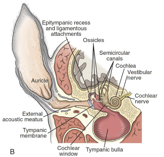
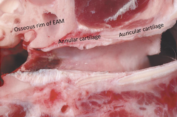
What structures make up the external ear, and how can it respond to injury?
The external ear consists of the auricle/pinna and the external acoustic meatus(ear canal), and it terminates at the tympanic membrane. Responses to injury include:
Inflammation
Epithelial and adnexal hyperplasia
Fibrosis
Osseous metaplasia/heterotopic ossification
Neoplasia
What is otitis externa and what is the histologic makeup of the external acoustic meatus?
Inflammation of the external acoustic meatus. The EAM is dermatologic tissue lined by:
Keratinized stratified squamous epithelium
Thin dermis
Sebaceous glands
Hair follicles
Ceruminous glands, which are more abundant medially than laterally
What categories of causes should be considered for otitis externa?
Predisposing causes
Primary causes
Secondary causes
What are the predisposing cause of otitis externa?
Conformation, excessive moisture, treatment effects, neoplasia and polyps, systemic disease
What are the primary causes of otitis externa?
Parasites, hypersensitivity reactions, keratinization disorders, foreign body, glandular disorders, autoimmune disease, vascular disease
What are the secondary causes of otitis externa?
Bacteria such as Staphylococcus, Proteus, Pseudomonas, E. coli, Klebsiella, and yeasts such as Malassezia and Candida albicans
What are the key features of Dermatophilus congolensis infection of the external ear?
Can affect any species
Zoonotic
Often transferred during nursing from dam
Traumatized inguinal skin of lactating dams can transmit infection to ears of nursing offspring
Flagellated zoospores invade epidermis, germinate into filamentous organisms, and damage/separate keratinocytes
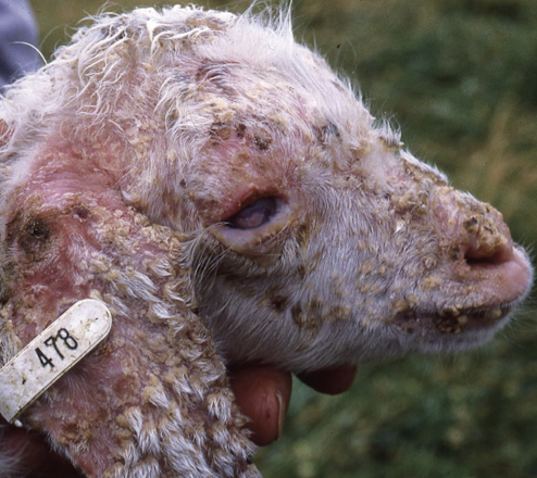
Histology of Dermatophilus congolensis infection of the external ear?
Intraepidermal pustular dermatitis
Superficial perivascular dermatitis
Folliculitis
What chronic responses to injury can occur in the external ear: Epithelial and adnexal hyperplasia?
Glandular change with increased ceruminous glands
Mentioned in Cocker Spaniels
What chronic responses to injury can occur in the external ear: Fibrosis?
Chronic lymphocytes
Fibroblasts
Collagen
Can lead to stenosis
What chronic responses to injury can occur in the external ear: Osseous metaplasia/heterotopic ossification?
Abrupt ossification arising from the auricular perichondral scaffold
What chronic responses to injury can occur in the external ear: Neoplasia?
Squamous cell carcinoma
Sebaceous gland tumors
Melanocytic tumors
Histiocytoma
Plasmacytoma
Mast cell tumor
Ceruminous gland tumor in cats
Trichoblastoma
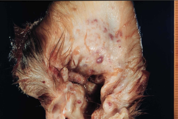
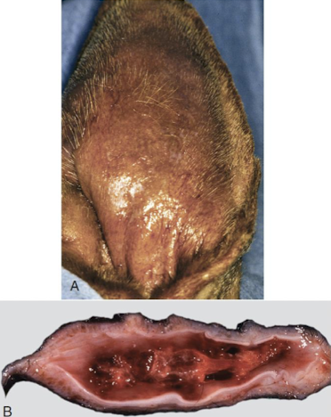
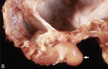
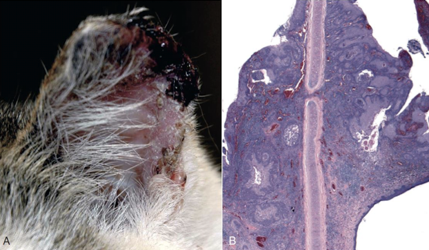
What is an aural hematoma?
Caused by ear discomfort leading to head shaking
Head shaking creates shearing forces on blood vessels
Can fracture auricular cartilage
Hemorrhage collects:
Intraparachondrially
Subparachondrially
Common in:
Pigs
Dogs
Cats
If untreated:
Heals by fibrosis
Causes a malformed auricle
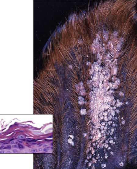
Aural plaques
Also called:
Equine ear papillomas
Papillary acanthoma
Hyperplastic dermatitis of the ear
Seen in horses under 1 year
Caused by Equus caballus papillomavirus (EcPV)
Spread by fly bite
Important histologic feature:
Koilocytes → giveaway that this is due to a virus
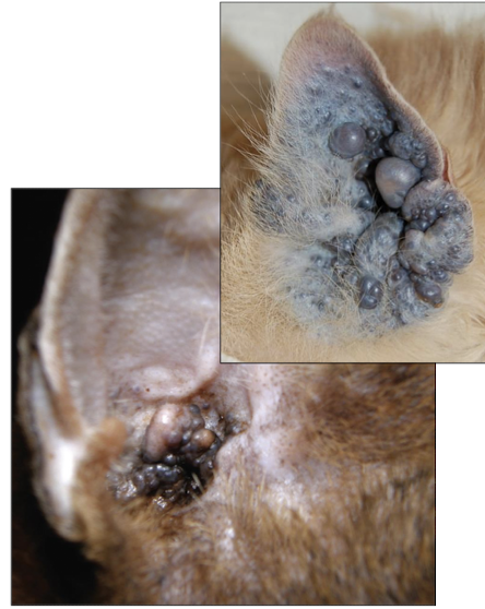
Feline ceruminous cystomatosis → “a cat has lots of little cysts coming from the ear wax glands.”
Benign lesion
Cystic, non-neoplastic proliferation of ceruminous glands
Located on:
Medial auricle
Ear base
May extend into the EAM
Especially seen in:
Abyssinian cats
Persian cats
Cause is unknown
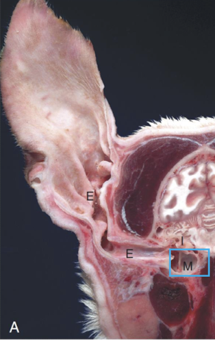
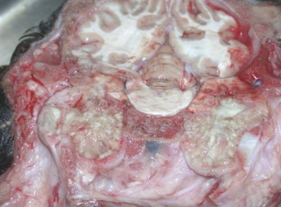
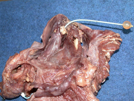
What is otognathia?
Rudimentary accessory mouth
Located at the base of the pinna
Seen in:
Sheep
Cattle
Orifice may be:
Blind
Continuous with the pharynx
May contain:
Rudimentary teeth
Mandible-like bones
Lateral tongue extensions
What are the main components and important features of the middle ear?
Tympanic membrane
Tympanic cavity
Air-filled
Surrounded by bone
Communicates with the pharynx via the auditory tube
Auditory tube
Allows migration of infectious organisms
Guttural pouches in horses
Enlarged diverticula of the auditory tube
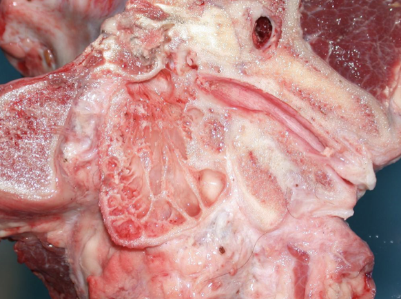
What species differences exist in the tympanic bulla and what is mucoperiosteum?
Septum bulla
Thin bony septum partially dividing the tympanic cavity
Cats
“Complete” septum
Dorsolateral and ventromedial compartments
Dogs
Incomplete septum
Single cavity
Bovids, goats, camelids
Septate
Multiple compartments
Horses
No prominent septum
Mucoperiosteum
Fused mucosa and periosteal lining of the bony middle ear surfaces
What ossicles are found in the middle ear?
Malleus
Incus
Stapes
What is otitis media?
Inflammation of the middle ear
Involves inflammation of the mucoperiosteum
How can otitis media develop, and what organisms are associated with it?
Descending infection
From the external auditory meatus
Often occurs from otitis externa in dogs
Systemic/hematogenous infection
Mycoplasmopsis bovis in cows
M. hyorhinis in pigs
Histophilus somni in cows
Ascending infection via the auditory tube
Trueperella pyogenes in pigs and cows
Pasteurella multocida in pigs and cows
What diseases affect the auditory tube and guttural pouch?
Salpinx / salpingitis
Guttural tympany (air)
Due to obstruction of outflow via the auditory tube
Empyema (pus)
Aspergillus infection
Especially on the dorsal and caudal wall
Carotid hemorrhage
Important complication
What are the key features of primary ciliary dyskinesia affecting the middle ear?
Genetic abnormal cilia movement
Seen in dogs under 1 year
Causes:
Cough
Persistent nasal discharge
Due to ineffective mucociliary clearance → cilia do not work
Ciliated epithelial cells normally clear fluid and debris in the auditory tube
Results in:
Unilateral otitis media
Bilateral otitis media
Sterile gelatinous material in the tympanic cavity
Aural (nasopharyngeal) polyp
Most common in cats under 2 years
Less frequent in dogs
Reported in one horse
Proposed causes:
Chronic upper respiratory infection
Otitis media
Ascending middle ear infection
Congenital defects
Gross appearance:
Pedunculated
Polypoid
Smooth surface
Develops from ciliated epithelium in the middle ear
May:
Remain confined to the middle ear
Extend through the auditory tube and obstruct the pharynx
Extend through a ruptured tympanic membrane into the EAM
In the EAM, may be visualized as a mass

Tympanokeratoma
Previously called:
Epidermoid inclusion cyst
Aural cholesteatoma
Seen in:
Dogs
Horses
Develops secondary to chronic inflammation
Chronic inflammation causes squamous metaplasia
Respiratory epithelium becomes stratified squamous epithelium
Contains:
Keratin debris
Cholesterol
What neoplasms can affect the middle ear or guttural pouch region?
Squamous cell carcinoma
Frequent in cats
Can arise in the guttural pouch of horses
Adenocarcinoma
What is otitis interna and what are its complications?
Also called labyrinthitis
Inflammation of the inner ear
May be accompanied by osteomyelitis of the petrous temporal bone
Usually extends from otitis media via the cochlear window
Sequelae include retrograde spread through the internal auditory meatus, causing:
Meningitis
Encephalitis
What are important causes of congenital hearing loss and deafness?
Albinotic congenital deafness
Due to dysfunction of intermediate cells
Causes abnormal endolymph
Leads to degeneration of cochlear hair cells
Can be:
Unilateral
Bilateral
Affects:
Up to 50% of white-coated, blue-eyed cats
About 30% of Dalmatians with MITF mutation
About 90% of horses with endothelin B receptor mutation in lethal white foal syndrome
What are acquired causes of hearing loss and deafness?
Acoustic noise trauma
Radiation
Ototoxicity
Aminoglycosides
Platinum-containing chemotherapy agents
Furosemide
Salicylates
Primary target:
Hair cells
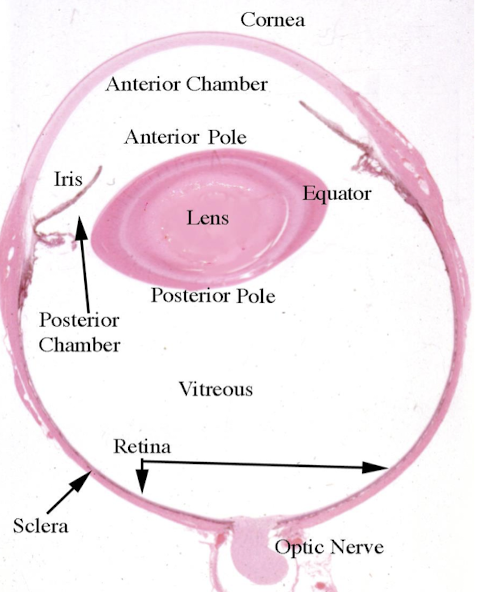
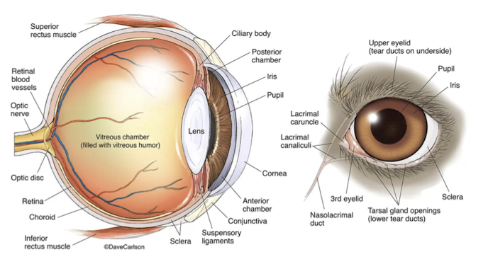
What are the major structures involved in eye form and function?
Globe (bulbi)
Optic nerve (CN II)
Accessory structures / adnexa:
Eyelids
Lacrimal apparatus
Sac
Duct
Orbital connective tissue
Extraorbital muscles
What are the major abnormalities of eyeball size?
Small eye
Microphthalmia = congenital
Phthisis bulbi = acquired
Large eye
Buphthalmus
Typically associated with glaucoma
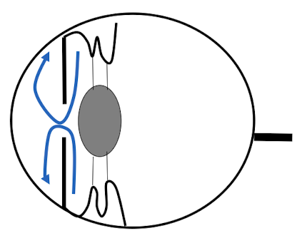
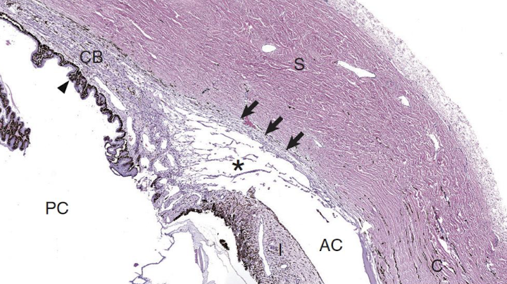
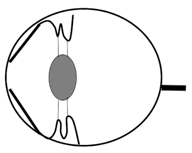
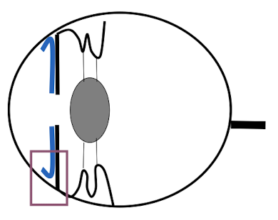
What is the normal flow of aqueous humor?
Produced by the ciliary body
Enters the posterior chamber
Passes through the pupil
Enters the anterior chamber
Drains through the iridocorneal / filtration angle
Then passes through:
Pectinate ligaments
Trabecular meshwork
Corneoscleral meshwork
Scleral vasculature
horses enter through posterior chamber
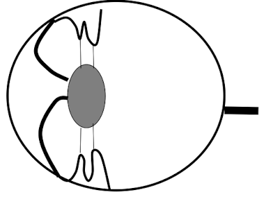
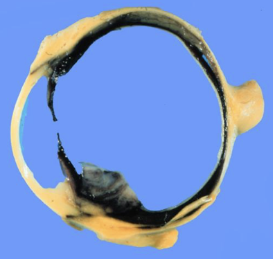
What is glaucoma?
Accumulation of aqueous humor
Causes increased intraocular pressure

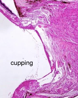
What are the major signs of glaucoma?
Buphthalmus
Retinal changes
Atrophy
Cupping of the optic disc
Degeneration of the optic nerve (in severe cases)
Uveal changes
Retraction and collapse of the filtration angle
Corneal changes
Edema from endothelial loss
Striae from breaks in Descemet’s membrane
Exposure keratitis
do not need all of these to diagnose glaucoma
What are the three major mechanisms of glaucoma?
Increased fluid production
Altered flow at the pupil
Reduced outflow
Most common mechanism
How can increased aqueous humor production cause glaucoma?
Inflammation, especially anterior uveitis, breaks down the blood-aqueous barrier
This increases ciliary body vascular permeability
Protein and fluid leak into aqueous humor
In dogs:
Ciliary body neoplasms can increase production
In cats:
Hypertension can increase ciliary body perfusion
What lesion can alter aqueous flow at the pupil and contribute to glaucoma?
Anterior synechia
What pupil-flow abnormalities can lead to glaucoma?
Posterior synechia with iris bombe
What is primary glaucoma due to obstructed outflow?
Most common
Can be congenital
Caused by goniodysgenesis
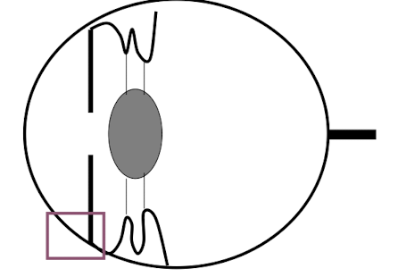
What is an important cause of secondary glaucoma due to obstructed outflow?
From inflammation
Preiridial fibrovascular membrane (PIFM)
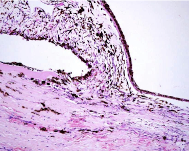
What are the causes and related lesions of PIFM?
Cytokine mediated
Especially associated with VEGF
Associated conditions:
Uveitis
Retinal detachment
Ciliary body tumor
Related lesion:
Retrocorneal fibrovascular membrane
Similar to PIFM
Occurs on the posterior limiting lamina / Descemet membrane
What findings may accompany obstructed outflow glaucoma with PIFM?
Preiridial fibrovascular membrane
Hyphema → inc blood in anterior chamber
Exudates → cellular debris
Plasmoid aqueous → too much protein, cells cannot settle
What are other causes of secondary glaucoma with obstructed outflow?
Neoplasia
Lens luxation
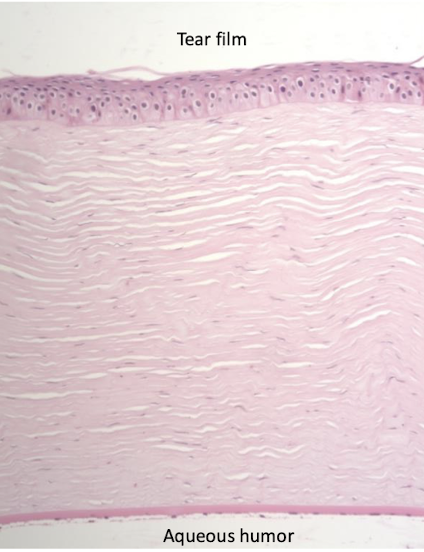
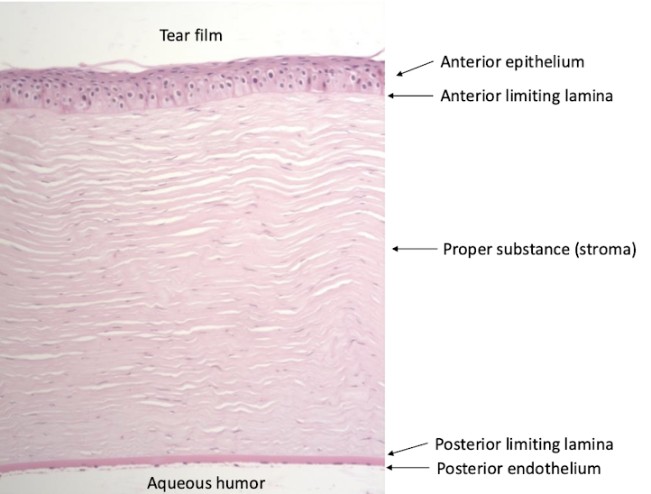
layers of the cornea
Anterior epithelium
Anterior limiting lamina
Proper substance / stroma
Posterior limiting lamina
Posterior endothelium
Tear film lies externally
Aqueous humor lies internally
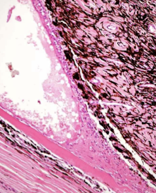
What are the major categories of corneal disease?
Keratitis
Ulceration
Neoplasia
What infectious causes of keratitis are discussed?
Infectious keratoconjunctivitis by Moraxella bovis
Bacterial adhesins attach to corneal epithelium
Cytotoxins cause epithelial ulceration
Causes neutrophilic inflammation
Feline herpesvirus-1
Viral replication causes epithelial necrosis
Causes ulceration
Can lead to chronic keratitis
Can cause corneal scarring
Sequela of FHV-1
Corneal sequestru = Necrosis of corneal stroma, Pigment accumulation
Forms a well-demarcated plaque
Corneal sequestrum in a cat → FHV-1
Necrosis of corneal stroma with accumulation pigment (well demarcated plaque)
Corneal ulceration Tears provide nutrition to the cornea
Minor epithelial defects heal by:
Epithelial sliding
Regeneration
Ulceration may lead to:
Vascularization
Pigmentation
Descemetocele
Bulging of Descemet’s membrane / posterior limiting lamina
Iris prolapse
Uncomplicated ulcer heals in 3-5 days
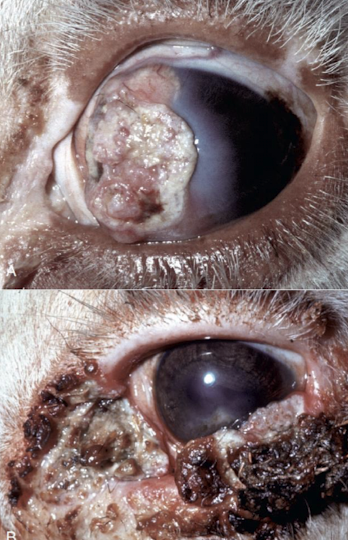
Corneal neoplasms
Squamous cell carcinoma (pictures)
Occurs in cattle, horses, cats, and dogs
Associated with chronic UV exposure
Most common at the corneoscleral / limbal junction
Limbal melanoma in dogs
Arises from melanocytes at the corneoscleral junction
Benign
What are the developmental and structural features of the lens?
Lens is a modified epithelial structure
Single layer of cuboidal epithelium is present only on the anterior lens
Equator remains mitotically active
Equator produces lens fibers throughout life
Lens fibers are elongated epithelial cells
Lens is avascular
Lens depends on aqueous humor for nutrients
Lens transparency depends on:
Highly ordered fibers
Proper dehydration state
Lack of organelles
Which part of the lens has no epithelium?
Posterior lens
What are lens fibers made up of?
elongated epithelial cells that lose nuceli + organelles
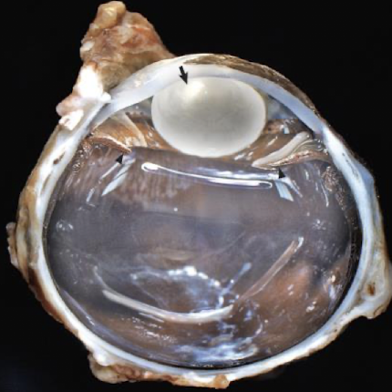
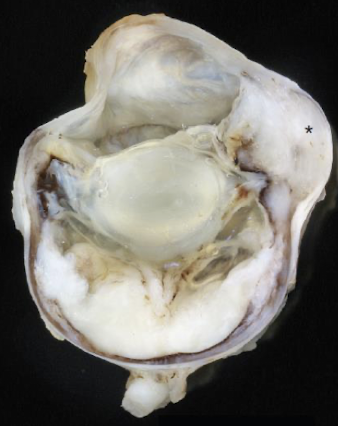
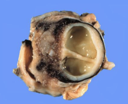
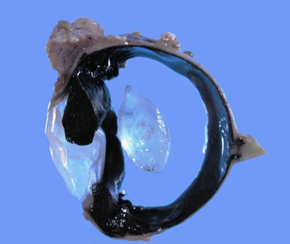
What is lens luxation?
Displacement of the lens due to zonular ligament failure
Anterior luxation of the lens
Lens moves into the anterior chamber
Emergency → leads to acute glaucoma
Posterior luxation of the lens
Lens falls into the vitreous
Causes of lens luxation
Primary
Breed-related zonular degeneration
Example: terriers
Secondary
Uveitis
Glaucoma
Trauma
Pathogenesis of lens luxation
Zonular breakdown → Loss of lens support → Increased lens mobility
Anterior lens luxation
How do diabetic cataracts develop?
Caused by osmotic injury
High glucose is converted to sorbitol via aldose reductase
Sorbitol cannot diffuse out
Sorbitol accumulates in the lens
Water enters the lens
Causes:
Fiber swelling
Fiber rupture
Opacity (cataract)
Features:
Onset in days to weeks
Usually bilateral
Common in dogs
Rare in cats
What is feline post-traumatic ocular sarcoma?
Also called:
Ocular posttraumatic lens tumor
FPTOS
Develops after lens capsule rupture exposes lens proteins
Chronic inflammation promotes neoplastic transformation
Likely arises from:
Lens epithelial cells
Mesenchymal transformation
Locally invasive:
Invades globe
Extends along optic nerve
Can metastasize
Histology:
Spindle cell sarcoma
May have lens rupture or lens remnants
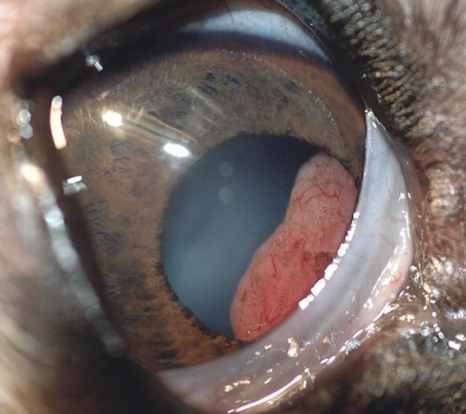
ocular posttraumatic lens tumor in a cat
wide range of where the neoplastic cells will go
What structures make up the uvea?
Uvea is the vascular tunic
Anterior uvea
Iris
Ciliary body
Posterior uvea
Choroid
Blood-eye barrier is formed by endothelial cells of iridial vessels
What are the major types of uveal inflammation?
Iritis
Iris
Cyclitis
Ciliary body
Anterior uveitis / iridocyclitis
Anterior segment
Choroiditis
Choroid
Panuveitis
Choroid + ciliary body + iris
Ophthalmitis
All parts of the eye
What inflammatory patterns and causes are associated with uveal disease?
Suppurative
Bacteria
FIP
Lymphocytic plasmacytic
Idiopathic in cats
Malignant catarrhal fever in cattle, sheep, and goats → blue eye
Granulomatous
Fungi
Yeasts
Lens-induced
Phacolytic
Lens protein leaks out of an intact capsule
Phacoclastic
Lens ruptures
What neoplasms can affect the uvea?
Anterior uveal melanoma
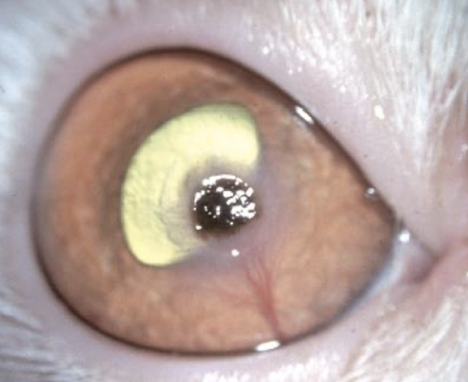
Diffuse iris melanoma in cats
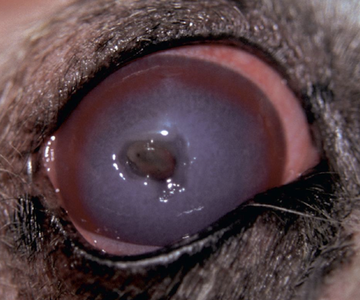
Iris melanoma in dogs
Ciliary body tumor
diffuse iris melanoma in a cat
iris melanoma in a dog
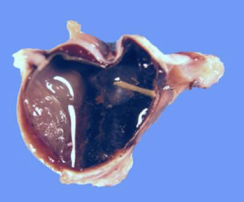
Ocular trauma caused by a foreign body
ciliary body tumor in a dog