Medsci 201_Alimentary system (stomach, SI, Colon, Liver)
1/93
There's no tags or description
Looks like no tags are added yet.
Name | Mastery | Learn | Test | Matching | Spaced | Call with Kai |
|---|
No analytics yet
Send a link to your students to track their progress
94 Terms
Changes from Pyloric and Duodenum transition zone are:
1. Gastric rugae --> intestinal plicae
2. Gastic pits & glands--> intestinal villi & crypts
3. Secretory epithelim--> simple columnar absorptive enterocytes + goblet cells+paneth cells (defense)
4. Enteroendocrine cells secrete different hormones (gastric--> cholecytokinin)
5. Brunner's glands
Chief cells produce
Pepsinogen production
Parietal cells produce
HCL & intrinsic factor
Intrinsic factor function?
secretion of parietal cells that help absorb B12 vitimin across intestine lining
Enteroendocrine cells produce
Gastrin
Structures found in the core of the intestinal villus
Lacteal
Blood vessels (venules & arterioles)
Functions of Microvilli in SMALL intestine
1. Increase surface area for absorption
2. Glycocalyx coating on top of microvilli bind desired moledules and barrier from undesirable molecules
3. Actin filaments connected to cytoskeleton - locased movement help to mix contents
Give the name of the nerve plexus located between the two layers of muscularis externa.
Myentric plexus
Is there skeletal muscle present in the muscularis externa?
In some regions of the gut tube skeletal muscle is present in the muscularis externa to enable voluntary movement (e.g. in upper oesophagus for swallowing, or the external anal sphincter for controlling defecation).
Origin of undifferentiated epithelial cells of oesophagus?
Undifferentiated (generative/stem) epithelial cells arise from the basal layer of the epithelium.
The muscularis mucosae consists of what type of muscles?
Smooth muscles
Adventitia vs. Serosa
Adventitia is located between two structures (eg. the trachea and the oesophagus).
Serosa lines a body cavity/peritoneal space.
The outermost layer of the abdominal oesophagus is called by ______, which is lined by _____.
Serosa, mesothelial cells
How many tunics in digestive system?
4 layers(tunics)
Name 4 tunics of digestive system
1. Mucosa (epithelium + Lamina propria + Muscularis mucosae)
2. Submucosa
3. Muscularis externa (inner circular + longitudinal outer)
4. Adventitia/Serosa
The outermost connective tissue covering of any organs, vessels or other structure?
Adventitia
Also Known as visceral peritoneum.. what is this?
Serosa
two layered slippery outer covering
Serosa
Outer________ of Serosa sitting on a bed of connective tissue.
mesothelium
Accumulation of fluid; abdominal swelling;distortion of visceral organs. What is this condition called?
Ascites
Complication from surgery, inflammation interferes with normal functioning
Peritonitis
General organization of the alimentary canal
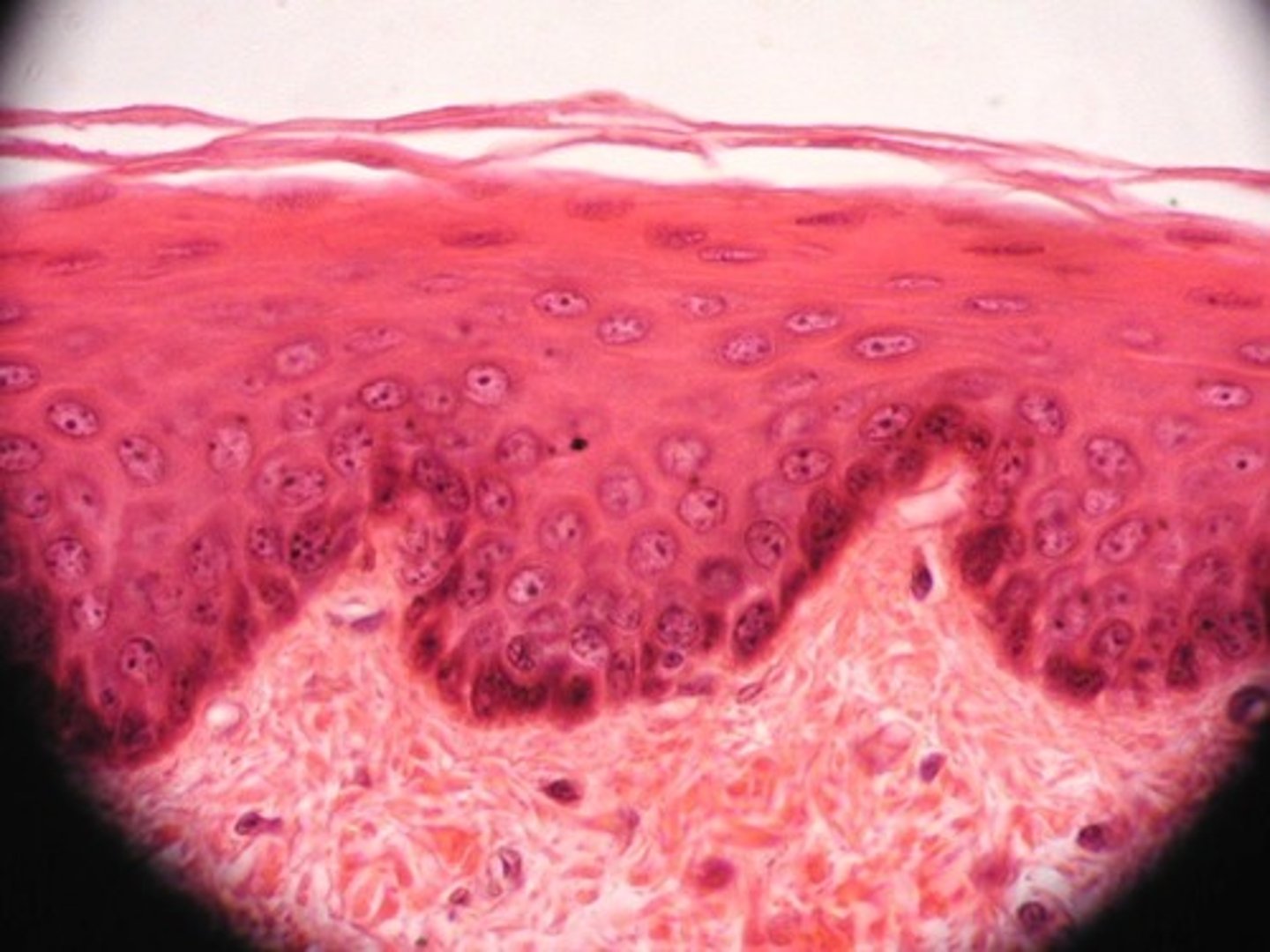
Epithelium of Oesophagus
Stratified squamous epithelium
Oesophagus function
Rapid transport (peristalsis) of food to stomach
Length Oesophgus
25cm long, straight tube, collapsed outline with folds of submucosa when empty
Epithelium of Oesophagus
Non-keratinised stratified squamous epithelium(6-8 layers)
Epithelium of Oesophagus transition to______towards stomach
Simple cuboidal/columnar
Muscularis mucosae in Oesophagus is_____.
Absent/rare but developed near stomach. Longitudinal smooth muscle discontinuous in places
Submucosa of Esophagus is_____________________.
Loose and irregular connective tissue.
Are there glands in Esophgus?
yes.
Muscularis externa of Esophagus move food by ____ and _____.
Peristalsis and Segmentation
Characteristics of Muscularis Externa of Esophagus
2 Thick coats (inner:spiral/oblique and outer:iregularly arranged)
Mostly smooth muscles but, towards mouth some skeletal.
Covering of Esophagus
Adventitia majority, except 1-2cm between diaphragm and stomach
Nerves of Esophagus
Enteric and autonomic nervous system
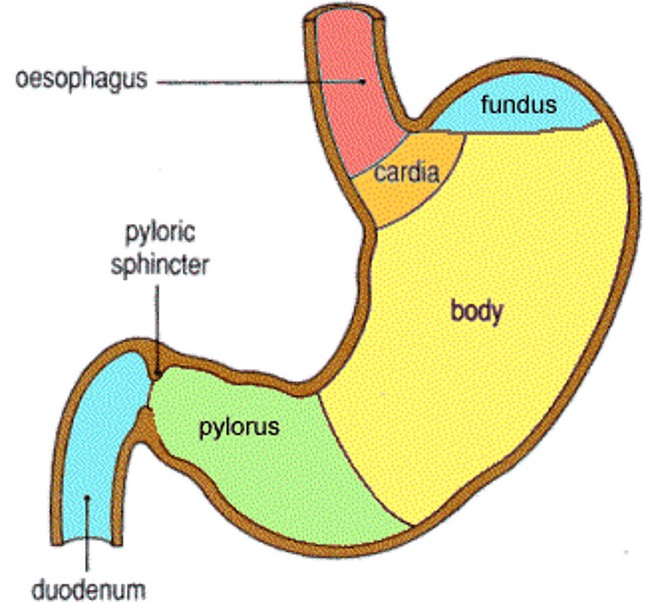
Storage tank, digestive processes of secretion and digestion and some absorption, what am I?
Stomach
3 regions of stomach
Mucus, acid, enzymes and hormones - Fundus/body
Mostly mucous - Cardia
Mucus, enzymes, hormone - Pylorus
3 muscle layers of stomach
Longitudinal
circular
Olbique (innermost)
Transient fold is called ----
Rugae
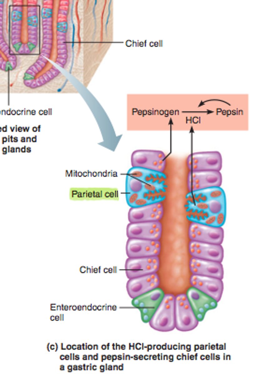
Mucous neck cells
produce mucus to lubricate the food entering the stomach
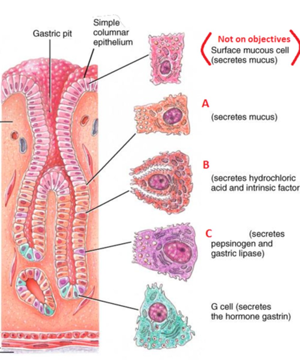
Parietal cells, function
Fried egg looking cell.
secretes HCL, denatures proteins, converts pepsinogen into pepsin
Chief cell, function
production and secretion of pepsinogen; secretion stimulated by ACh and HCl
G cell, function
hormone gastrin (regulatory function)
Simple columnar mucous cells (of surface and pits) of stomach, function
Secreates insoluble alkaline
Elongated nucleus
3 protective mechanisms of stomach cells
1. Mucous gel/surface coat (alkaline)
2. HCO3-
3. High cell turnover via undifferentiated stem cells
Mucous neck cell of gastric glands, function
Soluble, acidic glycoproteins secreted upon food.
Mucous granules less densely packed
Parietal cell
Cells found in gastric glands that secrete hydrochloric acid (for hydrolysis of ingested food) and gastric intrinsic factor (for absorption of vitamin B-12).
secrete HCl
OH- -------> HCO3- in exchange for CL-
HCO3- diffuses into venous blood leaving stomach
Helico Bacter Pyori
Ammonia produced attacks HCO3- of Mucous gel, surface coat.
Toxin blocks/attacks Mucous gel, surface coat.
What inhibits Prostaglandin E2 production?
NSAIDS - aspirin, non steroid anti-inflammatory drugs
Prostaglandin E2
Natural defense of the stomach
Stimulates Mucous gel/surface coat
TBC
Enteroendocrine cell
secrete hormones
Also called G-Cells. It is a secretory cell in the gastric gland of the stomach that secretes paracrine factors and the hormone Gastrin.
Located deep in the gastric glands.
Platelet Activating Factor produced by which bacteria?
Helico Bacter Pyloris
Parietal cell roles
1. Sterilise food
2. Acidify environment (important for Pepsinogen converting to Pepsin)
3.
Not enough acid--->
Gastritis
Intrinsic factor important for......
VtB12
VtB12 involved in
RBC Haemotopoiesis (RBC synthesis)
If low, Pernicious Anaemia
Overactive parietal cells
Exam question
Enteroendocrine cells (secretes hormone released from Basal Lamina) release granules containing _______. ______, _______.
Gastrin - stimulates all cells above (mucosal replacement, activates everything) ---> Activates ECL cells
Gastrin ----> Activates Somatostatin--->negative feedback inhibit ECL cells
Intrinsic factor important for......
Vt12
The fold in small intestine is called _____.
Plica (mucosal fold)
Cells in Villus region in small intestine that absorb small molecules resulting from digestion
Columnar absorptive cell (enterocytes)
Membrane of small intestine is studded with digestive enzymes such as
glucosidases (for breaking down carbohydrates)
Goblet cells in small intestine produce _________ granules filling apical cytolasm.
Mucinogen
Enteroendocrine cells in small intestine produces
Cholecytokinin
Gastrin, histamine, endorphins, serotonin, cholecystokinin, and somatostatin are hormones that are released directly into the lamina propria. Which of the following cell types synthesize and secrete these products?
Paneth cells in Small Intestine(SI), location?
Bottom of crypt of Liberkuhn (intestinal gland)
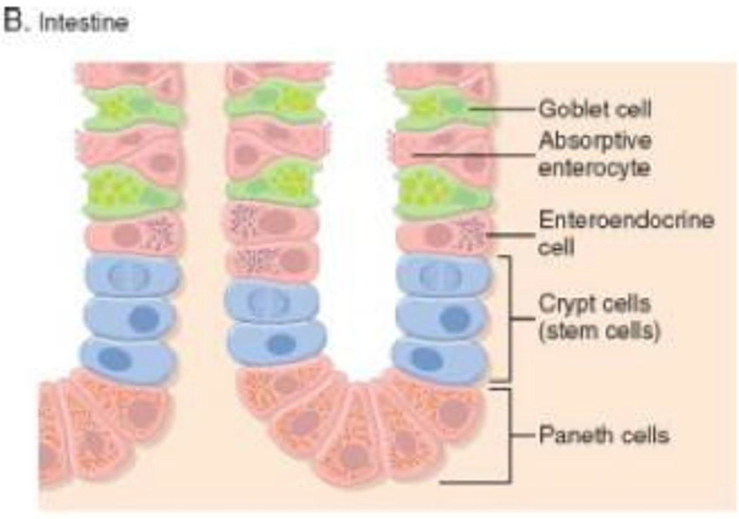
Paneth cells secret.. name 3;
TNF-alpha: produce inflammatory response
Lysozyme: bactericidal
Defensins: increase membrane permeability of invading organisms (causing cell leakage)
Duodenum: name 4 feathres
Brunner's glands-produce alkaline mucous
Lots of HCO3-
C-shaped
Shortest
pH 1-2
Jejunum: features
Lots of Plicae and Villi for absorption
Straight and 2-5m long
Suspended by mesentary
pH 7-8
Ileum
Peyer's patches (brown coloured lymphatic nodules)
Defence!
3-5m long
The ileocecal valve regulates the passage of materials into this expanded pouch. What is this?
Caecum
In colon, mucosa has plicae or villi like small intestine?
NO, in colon the mucosa does not have plicae or villi. However, mucosal glands (crypts of liberkuhn) are numerous.
Name 2 cell types of Colon mucosa
Columnar absorptive cells and Goblet cells
In Colon, Stem cells in Crypt (mucosal gland) replaces the cells above or below?
Above
In Colon, the Lamina propria contains a dense layer of _______ immediately beneath the surface epithelium(simple columnar).
Collagen
In Colon, the Teniae Coli is present in which part of muscularis externa?
Longitudinal outer
In colon, it is mostly covered by Serosa except for....
Posterior surface of large intestine, that area covered by adventitia.
Internal anal sphincter is composed of _____.
Smooth muscle
External anal sphincter is composed of ____.
Skeletal muscle
End of anal canal is
Anus
Esophagus: cell renewal location/ replacement rate
Basal part of epithelium/ days
Stomach: cell renewal location/ replacement rate
Near neck of gastric glands/ surface in days, deeper in months (as metabolically expensive to replace too often)
Small Intestine: cell renewal location/ replacement rate
Crypts of Liberkuhn/ surface in days, deeper in months
Large intestine: cell renewal location/ replacement rate
Lower 1/3 of glands (Crypts of liberkuhn)/ surface cells only and in days
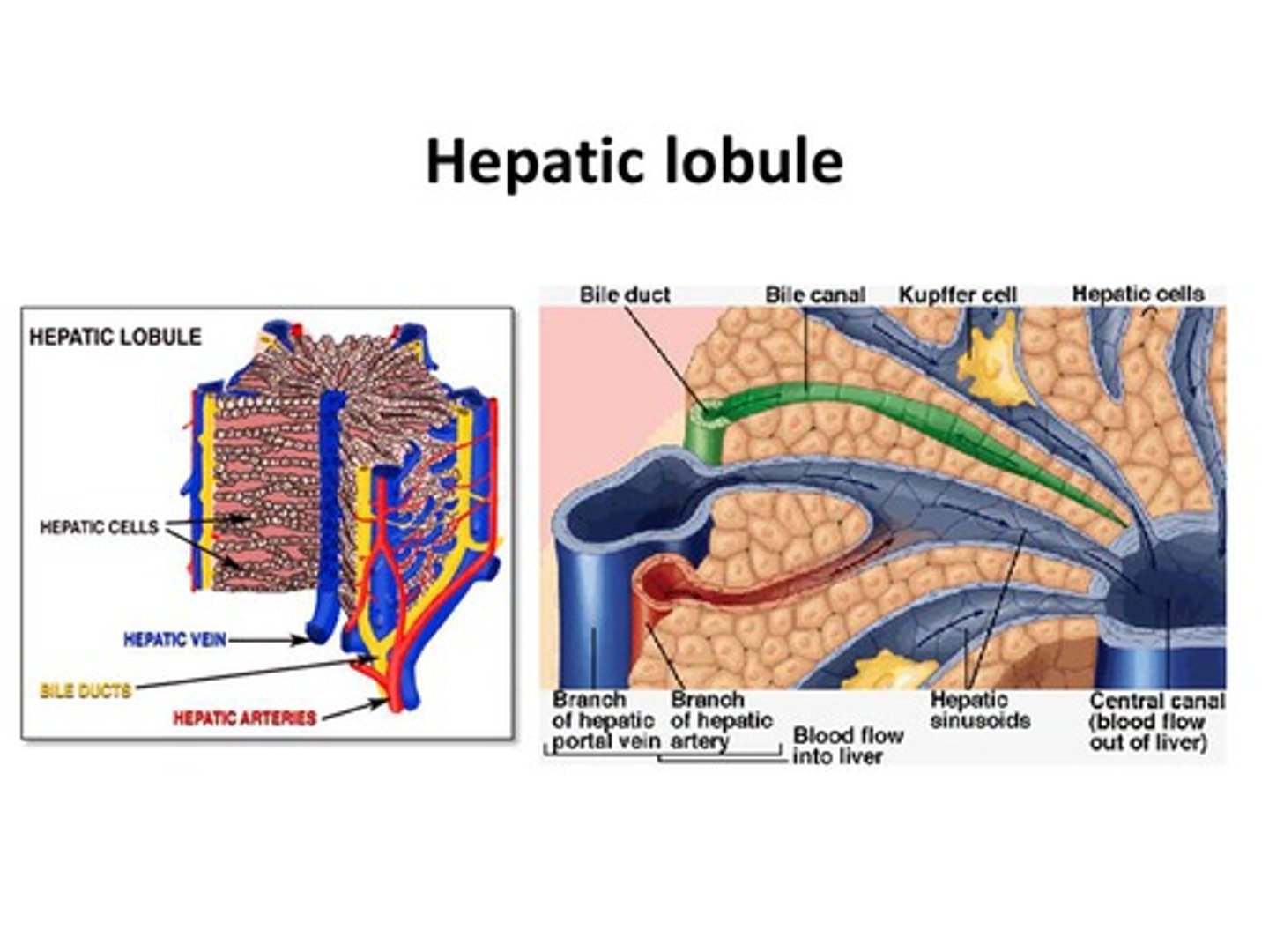
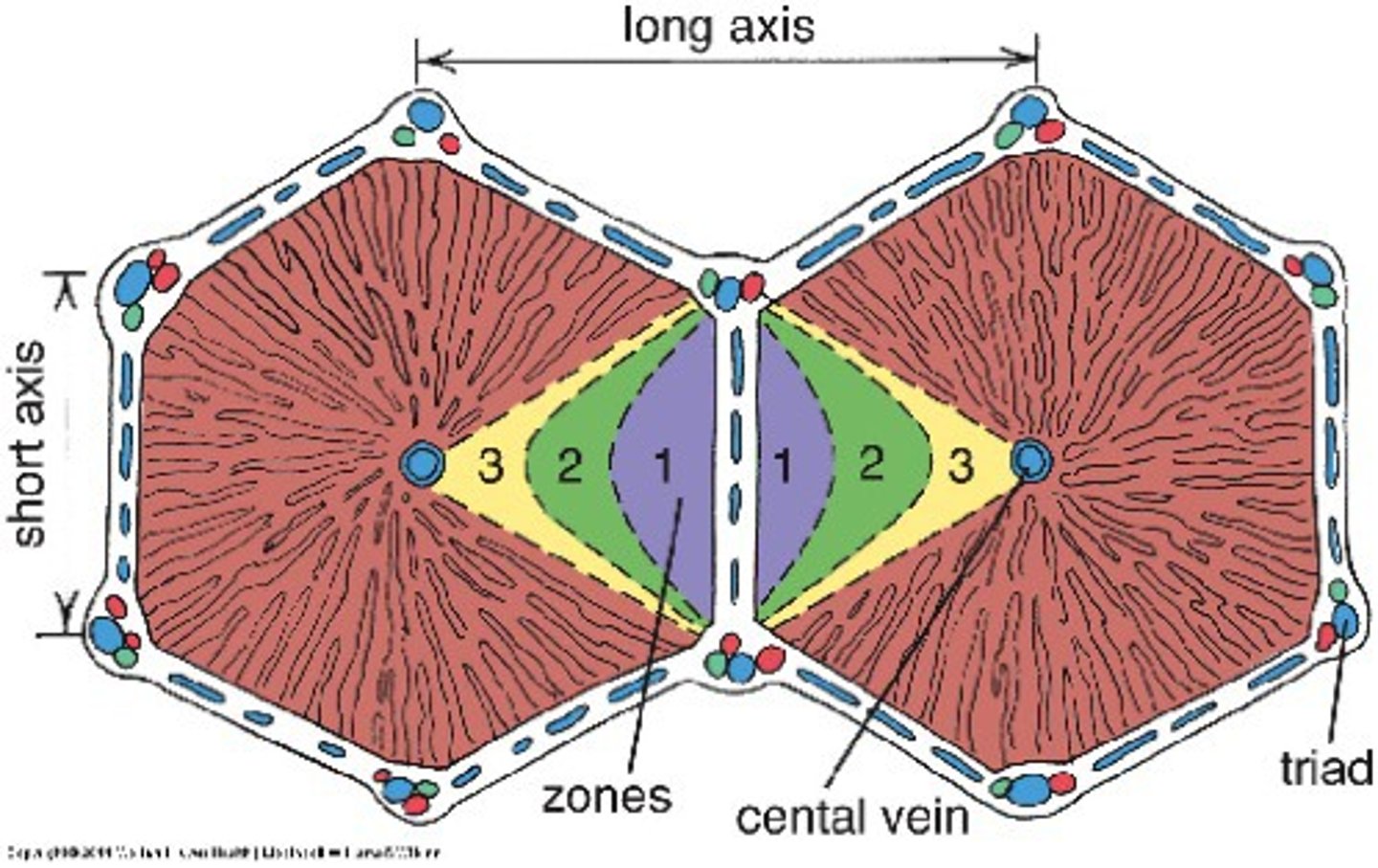
Anatomical unit of liver
Hepatic lobule
Portal lobule of liver
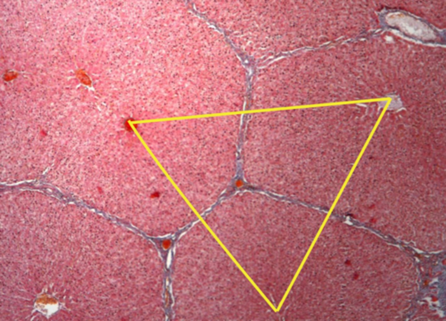
Liver acinus (functional unit)
diamond defined by two portal spaces and two central veins; supply area of a portal venule and hepatic arteriole
Branch of portal vein
High nutriet, Low O2, Low pressure
Branch of Hepatic artery
Low nutrient, High O2, High pressure
Away from Portal triad (towards Central Vein)
Decrease of Nutrients and O2 as they are used up by the liver cells
Cytological features of hepatocytes
Fat storing cells = in space of Disse, VtA metalolism
Endothelium = lines sinusoids, intercellular openings fenestrated
Kupffer cells = luminal, macrophage-like; prevents obstruction; antibacterial
Kupffer cell
(hepatic or stellate macrophages) in liver sinusoids remove debris and old RBCs
Kupffer cell in lumen of sinusoid function
Prevent obstruction and phagocytosis
Nucleus of Hepatocye is.........
Binucleate Polyploid (double DNA content)
Liver bile flows through.............
Bile canaliculi, Bile duct and Bile ductule
Still learning (12)
You've started learning these terms. Keep it up!