DNA structure and replication

0.0(0)
Studied by 1 person
call kaiCall Kai
learnLearn
examPractice Test
spaced repetitionSpaced Repetition
heart puzzleMatch
flashcardsFlashcards
GameKnowt Play
Card Sorting

1/31

encourage image

There's no tags or description

Looks like no tags are added yet.

Last updated 4:38 AM on 5/31/26
Name
Mastery
Learn
Test
Matching
Spaced
Call with Kai

No analytics yet

Send a link to your students to track their progress

32 Terms

1
New cards

Role of DNA

  • life depends on the ability of cells to store, retrieve and translate the instructions required to make and maintain a living organism

  • hereditary - passed on from a cell to its daughter cells at somatic cell division, and from one generation of an organism to the next through the organism’s gametes

  • stored within every living cell as its genes, information-containing elements that determine the characteristics of a species as a whole and of the individuals within it

Characteristic properties:

  • high capacity for information storage and be chemically stable to be able to encode information without fail

  • replicate accurately

  • be capable of variation (different traits)

2
New cards

DNA as hereditary material

  • chromosome analysis

  • metabolic stability of DNA

  • constancy of DNA within a cell

  • correlation btw mutagens and their effects on DNA

  • *Griffth’s Experiment

  • *Avery’s Experiments

  • *Hershey-Chase Experiment

3
New cards

Structure of DNA

Nucleotides - building blocks of nucleic acids

  1. Nucleic acids are macromolecules that exist as polymers called polynucleotides

  2. each nucleotide is composed of

a. five-carbon sugar - pentose

b. nitrogenous base

c. phosphate group

pentose + nitrogenous base = nucleoside

  1. 2 types of nucleic acids

a. Deoxyribonucleic acid (DNA) - pentose sugar is deoxyribose

—> deoxyribonucleotides are the monomers of DNA

b. Ribonucleic acid (RNA) - penrose sugar is ribose

—> ribonucleotides are the monomers of RNA
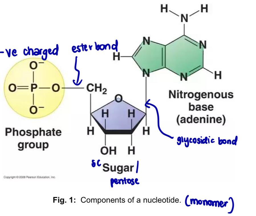
<p>Nucleotides - building blocks of nucleic acids</p><ol><li><p>Nucleic acids are macromolecules that exist as polymers called <strong>polynucleotides</strong></p></li><li><p>each nucleotide is composed of</p></li></ol><p>a. <mark data-color="yellow" style="background-color: yellow; color: inherit;">five-carbon sugar - pentose</mark></p><p>b. <mark data-color="blue" style="background-color: blue; color: inherit;">nitrogenous base</mark></p><p>c. <mark data-color="red" style="background-color: red; color: inherit;">phosphate group</mark></p><p>pentose + nitrogenous base = nucleoside</p><ol start="3"><li><p>2 types of nucleic acids</p></li></ol><p>a. Deoxyribonucleic acid (DNA) - pentose sugar is deoxyribose</p><p>—&gt; deoxyribonucleotides are the monomers of DNA</p><p>b. Ribonucleic acid (RNA) - penrose sugar is ribose</p><p>—&gt; ribonucleotides are the monomers of RNA</p><p></p>
4
New cards

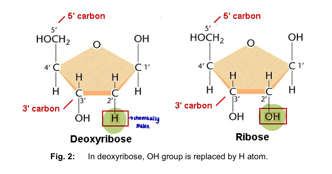
Pentose sugar

  • 5-carbon sugars and occur as ring forms

  • in nucleic acids, the 5’ carbon is linked in an ester bond to the phosphate group

  • the 1’ carbon is linked in a glycosidic bond to the nitrogenous base

  • difference between ribose and deoxyribose sugars

  1. (OH or H) at the 2’ carbon of deoxyribose, the hydroxyl group (-OH) is replaced by a hydrogen atom (H)

  2. led to significant differences in structure and functions

—> (DNA coils more than RNA) partial negative charge of the -OH group in ribose repels the -ve charge of the phosphate, preventing the RNA chain from coiling in as tight as a helix as it does in DNA

—> RNA is more susceptible to chemical and enzyme degradation
<ul><li><p>5-carbon sugars and occur as <strong>ring</strong> forms</p></li><li><p>in nucleic acids, the<strong> 5’ carbon</strong> is linked in an <strong>ester bond</strong> to the <strong>phosphate group </strong></p></li><li><p>the<strong> 1’ carbon</strong> is linked in a <strong>glycosidic bond</strong> to the <strong>nitrogenous base </strong></p></li><li><p><u>difference</u> between <u>ribose</u> and <u>deoxyribose</u> sugars </p></li></ul><ol><li><p>(OH or H) at the<strong> 2’ carbon</strong> of deoxyribose, the <strong>hydroxyl</strong> group (-OH) is <u>replaced</u> by a <strong>hydrogen</strong> atom (H) </p></li><li><p>led to significant differences in <u>structure</u> and <u>functions </u></p></li></ol><p>—&gt; (DNA coils more than RNA) partial negative charge of the <strong>-OH group</strong> in ribose <strong>repels</strong> the -ve charge of the <strong>phosphate</strong>, <strong>preventing the RNA chain from coiling in</strong> as tight as a helix as it does in DNA </p><p>—&gt; <strong>RNA</strong> is <u>more</u> susceptible to chemical and enzyme <strong>degradation </strong></p><p></p><p></p>
5
New cards

Nitrogenous bases

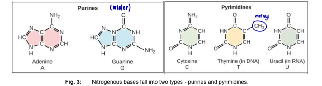
  • nitrogenous base has a nitrogen-containing ring structure

  • 2 types: purines & pyrimidines

Purines

—> 6-membered ring fused to a 5-membered ring

DNA: Adenine (A) and Guanine (G)

RNA: A and G

Pyrimidines

—> 6-membered ring

DNA: Cytosine (C) and Thymine (T)

RNA: Cytosine (C) and Uracil (U)

*pyramid = 1 = single ring

*Pure As Gold

Cut The Py

  • difference between T and U is the presence of a methyl substituent at the 5-carbon
<ul><li><p>nitrogenous base has a nitrogen-containing ring structure</p></li><li><p>2 types: purines &amp; pyrimidines</p></li></ul><p><strong>Purines</strong></p><p>—&gt; 6-membered ring fused to a 5-membered ring</p><p>DNA: Adenine (A) and Guanine (G)</p><p>RNA: A and G</p><p></p><p><strong>Pyrimidines</strong></p><p>—&gt; 6-membered ring</p><p>DNA: Cytosine (C) and Thymine (T)</p><p>RNA: Cytosine (C) and Uracil (U)</p><p></p><p>*pyramid = 1 = single ring</p><p>*<strong><u>P</u></strong>ure <strong><u>A</u></strong>s <strong><u>G</u></strong>old</p><p><strong><u>C</u></strong>ut <strong><u>T</u></strong>he <strong><u>P</u></strong>y</p><p></p><ul><li><p>difference between T and U is the presence of a methyl substituent at the 5-carbon </p></li></ul><p></p>
6
New cards

Nucleoside

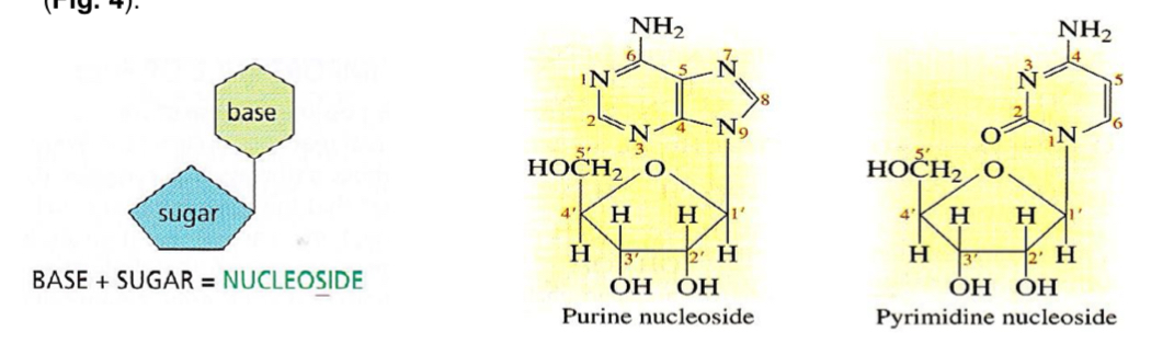
nucleoside = pentose + nitrogenous base

  • occurs with the elimination of water (condensation)

  • 1’ carbon of the pentose is linked in a glycosidic bond to the nitrogenous base

  • 2 types: ribonucleoside, deoxyribonucleoside
<p><strong><mark data-color="yellow" style="background-color: yellow; color: inherit;">nucleoside</mark> = <mark data-color="blue" style="background-color: blue; color: inherit;">pentose</mark> + <mark data-color="red" style="background-color: red; color: inherit;">nitrogenous base</mark></strong></p><ul><li><p>occurs with the elimination of water (<strong>condensation</strong>)</p></li><li><p><strong>1’ carbon</strong> of the pentose is linked in a <strong>glycosidic bond </strong>to the <strong>nitrogenous base</strong></p></li><li><p>2 types: ribonucleoside, deoxyribonucleoside</p></li></ul><p></p>
7
New cards

Nucleotide

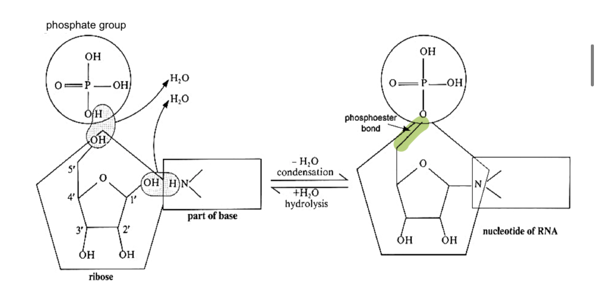
nucleotide = nucleoside + phosphate group

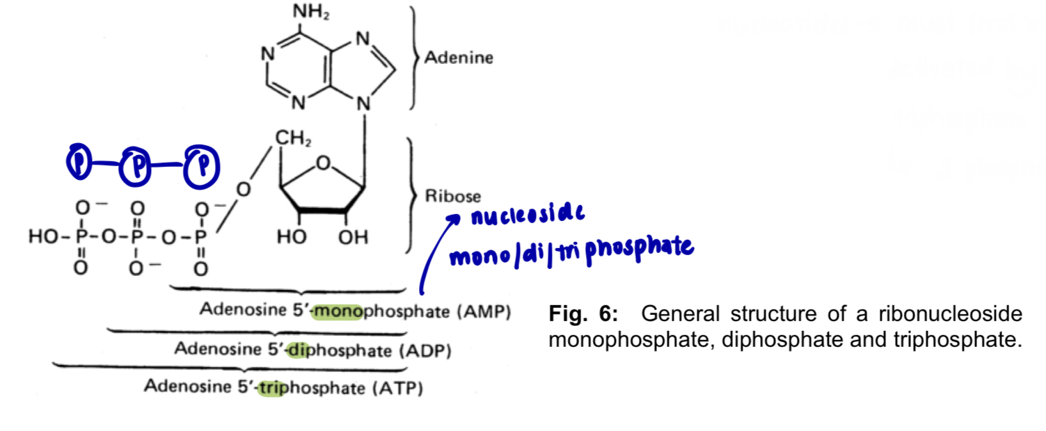
  • formed by further condensation between the nucleoside and phosphate group, forming a phosphoester bond btw the 5’ carbon of pentose and phosphate group

  • number of phosphate groups linked to pentose sugar varies from 1 to 3

1 phosphate group —> nucleoside monophosphate e.g. AMP, adenosine monophosphate

2 phosphate groups —> nucleoside diphosphate e.g. ADP, adenosine diphosphate

3 phosphate groups —> nucleoside triphosphate

e.g. ATP, adenosine triphosphate

*nucleotides must first be activated by triphosphate (2 phosphates are released)
<p><strong><mark data-color="purple" style="background-color: purple; color: inherit;">nucleotide</mark> = <mark data-color="yellow" style="background-color: yellow; color: inherit;">nucleoside</mark> + <u><mark data-color="blue" style="background-color: blue; color: inherit;">phosphate group</mark></u></strong></p><ul><li><p>formed by further <strong>condensation</strong> between the nucleoside and phosphate group, forming a <strong>phosphoester bond </strong>btw the <strong>5’ carbon</strong> of <strong>pentose</strong> and <strong>phosphate group</strong></p></li><li><p>number of phosphate groups linked to pentose sugar varies from 1 to 3 </p></li></ul><p>1 phosphate group —&gt; nucleoside <u>mono</u>phosphate e.g. AMP, adenosine monophosphate </p><p>2 phosphate groups —&gt; nucleoside <u>di</u>phosphate e.g. ADP, adenosine diphosphate </p><p>3 phosphate groups —&gt; nucleoside <u>tri</u>phosphate </p><p>e.g. ATP, adenosine triphosphate </p><p></p><p>*nucleotides must first be activated by triphosphate (2 phosphates are released) </p>
8
New cards

mono/di/triphosphate groups

knowt flashcard image
9
New cards

Formation of dinucleotides and polynucleotides

  • two nucleotides join to form a dinucleotide by condensation btw the 5’-phosphate group of one nucleotide and the 3’ hydroxyl group of the other to form a phosphodiester bond

*phosphoester bond btw 2 nucleotides

VS phosphodiester bond (the whole sugar phosphate backbone)

  • condensation rxn btw nucleotides is repeated several million times to form a polynucleotide

  • phosphodiester bonds btw 5’ phosphate and 3’ hydroxyl groups of nucleotides form a linear, unbranched sugar-phosphate backbone

  • phosphodiester bonds are strong and covalent bonds

—> confer strength and stability on the polynucleotide chain - prevents breakage of the chain during DNA replication
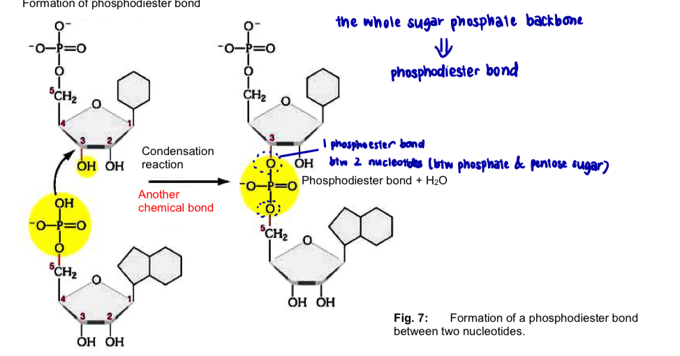
<ul><li><p>two nucleotides join to form a <strong>dinucleotide</strong> by <strong>condensation</strong> btw the <strong>5’-phosphate group </strong>of one nucleotide and the <strong>3’ hydroxyl group</strong> of the other to form a <strong>phosphodiester</strong> bond </p></li></ul><p></p><p>*phosphoester bond btw 2 nucleotides </p><p>VS phosphodiester bond (the whole sugar phosphate backbone) </p><p></p><ul><li><p>condensation rxn btw nucleotides is repeated several million times to form a polynucleotide </p></li><li><p>phosphodiester bonds btw 5’ phosphate and 3’ hydroxyl groups of nucleotides form a <strong>linear, unbranched sugar-phosphate backbone </strong></p></li><li><p>phosphodiester bonds are <strong>strong and covalent bonds</strong></p></li></ul><p>—&gt; confer <strong>strength</strong> and <strong>stability</strong> on the polynucleotide chain -<strong> prevents breakage</strong> of the chain during DNA replication </p><p></p>
10
New cards

Polarity/directionality in a polynucleotide

  • the manner in which deoxyribonucleoside triphosphate are added to the 3’ end of a growing chain has resulted in a polynucleotide molecule that has polarity or directionality

  • each DNA/RNA strand/chain has 2 free ends that are chemically different from each other

    • 5’ end with a free 5’ carbon carrying a phosphate group

    • 3’ end with a free 3’ carbon carrying a hydroxyl group

  • DNA or RNA base sequence is read in a 5’ to 3’ direction —> directionality/polarity
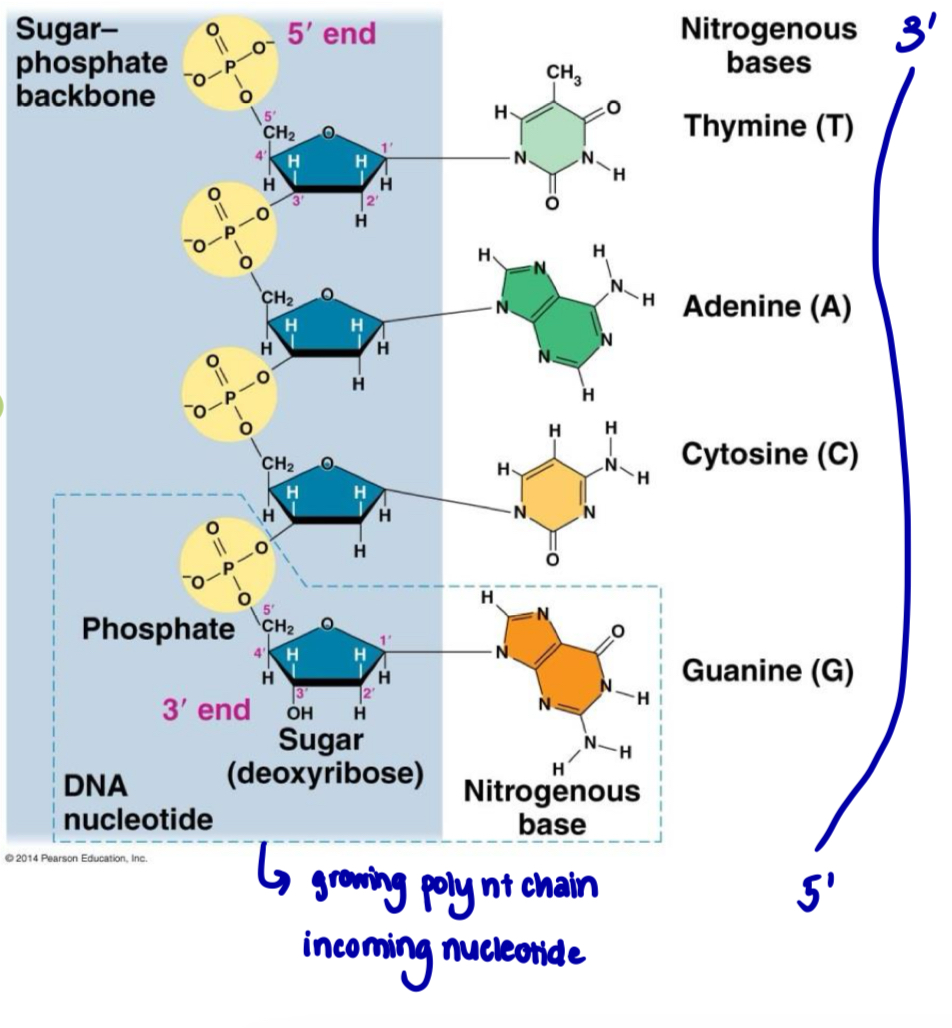
<ul><li><p>the manner in which deoxyribonucleoside triphosphate are added to the 3’ end of a growing chain has resulted in a polynucleotide molecule that has polarity or directionality </p></li><li><p>each DNA/RNA strand/chain has 2 free ends that are chemically different from each other </p><ul><li><p><strong>5’ end</strong> with a free 5’ carbon carrying a phosphate group </p></li><li><p><strong>3’ end</strong> with a free 3’ carbon carrying a hydroxyl group </p></li></ul></li><li><p>DNA or RNA base sequence is read in a <strong>5’ to 3’ direction —&gt; directionality/polarity </strong></p></li></ul><p></p><p></p>
11
New cards

Physical structure of DNA

  • a DNA molecule is double helix comprising 2 DNA strands

  • based on X-ray diffraction data, DNA is

    • long, thin molecule, diameter 2nm

    • 2 strands

    • coiled in the form of a double helix, the helix making one complete twist every 3.4 nm

    • has 10 bases to each complete turn of the helix

  • ATGC base composition

    • base composition of DNA of an organism is constant throughout all the somatic cells and is characteristic for a given species

    • no. of A = no. of T; no. of G = no. of C

    • there’s always an equal proportion of purines (A+G) and pyrimidines (C+T)

  • specific complementary base pairing between A on one DNA strand and T on the other, btw G and C
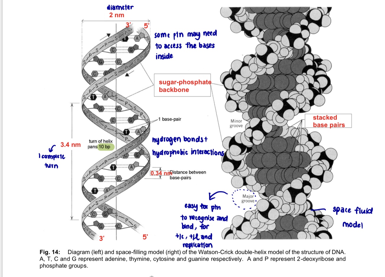
<ul><li><p>a DNA molecule is <strong>double helix</strong> comprising 2 DNA strands</p></li><li><p>based on X-ray diffraction data, DNA is</p><ul><li><p>long, thin molecule, <strong>diameter 2nm</strong></p></li><li><p>2 strands</p></li><li><p>coiled in the form of a double helix, the helix making <strong>one complete twist every 3.4 nm</strong></p></li><li><p>has <strong>10 bases</strong> to each complete turn of the helix</p></li></ul></li><li><p>ATGC base composition</p><ul><li><p>base composition of DNA of an organism is constant throughout all the somatic cells and is characteristic for a given species</p></li><li><p><strong>no. of A = no. of T</strong>; <strong>no. of G = no. of C</strong></p></li><li><p>there’s always an equal proportion of purines (A+G) and pyrimidines (C+T)</p></li></ul></li><li><p>specific complementary base pairing between A on one DNA strand and T on the other, btw G and C</p></li></ul><p></p>
12
New cards

The Double helix of DNA

  1. DNA consists of 2 polynucleotide strands/chains

  • each strand forms a right-handed helix and the two strands coil around each other to form a double helix

1 DNA molecule = 1 DNA double helix = 2 polynucleotide strands

  1. the diameter of the helix is uniformly 2nm (Just enough space for 1 purine + 1 pyrimidine in the centre of the double-helix)

  2. strands run in opposite directions i.e. antiparallel

  • 5’ to 3’, the other is 3’ to 5’

  1. each strand has a sugar-phosphate backbone with

  • phosphate groups that project outside the double helix since they are hydrophilic

  • nitrogenous bases that orientate inwards toward the central axis at almost right angles

  1. bases of the opposite strands are bonded together by relatively weak hydrogen bonds

  2. base pairs are stacked 0.34 nm apart along the central axis of the helix as a result of hydrophobic interactions contributing to overall stability of the molecule

  3. double helix makes a complete turn every 10 base pairs, so each turn is 3.4 nm

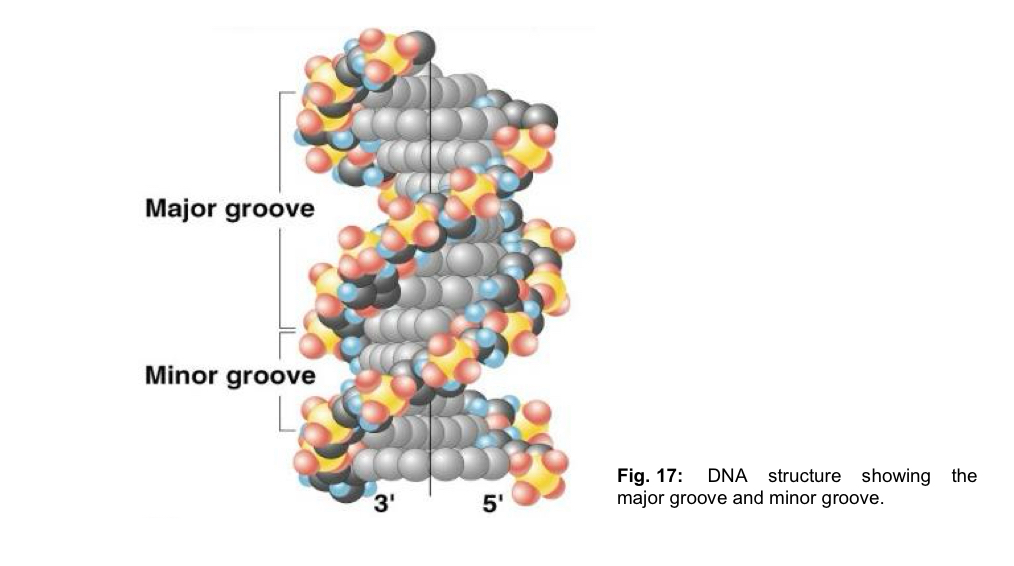
  4. there are grooves of unequal sizes btw the sugar phosphate backbones - major grooves and minor grooves

  • both these grooves are large enough to allow protein molecules to gain access and make contact with the bases
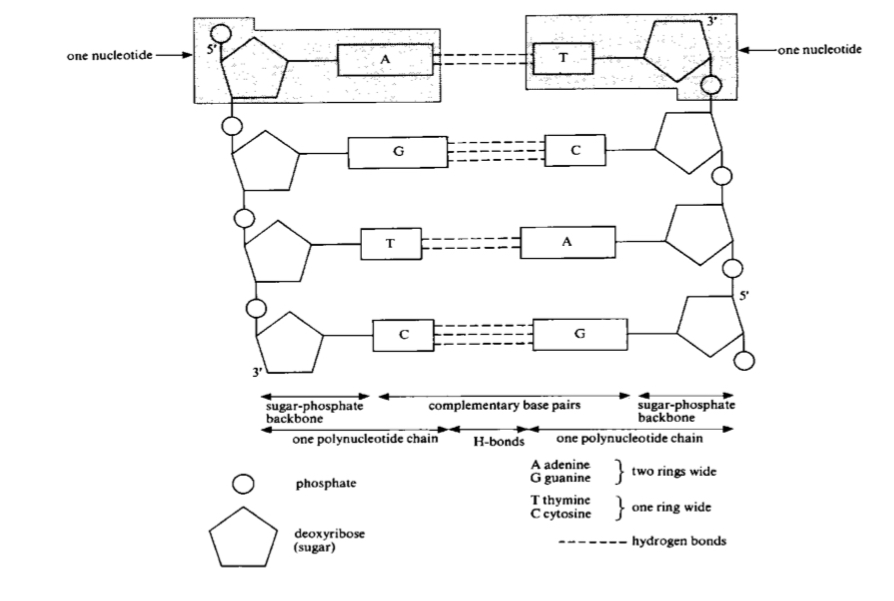
<ol><li><p>DNA consists of <mark data-color="yellow" style="background-color: yellow; color: inherit;">2 polynucleotide strands/chains</mark></p></li></ol><ul><li><p>each strand forms a right-handed helix and the two strands coil around each other to form a double helix</p></li></ul><p>1 DNA molecule = 1 DNA double helix = 2 polynucleotide strands</p><ol start="2"><li><p><mark data-color="blue" style="background-color: blue; color: inherit;">the diameter of the helix is uniformly 2nm </mark>(Just enough space for 1 purine + 1 pyrimidine in the centre of the double-helix)</p></li><li><p>strands run in opposite directions i.e. <mark data-color="red" style="background-color: red; color: inherit;">antiparallel</mark></p></li></ol><ul><li><p>5’ to 3’, the other is 3’ to 5’</p></li></ul><ol start="4"><li><p><mark data-color="purple" style="background-color: purple; color: inherit;">each strand has a sugar-phosphate backbone with</mark></p></li></ol><ul><li><p><strong>phosphate groups</strong> that project <strong>outside</strong> the double helix since they are <strong>hydrophilic</strong></p></li><li><p><strong>nitrogenous bases that orientate inwards </strong>toward the central axis at almost right angles</p></li></ul><ol start="5"><li><p>bases of the opposite strands are bonded together by relatively <mark data-color="green" style="background-color: green; color: inherit;">weak hydrogen bonds</mark></p></li><li><p><mark data-color="yellow" style="background-color: yellow; color: inherit;">base pairs are stacked 0.34 nm apart</mark> along the central axis of the helix as a result of <mark data-color="yellow" style="background-color: yellow; color: inherit;">hydrophobic interactions contributing to overall stability</mark> of the molecule </p></li><li><p>double helix makes a <mark data-color="blue" style="background-color: blue; color: inherit;">complete turn every 10 base pairs</mark>, so each turn is 3.4 nm </p></li><li><p>there are grooves of unequal sizes btw the sugar phosphate backbones - <mark data-color="red" style="background-color: red; color: inherit;">major grooves and minor grooves </mark></p></li></ol><ul><li><p>both these grooves are large enough to allow <strong>protein molecules to gain access</strong> and make contact with the <strong>bases</strong> </p></li></ul><p></p><p></p>
13
New cards

Complementary base pairing between DNA strands

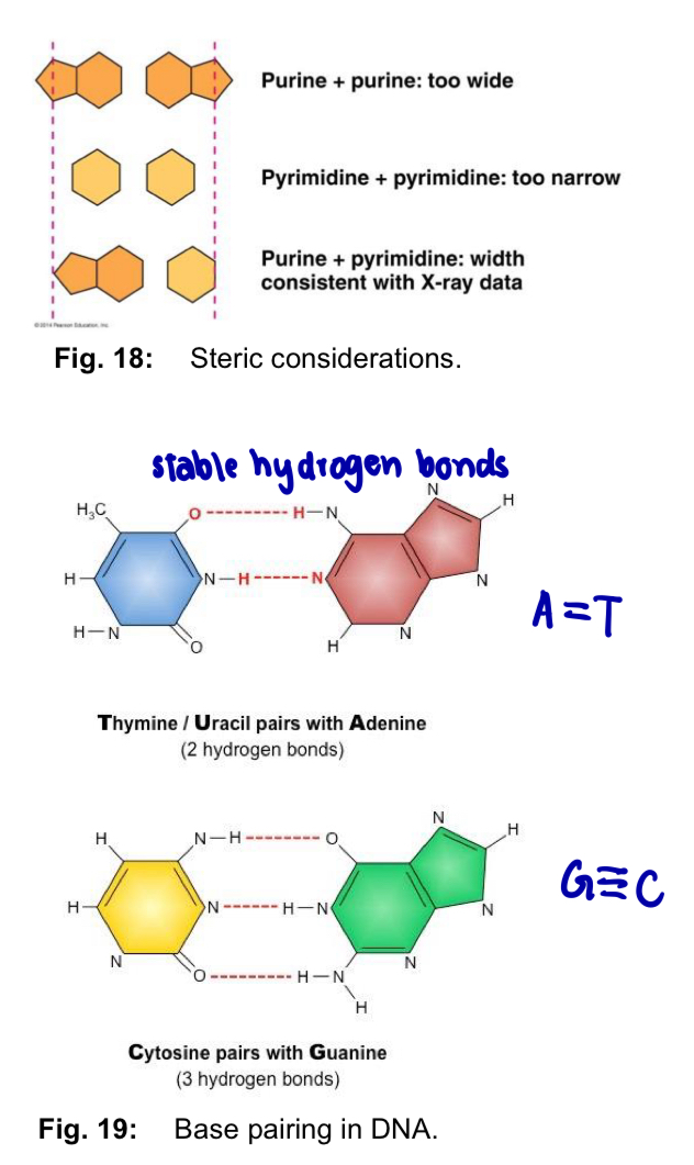
  • complementary base pairing occurs btw A and T (2 hydrogen bonds) and btw C and G (3 hydrogen bonds)

  • base sequence in one strand determines the base sequence in the complementary strand

  • weak hydrogen bonds make it relatively easy to separate the two strands of the DNA e.g. by heating

  • separating the A-T pair is easier than separating the G-C pair, since A-T pair only involves 2 H bonds while G-C involves 3 H bonds

14
New cards

Grooves

knowt flashcard image
15
New cards

Why form complementary base pairs?

  1. Steric restrictions

  • sugar-phosphate backbone of each polynucleotide chain has a regular helical structure

  • DNA double-helix has a uniform diameter of 2nm

  • T and C are pyrimidines - single ring

  • A and G are purines - twice as wide as pyrimidines

  • pair purine with pyrimidine = fits diameter

purine + purine = too wide

pyrimidine + pyrimidine = too narrow

  1. Hydrogen bond factors

  • each nitrogenous base has chemical side groups (H, N and O) - hydrogen bonds with its appropriate partner

  • such chemical side groups in purines and pyrimidines have well defined positions

  • A is capable of forming 2 hydrogen bonds with T, while G is capable of forming 3 hydrogen bonds with C

Important notes on CBP

  1. significance of CBP - since the 3D structure of DNA is only stable when the base pairs are complementary = base sequence of one strand could determine the base sequence of its complementary strand —> **necessary in DNA replication and transmission of the genetic information stored

  2. variation of linear base sequence - linear sequence of the 4 bases can be varied

  • 1 nucleotide —> 4 combinations

  • 10 nucleotides —> 4^10 combinations

  • humans have 3 × 10^9 nucleotide pairs —> 1 048 576 combinations

  • each gene has a unique base sequence

<ol><li><p><mark data-color="yellow" style="background-color: yellow; color: inherit;">Steric restrictions </mark></p></li></ol><ul><li><p>sugar-phosphate backbone of each polynucleotide chain has a <strong>regular helical structure </strong></p></li><li><p>DNA double-helix has a uniform diameter of 2nm </p></li><li><p>T and C are pyrimidines - single ring </p></li><li><p>A and G are purines - twice as wide as pyrimidines </p></li><li><p>pair purine with pyrimidine = fits diameter </p></li></ul><p>purine + purine = too wide </p><p>pyrimidine + pyrimidine = too narrow </p><p></p><ol start="2"><li><p><mark data-color="blue" style="background-color: blue; color: inherit;">Hydrogen bond factors </mark></p></li></ol><ul><li><p>each nitrogenous base has chemical side groups (H, N and O) - hydrogen bonds with its appropriate partner </p></li><li><p>such chemical side groups in purines and pyrimidines have well defined positions </p></li><li><p>A is capable of forming 2 hydrogen bonds with T, while G is capable of forming 3 hydrogen bonds with C </p></li></ul><p><strong><u>Important notes on CBP </u></strong></p><ol><li><p>significance of CBP - since the 3D structure of DNA is only <strong>stable</strong> when the <strong>base pairs are complementary</strong> = base sequence of one strand could determine the base sequence of its complementary strand —&gt; **necessary in <strong>DNA replication and transmission of the genetic information </strong>stored </p></li><li><p><strong>variation of linear base sequence -</strong> linear sequence of the 4 bases can be varied </p></li></ol><ul><li><p>1 nucleotide —&gt; 4 combinations </p></li><li><p>10 nucleotides —&gt; 4^10 combinations </p></li><li><p>humans have 3 × 10^9 nucleotide pairs —&gt; 1 048 576 combinations </p></li><li><p>each gene has a unique base sequence </p></li></ul><p></p>
16
New cards

Packing of DNA in eukaryotic chromosomes

  • human cell contains 46 chromosomes, each of which is a single DNA molecule bundled up with various ptn

  • inherit 23 chromosomes from each parent

  • each set of 23 chromosomes encodes a complete copy of our genome, made up of 6 × 10^9 nucleotides

  • chromosomes must condense intro structures that are more easily managed = properly distributed to each daughter cell during cell and nuclear division

  • DNA packing —> proteins helping in the folding and condensation of DNA (into highly compact chromosome)

Level 1: DNA double helix

Level 2: beads on a string form chromatin

Level 3: 30nm chromatin fibre of packed nucleosomes

Level 4: chromosome in an extended form

Level 5: metaphase chromosome
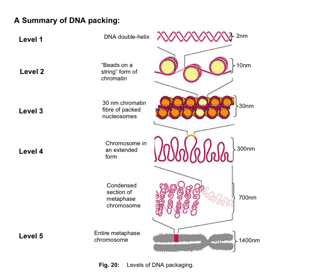
<ul><li><p>human cell contains 46 chromosomes, each of which is a single DNA molecule <u>bundled up with various ptn </u></p></li><li><p>inherit 23 chromosomes from each parent </p></li><li><p>each set of 23 chromosomes encodes a complete copy of our genome, made up of 6 × 10^9 nucleotides </p></li><li><p>chromosomes must condense intro structures that are more easily managed = properly distributed to each daughter cell during cell and nuclear division </p></li><li><p>DNA packing —&gt; proteins helping in the folding and condensation of DNA (into highly compact chromosome) </p></li></ul><p>Level 1: DNA double helix </p><p>Level 2: beads on a string form chromatin </p><p>Level 3: 30nm chromatin fibre of packed nucleosomes </p><p>Level 4: chromosome in an extended form </p><p>Level 5: metaphase chromosome </p>
17
New cards

Stable, Invariant storage of genetic information

  1. genetic information that must be stored/preserved lies in the specific order of the base pairs i.e. base sequence must be stable and invariant

  2. DNA is relatively resistant to spontaneous changes (mutations) = stable storage

  3. structural features that stabilise the DNA double-helix

  • extensive hydrogen bonds btw base pairs

  • hydrophobic interactions btw the stacked base pairs

  • exposure to outside influences of only the sugar-phosphate backbone

  • nitrogenous bases being safely tucked inside the double-helix

  • eukaryotes only - DNA double helix being tightly wound around histones to form a repeating array of nucleosomes —> folded into chromosome, in which DNA is prevented from thermal and physical damage

  1. structural features —> invariant base sequence

  • specific, complementary base pairing btw DNA strands

  • genetic info is redundant (present more than once) in the DNA

  • if base sequence in one of the two strands is accidentally altered, cell discards the damaged strand, makes a perfectly good strand by using remaining intact strand as a template

  • redundancy of genetic info helps to maintain its integrity

18
New cards

Replication of DNA

WHY

  • to ensure that the genetic information is passed down the progeny faithfully, there must be a high degree of accuracy during the copying process

  • daughter DNA molecules must be exactly the same as parental DNA molecule in terms of sequence information

DNA strand templates for replication

  • two strands of DNA are complementary

  • when a cell copies a DNA molecule, each strand serves as a template for ordering nucleotides into a new, complementary strand

19
New cards

Models of DNA replication - semi-conservative model

  • *Semi-conservative model

  1. the two DNA strands unwind and separate from each other, hydrogen bonds btw CBP are broken

  2. each DNA strand then acts as a template for the assembly of a complementary strand

  3. nucleotides line up singly along the template DNA strand according to CBP

  4. DNA polymerases join the nucleotides together at their sugar-phosphate moieties

  5. upon completion of DNA replication, 2 identical daughter DNA molecules are produced from a single* parental DNA molecule

  6. ***each of the two daughter DNA molecules consists of 1 parental DNA strand + 1 newly-synthesised daughter DNA strand

  • other proposed models

—> Conservative model: parental DNA molecule emerges from replication process intact (it is conserved) and generates DNA copies consisting of entirely new molecules

—> Dispersive model: all 4 strands of DNA following replication have a mixture of old and new DNA
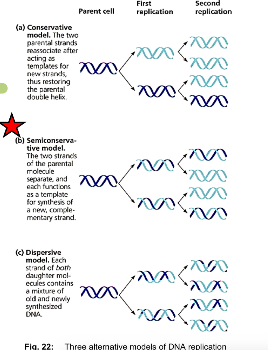
<ul><li><p>*Semi-conservative model </p></li></ul><ol><li><p>the two DNA strands <strong>unwind and separate </strong>from each other, <strong>hydrogen bonds </strong>btw CBP are <u>broken</u> </p></li><li><p>each <strong>DNA strand</strong> then acts as a <strong>template</strong> for the assembly of a <strong>complementary</strong> strand </p></li><li><p>nucleotides line up singly along the template DNA strand according to CBP </p></li><li><p><strong>DNA polymerases</strong> <strong>join the nucleotides together </strong>at their sugar-phosphate moieties </p></li><li><p>upon completion of DNA replication, <strong><u>2</u> identical daughter DNA molecules </strong>are produced from a <strong><u>single*</u></strong> <strong>parental DNA molecule </strong></p></li><li><p><strong>***</strong>each of the two daughter DNA molecules consists of <strong><mark data-color="yellow" style="background-color: yellow; color: inherit;">1 </mark><u><mark data-color="yellow" style="background-color: yellow; color: inherit;">parental</mark></u><mark data-color="yellow" style="background-color: yellow; color: inherit;"> DNA strand </mark>+ <mark data-color="blue" style="background-color: blue; color: inherit;">1 </mark><u><mark data-color="blue" style="background-color: blue; color: inherit;">newly-synthesised daughter</mark></u><mark data-color="blue" style="background-color: blue; color: inherit;"> DNA strand</mark> </strong></p></li></ol><ul><li><p>other proposed models </p></li></ul><p>—&gt; Conservative model: parental DNA molecule emerges from replication process intact (it is conserved) and generates DNA copies consisting of entirely new molecules </p><p>—&gt; Dispersive model: all 4 strands of DNA following replication have a mixture of old and new DNA </p><p></p><p></p>
20
New cards

Meselson-Stahl Experiment: evidence for semi-conservative replication

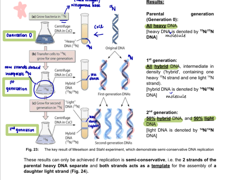
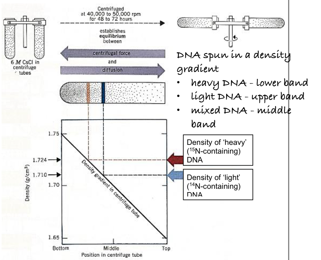
  • used isotopes of nitrogen 14N & 15N

  1. for many generations, cells of the bacterium (Escherichia coli) were grown on medium containing only the heavy isotope of nitrogen 15N - 15N was incorporated into all the nitrogenous bases —> heavy DNA

  2. bacteria were then transferred to medium containing only the light isotope of nitrogen 14N and allowed to divide just once (1st generation of bacteria)

  3. density-gradient centrifugation was performed on a DNA extract from the bacteria

  • DNA is separated on the basis of density

    • heavier 15N DNA molecules are denser than the 14N DNA molecules, hence 15N DNA molecules are spun further down in the centrifuge tube where CsCl were denser

    • lighter 14N DNA molecules were closer to the top where CsCl was less dense

density-gradient centrifugation:

  • rapid rotations to separate particles

  • a CsCl density gradient with increasing density towards the bottom of the centrifugation tube is formed because of the equilibrium btw

    • sedimentation of the CsCl to the bottom of the spinning tube as a result of centrifugal forces

    • diffusion of CsCl towards the top of the tube

  • DNA molecules move to the position where their density equals that of CsCl and floats at that position

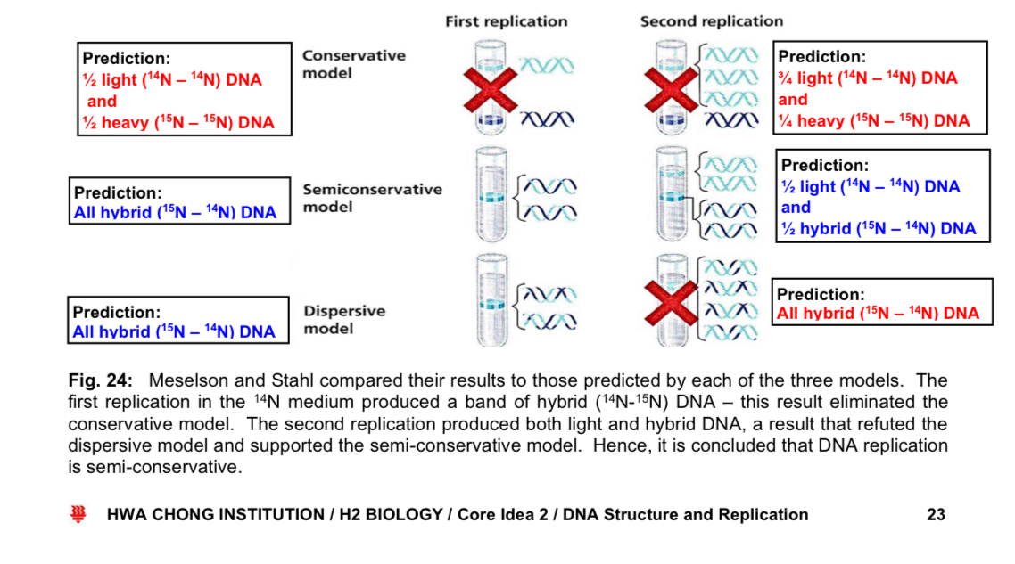
  1. these bacteria were allowed to undergo a second round of replication and binary fission (in the presence of 14N) producing the 2nd generation

  • DNA was again separated by density centrifugation
<ul><li><p>used isotopes of nitrogen 14N &amp; 15N </p></li></ul><ol><li><p>for many generations, cells of the bacterium (Escherichia coli) were grown on medium containing <strong><u>only</u> the heavy isotope of nitrogen 15N</strong> - 15N was incorporated into all the nitrogenous bases —&gt; heavy DNA </p></li><li><p>bacteria were then transferred to medium containing <strong>only the light isotope of nitrogen </strong>14N and allowed to <strong>divide just once (1st generation of bacteria) </strong></p></li><li><p><strong>density-gradient centrifugatio</strong>n was performed on a DNA extract from the bacteria </p></li></ol><ul><li><p>DNA is separated on the<strong> basis of density </strong></p><ul><li><p>heavier 15N DNA molecules are denser than the 14N DNA molecules, hence <u>15N DNA molecules are spun further down in the centrifuge tube</u> where <u>CsCl were denser </u></p></li><li><p>lighter 14N DNA molecules were closer to the top where CsCl was less dense </p></li></ul></li></ul><p>density-gradient centrifugation: </p><ul><li><p>rapid rotations to separate particles </p></li><li><p>a CsCl density gradient with increasing density towards the bottom of the centrifugation tube is formed because of the equilibrium btw </p><ul><li><p><em>sedimentation</em> of the CsCl to the bottom of the spinning tube as a result of centrifugal forces </p></li><li><p>diffusion of CsCl towards the top of the tube </p></li></ul></li><li><p>DNA molecules move to the position where their density equals that of CsCl and floats at that position </p></li></ul><p></p><ol start="4"><li><p>these bacteria were allowed to undergo a <strong>second round of replication </strong>and<strong> binary fission</strong> (in the presence of 14N) producing the <strong>2nd generation </strong></p></li></ol><ul><li><p>DNA was again separated by density centrifugation </p></li></ul><p></p>
21
New cards

Semi-conservative model illustration

knowt flashcard image
22
New cards

prediction of the DNA strands with the different models
knowt flashcard image
23
New cards

Mechanism of DNA replication

actual process

  1. is very complex, timing of steps is very precise —> double helix must unwind while the replication machinery copies the two anti parallel strands simultaneously

  2. is very fast - each human cell has approximately 3 × 10^9 base pairs, takes the cell just a few hours to copy all these DNA

  3. is very accurate

  4. requires the cooperation of a large team of enzymes and other proteins, as well as ATP

24
New cards

Diagram of DNA replication

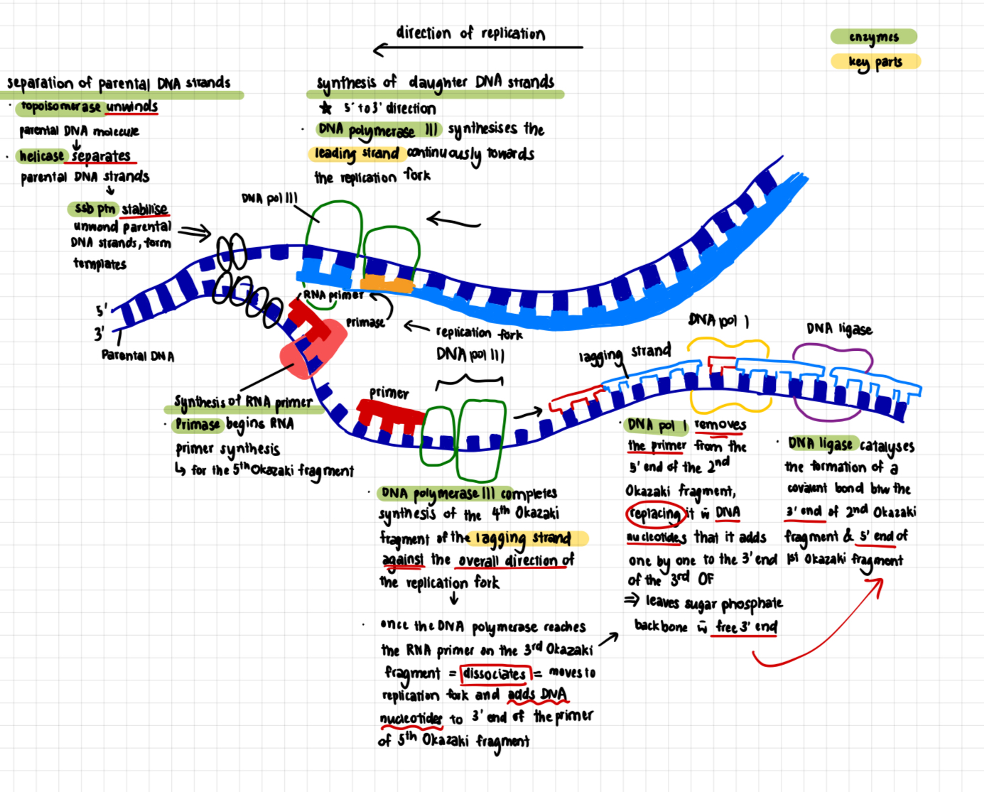
knowt flashcard image
25
New cards

Locations of origins of replication

  • DNA replication begins at one or more sites on the DNA molecule called origins of replication (oriR)

  • each oriR is a specific sequence of nucleotides, which is generally A-T rich (easier to disrupt the bonds cos only 2 hydrogen bonds btw A and T)

  1. proteins that initiate DNA replication (initiator proteins) recognise this sequence and bind to the oriR sequence - DNA double helix is separated into two strands —> replication bubble

  2. length of DNA unwound to initiate replication is typically ~ 50bp, ATP required

  3. at each end of a replication bubble Y-shaped structure called a replication fork, where the new strands of DNA are synthesised

  • 2 replication forks move away from the oriR as replication proceeds bidirectionally, until the entire DNA molecule is separated
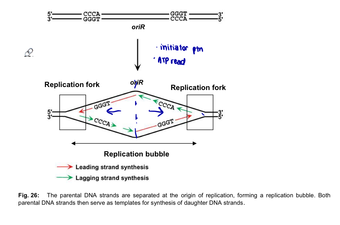
<ul><li><p>DNA replication begins at one or more sites on the DNA molecule called <strong>origins of replication</strong> (oriR) </p></li><li><p>each oriR is a <u>specific sequence of nucleotides,</u> which is generally A-T rich (easier to disrupt the bonds cos only 2 hydrogen bonds btw A and T) </p></li></ul><ol><li><p>proteins that initiate DNA replication (<strong>initiator proteins</strong>) <strong>recognise</strong> this sequence and <strong>bind</strong> to the oriR sequence - DNA double helix is <u>separated</u> into two strands —&gt; <strong>replication bubble </strong></p></li><li><p>length of DNA unwound to <strong>initiate replication </strong>is typically ~<strong> 50bp</strong>, ATP required </p></li><li><p>at each <u>end of a replication bubble</u> Y-shaped structure called a <strong>replication fork</strong>, where the new strands of DNA are synthesised </p></li></ol><ul><li><p>2 replication forks <u>move away from the oriR </u>as replication proceeds <u>bidirectionally</u>, until the entire DNA molecule is <u>separated</u> </p></li></ul><p></p><p></p>
26
New cards

Prokaryotes (bacteria)

  • prokaryotic chromosome is a small circular DNA molecule, with a single origin of replication

  • DNA replication proceeds bidirectionally from the origin of replication to a termination site located approximately halfway around the circular chromosome —> synthesis of 2 daughter DNA molecules
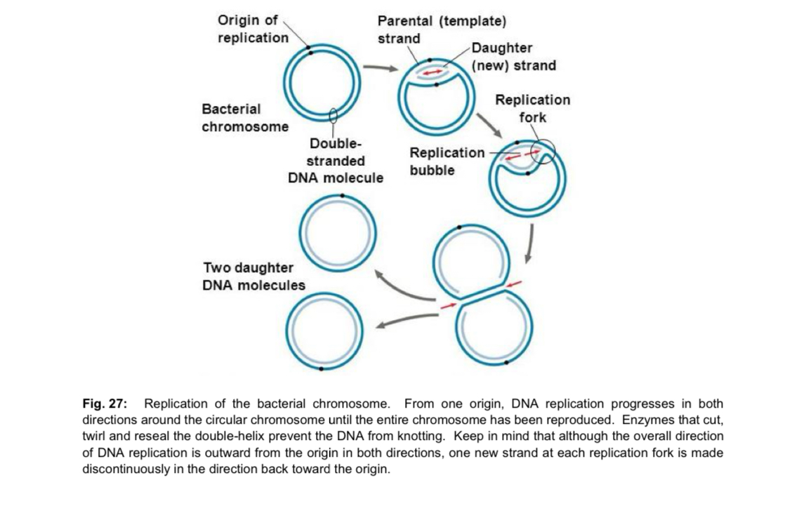
<ul><li><p>prokaryotic chromosome is a small <strong>circular</strong> DNA molecule, with a <strong>single origin of replication </strong></p></li><li><p>DNA replication proceeds <strong>bidirectionally</strong> from the origin of replication to a termination site located approximately halfway around the circular chromosome —&gt; synthesis of 2 daughter DNA molecules </p></li></ul><p></p>
27
New cards

Eukaryotes

  • eukaryotic chromosome is much larger and consists of a linear DNA molecule, with multiple origins of replication

    • advantage of having multiple oriR: multiple replication bubbles form and eventually fuse = speed up the copying of very long DNA molecules

    • replication takes approximately 8 hours in human cells with multiple oriR; if only 1 oriR, 100 times longer

    • impt given the much larger size of eukaryotic chromosome
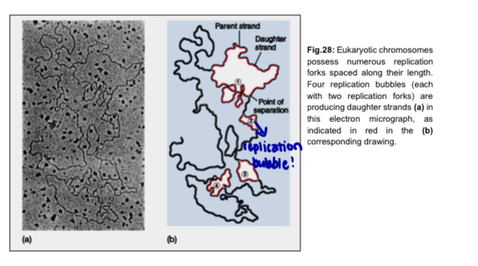
<ul><li><p>eukaryotic chromosome is much larger and consists of a <strong>linear</strong> DNA molecule, with <strong>multiple origins of replication </strong></p><ul><li><p>advantage of having multiple oriR: <strong>multiple replication bubbles form </strong>and eventually <strong>fuse</strong> = <strong>speed up</strong> the <strong>copying</strong> of very long DNA molecules </p></li><li><p>replication takes approximately 8 hours in human cells with multiple oriR; if only 1 oriR, 100 times longer </p></li><li><p>impt given the much larger size of eukaryotic chromosome </p></li></ul></li></ul><p></p>
28
New cards

Replication of eukaryotic chromosome

  1. Replication begins at multiple origins of replication, where the two parental strands separate to form replication bubbles

  2. the bubbles expand laterally as DNA replication proceeds bidirectionally

  3. eventually, the replication bubbles fuse and synthesis of the daughter strands is complete
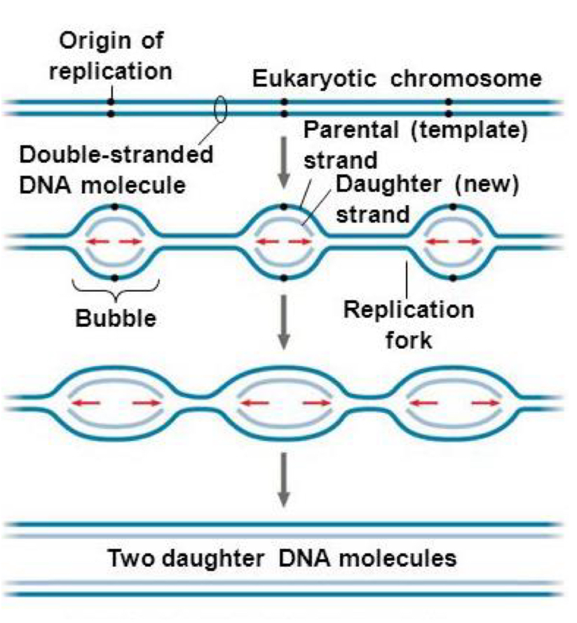
<ol><li><p>Replication begins at <strong>multiple origins of replication</strong>, where the two parental strands separate to form <strong>replication bubbles </strong></p></li><li><p>the <strong>bubbles expand laterally </strong>as DNA replication proceeds <strong>bidirectionally</strong> </p></li><li><p>eventually, the replication bubbles <strong>fuse</strong> and synthesis of the daughter strands is complete </p></li></ol><p></p>
29
New cards

Separation of parental DNA strands

  • separate base pairs of parental DNA molecules so that both DNA strands can act as templates for the synthesis of daughter DNA strands

  • origin of replication - where separation initially occurs

  • 3 proteins involved

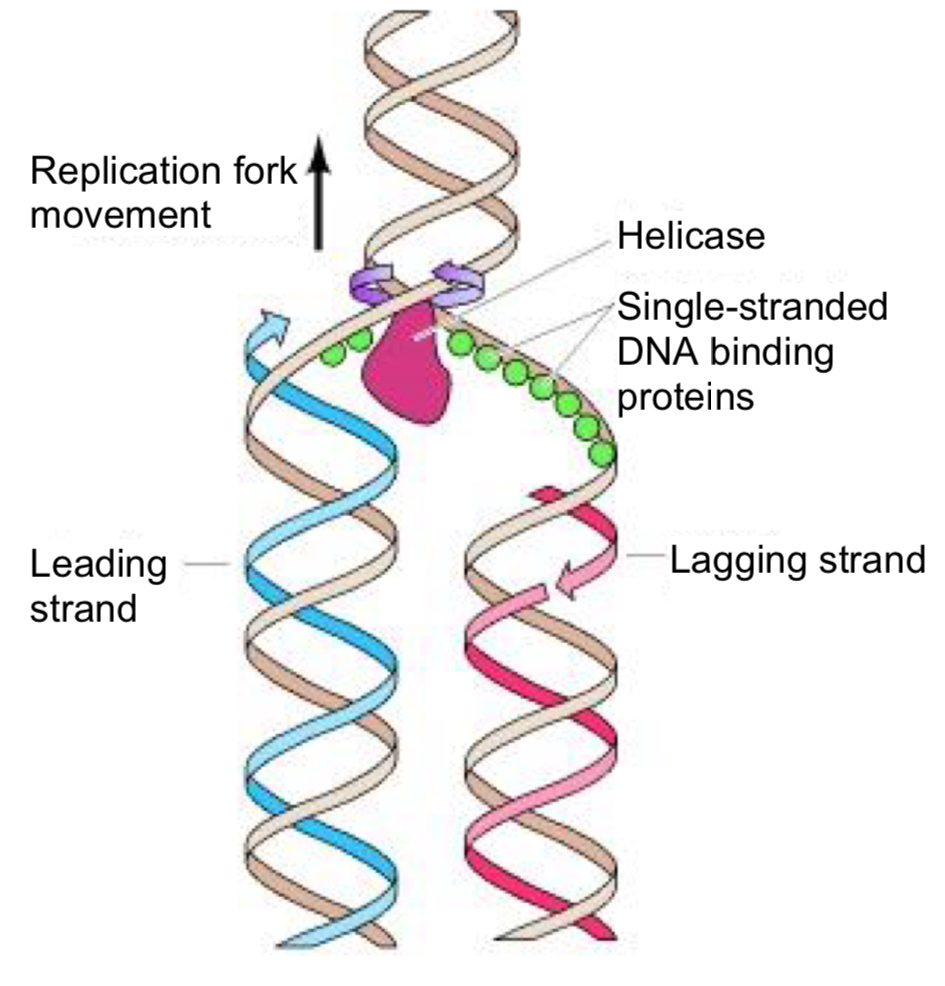
  1. helicases (unwind and separate)

  • after initiation, helicase bind to one strand of the DNA molecule

  • using ATP as energy source, helicases break the hydrogen bonds holding the two strands of DNA together = unwind the DNA double-helix and separates the parental DNA strands at the region of the replication fork

  • each of the 2 parental DNA strands serve as the template for the synthesis of a new DNA strand

  1. single-strand DNA binding proteins (ssb proteins)

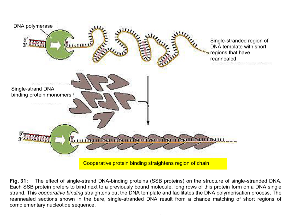
  • unwound single-stranded portion of the DNA double-helix is temporarily stabilised by the binding of single-strand DNA-binding proteins

  • prevents the single-stranded (ss) DNA from re-annealing to reform the duplex

  • keeps the two parental strands in the appropriate single-stranded condition to act as template

  • protects the ssDNA, which is very unstable, from being degraded

  1. topoisomerases

  • unwinding causes tighter twisting/supercoiling ahead of the replication fork, resulting in tension/torque

  • topoisomerases cleave a strand of the helix to create a transient single-stranded nick

  • relieves strain on the DNA molecule by allowing free rotation (swiveling) around the intact strand, and then reseal the broken strand
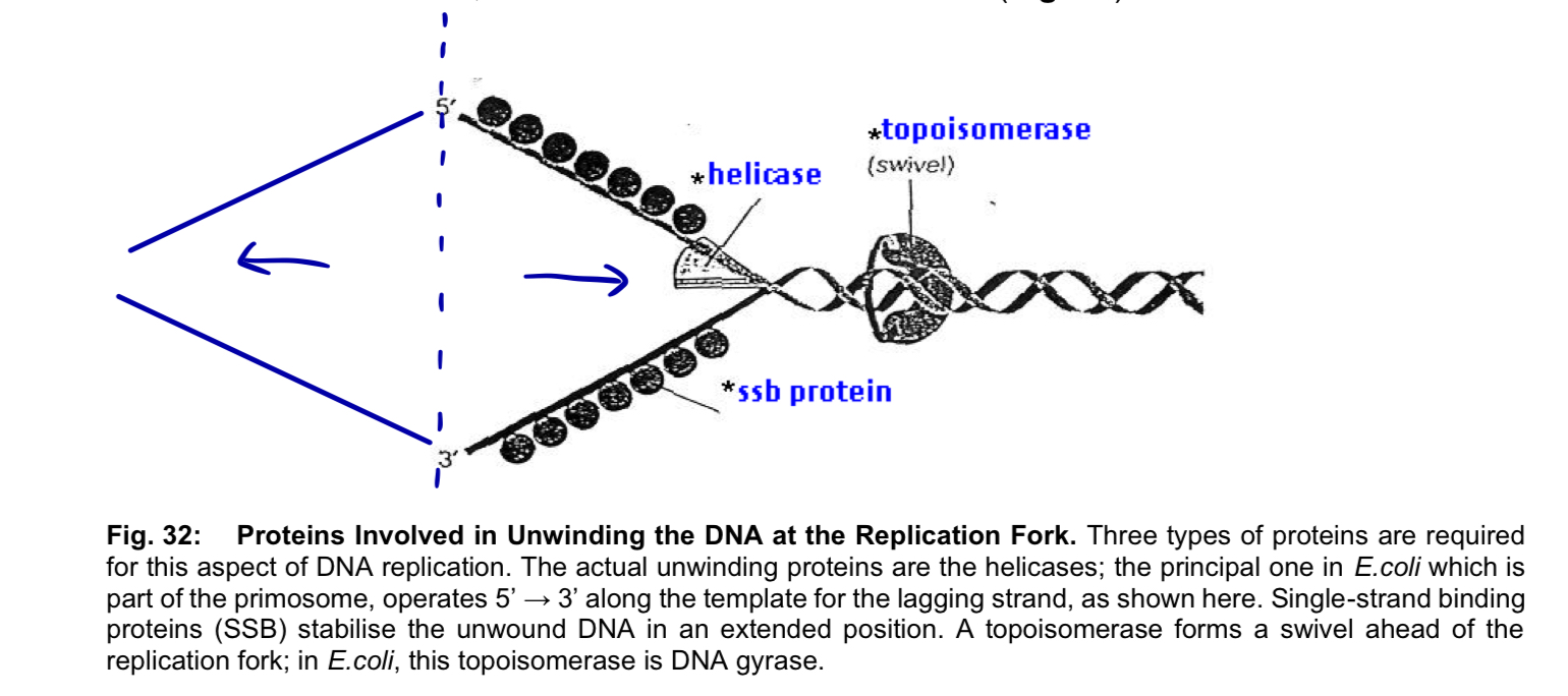
<ul><li><p>separate base pairs of parental DNA molecules so that both DNA strands can act as templates for the synthesis of daughter DNA strands</p></li><li><p>origin of replication - where separation initially occurs</p></li><li><p>3 proteins involved</p></li></ul><ol><li><p><mark data-color="purple" style="background-color: purple; color: inherit;">helicases</mark> (unwind and separate)</p></li></ol><ul><li><p>after initiation, helicase <strong>bind to one strand of the DNA molecule</strong></p></li><li><p>using <strong>ATP</strong> as energy source, helicases <strong>break the hydrogen bonds</strong> holding the two strands of DNA together = <strong>unwind</strong> the DNA double-helix and <strong>separates</strong> the parental DNA strands at the region of the replication fork</p></li><li><p>each of the 2 parental DNA strands serve as the <strong>template</strong> for the <strong>synthesis of a new DNA strand</strong></p></li></ul><ol start="2"><li><p><mark data-color="blue" style="background-color: blue; color: inherit;">single-strand DNA binding proteins</mark> (ssb proteins)</p></li></ol><ul><li><p>unwound single-stranded portion of the DNA double-helix is temporarily <strong>stabilised</strong> by the binding of <strong>single-strand DNA-binding proteins</strong></p></li><li><p><strong>prevents</strong> the single-stranded (ss) DNA from <strong>re-annealing</strong> to reform the duplex</p></li><li><p>keeps the two parental strands in the appropriate single-stranded condition to act as <strong>template</strong></p></li><li><p><strong>protects</strong> the <strong>ssDNA</strong>, which is very <strong>unstable</strong>, from being <strong>degraded</strong></p></li></ul><ol start="3"><li><p><mark data-color="red" style="background-color: red; color: inherit;">topoisomerases</mark></p></li></ol><ul><li><p>unwinding causes <strong>tighter twisting/supercoiling </strong>ahead of the replication fork, resulting in <strong>tension/torque </strong></p></li><li><p>topoisomerases <strong>cleave a strand </strong>of the helix to create a <strong>transient single-stranded nick </strong></p></li><li><p><strong>relieves strain</strong> on the DNA molecule by allowing <strong>free rotation (swiveling)</strong> around the intact strand, and then<strong> reseal the broken strand </strong></p></li></ul><p></p><p></p>
30
New cards

Helicase diagram

knowt flashcard image
31
New cards

SSB proteins

knowt flashcard image
32
New cards

Synthesis of RNA primers

limitation 1 of DNA polymerases: none of the DNA polymerases can initiate the synthesis of a DNA strand on its own. DNA synthesis cannot occur de novo.

solution: RNA primer to initiate synthesis of a DNA strand

  1. a portion of the parental DNA strand serves as template for making the RNA primer with the complementary base sequence

  2. an enzyme called primate joins the ribonucleotides to make the primer.

  • primer is about 10 nucleotides losing in eukaryotes