anatomy: knee & leg
1/151
There's no tags or description
Looks like no tags are added yet.
Name | Mastery | Learn | Test | Matching | Spaced | Call with Kai |
|---|
No analytics yet
Send a link to your students to track their progress
152 Terms
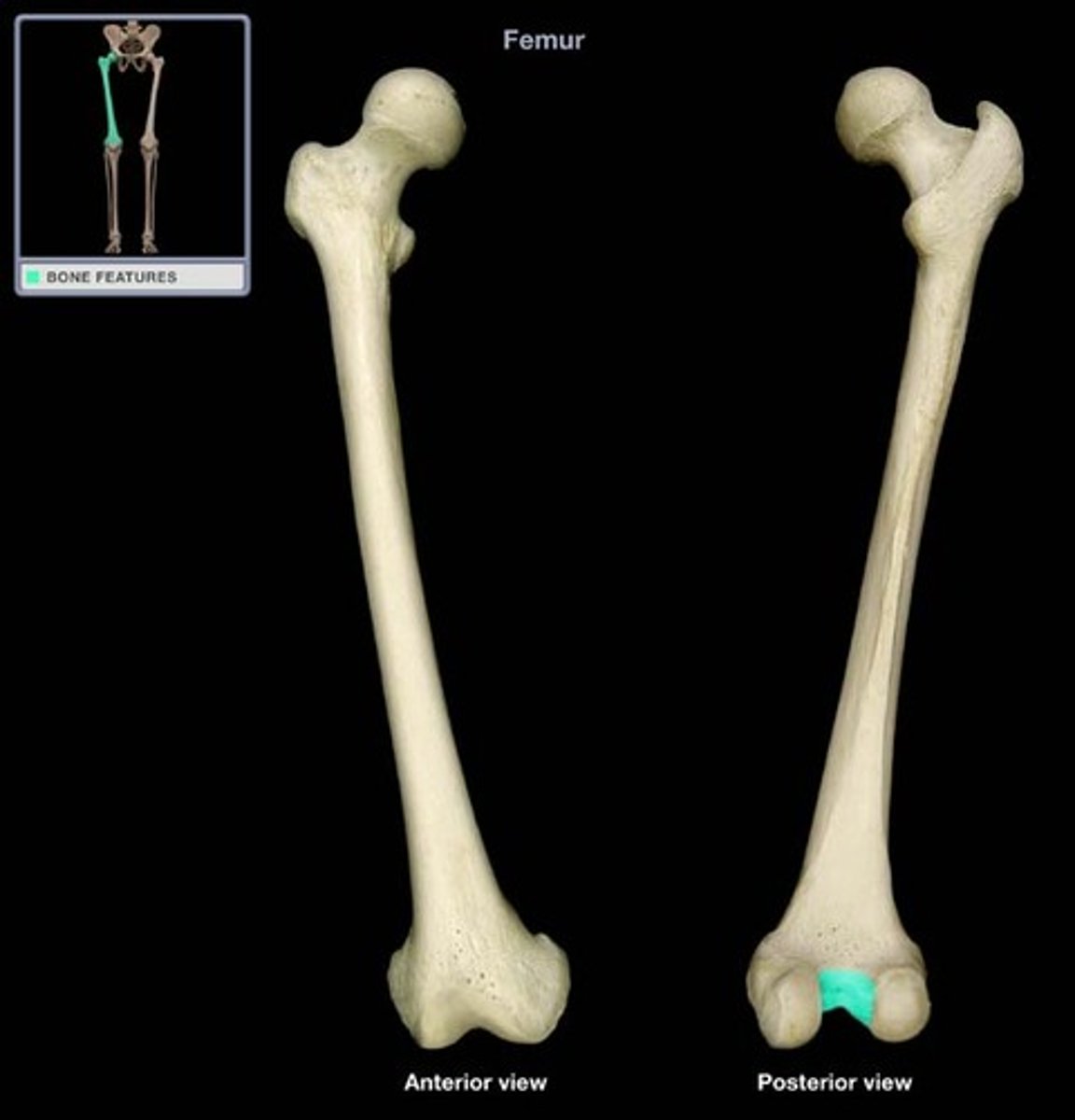
The femoral condyles are separated posteriorly and inferiorly by
intercondylar fossa
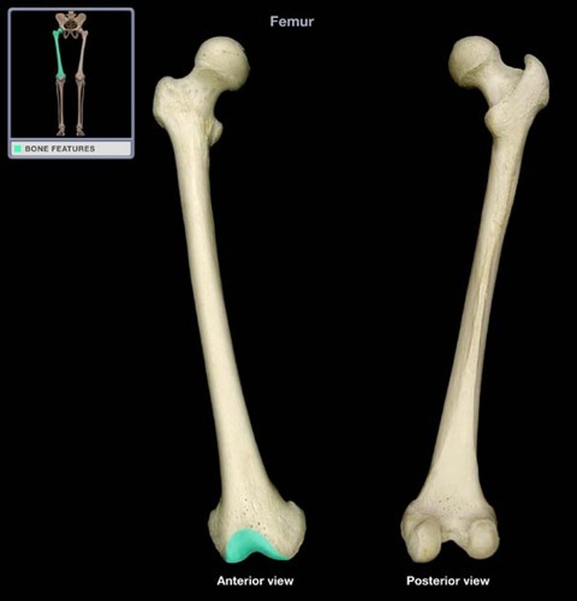
In the patellar surface, condyles merge ___, forming ___, which articulates with the patella.
anteriorly; depression
Three surfaces and borders of the tibia
medial, lateral/interosseous, posterior
The distal end of the tibia extends into the
medial malleolus
Tibia articulations (select all that apply)
- condyles of the femur superiorly
- talus inferiorly
- fibula via interosseous membrane
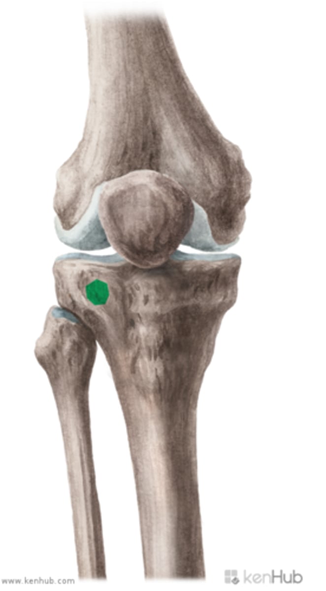
Gerdy's tubercle provides the distal attachment for the ___, adding ___ to the knee joint.
IT band; stability
Gerdy's tubercle
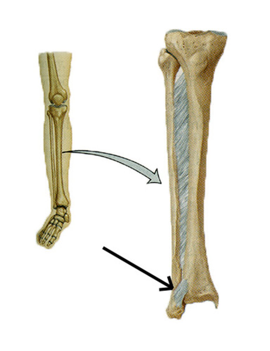
The distal end of the fibula becomes the
lateral malleolus
The fibula provides a distal attachment for ___ muscle and proximal attachment for ___ muscles.
1; 8
The patella is a
large triangular sesamoid bone
The patella is formed in the tendon of the ___ ____ birth.
quads; after
The subcutaneous anteiror surface of the patella is
convex
Function of patella
provides a mechanical advantage to the quads in extending the leg at the knee
The base of the patella is ___ and the apex is ___.
superior; inferior
The knee is a ___ joint.
hinge type synovial
Knee articulations (select all that apply)
- two femorotibial articulations (between femoral and tibial condyles)
- one patellofemoral articulation between the patella and femur
Patellofemoral joint type
planar/gliding joint
The stability of the knee joint depends on (select all that apply)
the strength and actions of the surrounding muscles and their tendons (distal fibers of vastus medialis & lateralis), the ligaments that connect the femur and tibia
The knee joint capsule is made up of an
external fibrous capsule and internal synovial membrane
5 major ligaments of the knee joint (select all that apply)
patellar, fibular collateral, tibial collateral, oblique popliteal, arcuate popliteal
The oblique popliteal ligament is formed from
semimembranosus
Medial and lateral patellar retinacula
aponeurotic expansions of the vastus medialis and lateralis and overlying deep fascia, making up the joint capsule of the knee on each side of patella
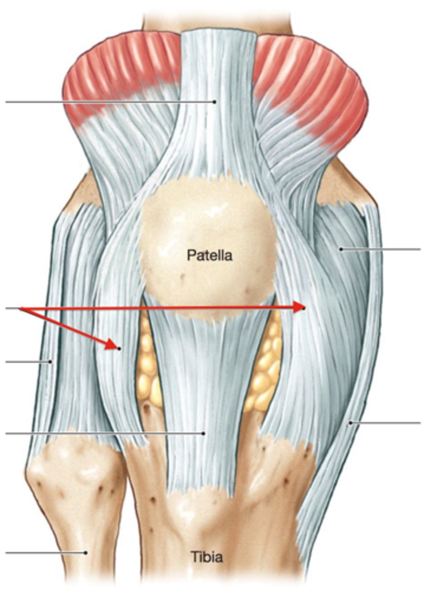
Function of the medial and lateral patellar retinacula
maintains alignment of the patella relative to the patellar articular surface of the femur
Collateral ligaments of the knee
FCL, TCL
The collateral ligaments of the knee are taut in ___, allowing ___ while standing.
full extension; stability
The collateral ligaments of the knee are slack in ___, allowing ___ of the knee.
flexion; rotation
The fibular collateral ligament is a ___ ligament.
cord-like extracapsular
Fibular collateral ligament
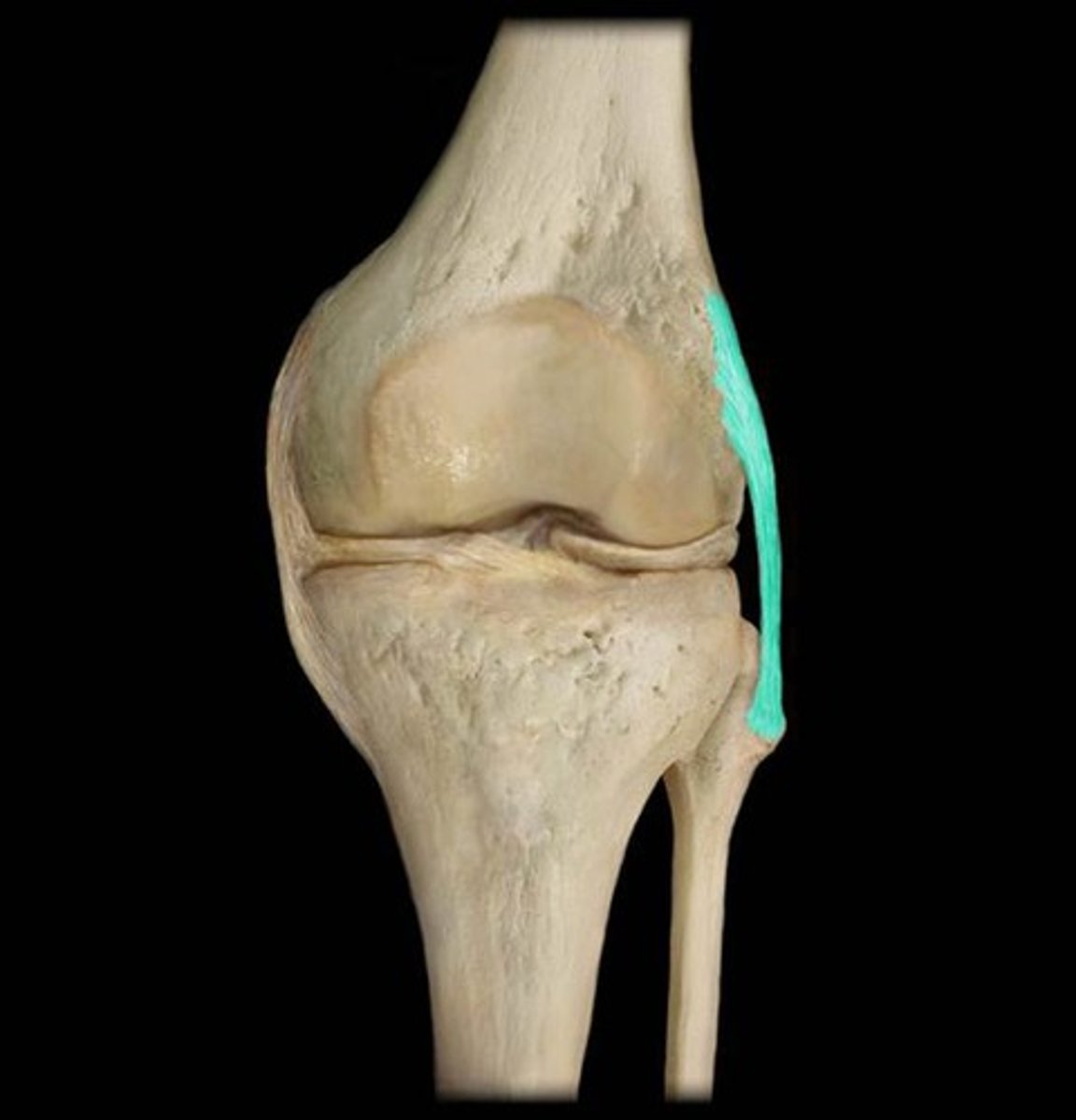
The fibular collateral ligament limits
genu varus
The tibial collateral ligament is a ___ ligament.
strong capsular
Tibial collateral ligament
connects medial femoral and medial tibial condyle
The tibial collateral ligament limits
genu valgus
The oblique popliteal ligament reinforces the joint capsule ___.
posteriorly
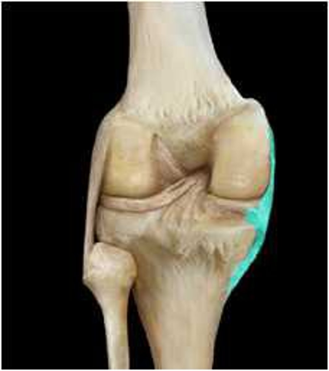
Which ligament(s) of the knee are extracapsular
fibular collateral ligament, oblique popliteal ligament, arcuate popliteal ligament,
Oblique popliteal ligament
runs posterior to medial tibial condyle and passes superolaterally towards lateral femoral condyle
The arcuate popliteal ligament strengthens the joint capsule
posterolaterally
The intra-articular knee has (select all that apply)
cruciate ligaments, menisci, tendon of the popliteus is also intra-articular during part of its course
The cruciate ligaments ___ within the joint capsule of the joint but ___ the synovial cavity.
crisscross; outside
During medial rotation of the tibia on the femur
cruciate ligaments wind around each other
During lateral rotation of the tibia on the femur
cruciate ligaments unwind
The ___ orientation of the cruciate ligaments causes one or both ligaments to ___ in every position
oblique; tense
Cruciate ligaments maintain contact with the ______ during ___ of the knee.
femur and tibial; flexion
The ACL is the ___ of the two cruciate ligaments.
weaker
The ACL extends
superiorly, posteriorly, laterally
The ACL limits (select all that apply)
- posterior rolling of femoral condyles on tibial plateau during flexion
- posterior displacement of femur on tibia
- hyperextension of the knee joint
Intercondylar fossa
depression between the condyles
Patellar surface
articulates with patella
Tibial plateau
- flat articular surface
- articulates with femur
- separated by intercondylar eminence
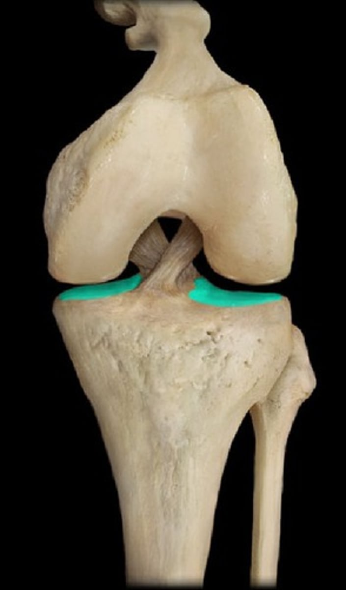
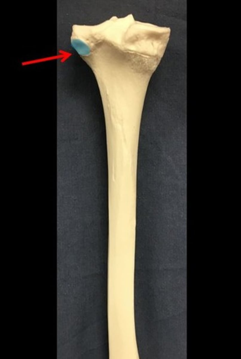
Fibular facet
on proximal tibia; articulation site for head of fibula
The ___ is important for the stability of the ankle joint.
fibula
The fibula attaches to the tibia via
tibiofibular syndesmosis
The fibula is a ___ joint.
syndesmotic
Which ligament(s) of the knee are capsular
tibial collateral ligament
Arcuate popliteal ligament
runs from posterior aspect of fibular head and superomedially to over the tendon of popliteus
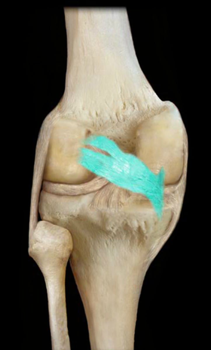
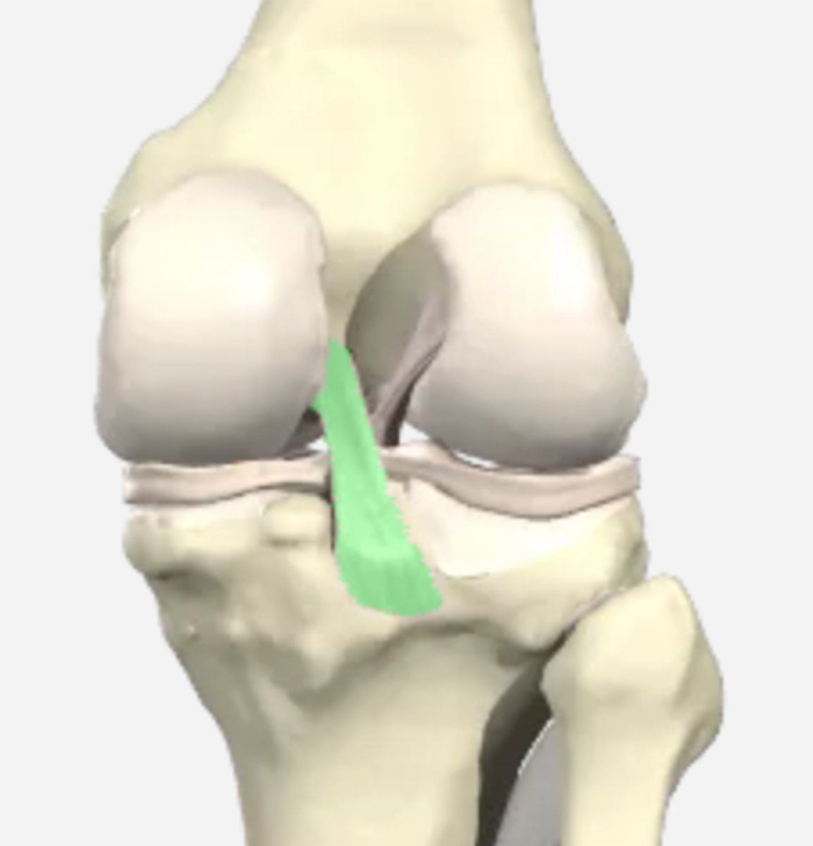
Anterior cruciate ligament
runs from anterior intercondylar tibia, posterior to attachment of medial meniscus to posterior part of medial side of lateral condyle of femur
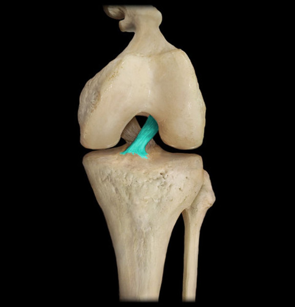
Posterior cruiciate ligament
runs from posterior intercondylar area and attaches at anterior lateral surface of medial condyle
Which muscle performs medial and lateral rotation of the tibia
hamstrings
ACL attachments
anterior intercondylar area of tibia, posterior to attachment of medial meniscus to posterior part of medial side of lateral condyle of femur
The PCL is the ___ of the two cruciate ligaments.
stronger
The PCL limits (select all that apply)
- anterior rolling of the femur on the tibial plateau during extension, converting it to spin
- anterior displacement of femur on tibia or posterior displacement of tibia on femur
- hyperflexion of knee joint
In the weight-bearing flexed knee, the ___ is the main stabilizing factor for the femur (e.g. when walking downhill).
PCL
When the joint is flexed at a right angle, the tibia cannot be pulled anteriorly because it is held by the ___.
ACL
The PCL runs
superior, anterior, medial
The ACL runs
superior, lateral, anterior, posterior
Function of the menisci
deepens the surface, shock absorption
The menisci are attached via
coronary ligaments
The menisci are made up of
fibrocartilage on the articular surface of the tibia
The medial meniscus is ___ and broader ___ than ___.
C-shaped; posteriorly; anteriorly
The lateral meniscus is ___ and ___ than the medial meniscus.
nearly circular; smaller
The lateral meniscus is ___ movable than the medial meniscus.
more freely
The lateral meniscus is an attachment site for the
popliteus
The lateral meniscus attaches to the ___ and ___ via the ___.
PCL; medial femoral condyle; posterior meniscofemoral ligament
Movements of the knee joint
flexion, extension, some rotation when flexed
Muscles that extend the knee
quadriceps femoris
Muscles that flex the knee
hamstrings, sartorius, gracillis, gastroch, popliteus
Muscles that medially rotate the knee
semitendinosus, semimembranosus, gracillis, sartorius, popliteus
Muscles that laterally rotate the knee
biceps femoris
Suprapatellar bursa
anterior bursae underneath the quad tendon
Prepatellar and infrapatellar bursae
between skin and patella/patellar tendon ot allow smooth movement
Deep infrapatellar bursa
underneath patellar tendon
Posterior bursae include
popliteus, gastrocnemius, semimembranosus, anserine
Popliteus bursa
deep to popliteus tendon
Gastrochnemius bursa
deep to medial and lateral heads of gastrochnemius
Semimembranosus bursa
deep to the semimembranosus
Anserine bursa
deep to the attachment of sartorius, gracilis, and semitendinosus
The plica is formed by a
sharp margin of synovial membrane
Inflammation of the plica can cause
synovitis and erosion of cartilage
The plica can lose ___ and become more fibrous with wear and inflammation.
elasticity
Superolateral border of the popliteal fossa
biceps femoris
superomedial border of popliteal fossa
semimembranosus
Inferolateral & inferomedial borders of the popliteal fossa
lateral and medial heads of the gastrocnemius (respectively)
Deep superior boundaries of the popliteal fossa
medial and lateral supracondylar lines of the femur
Deep inferior boundary of the popliteal fossa
soleal line of the tibia
Deep floor (anterior wall) of the popliteal fossa
popliteal surface of the femur superiorly and posterior aspect of the joint capsule of the knee joint centrally
Nerves of the popliteal fossa
- common fibular nerve
- inferior branches of posterior cutaneous nerve of thigh
- tibial nerve
- sural nerve
The common fibular nerve gives off
sural communicating branch
The inferior branches of the posterior cutaneous nerve of thigh supplies
skin over the popliteal fossa
Tibial nerve gives branches to
leg musculature and medial sural cutaneous nerve
The sural nerve is made up of
medial sural cutaneous nerve joined by sural communicating branch of fibular nerve
The popliteal artery ends at the ___ border of popliteus by dividing into the _______.
inferior; anterior and posterior tibial arteries