PSYC 301 - Split-Brain Patients
1/10
There's no tags or description
Looks like no tags are added yet.
Name | Mastery | Learn | Test | Matching | Spaced | Call with Kai |
|---|
No analytics yet
Send a link to your students to track their progress
11 Terms
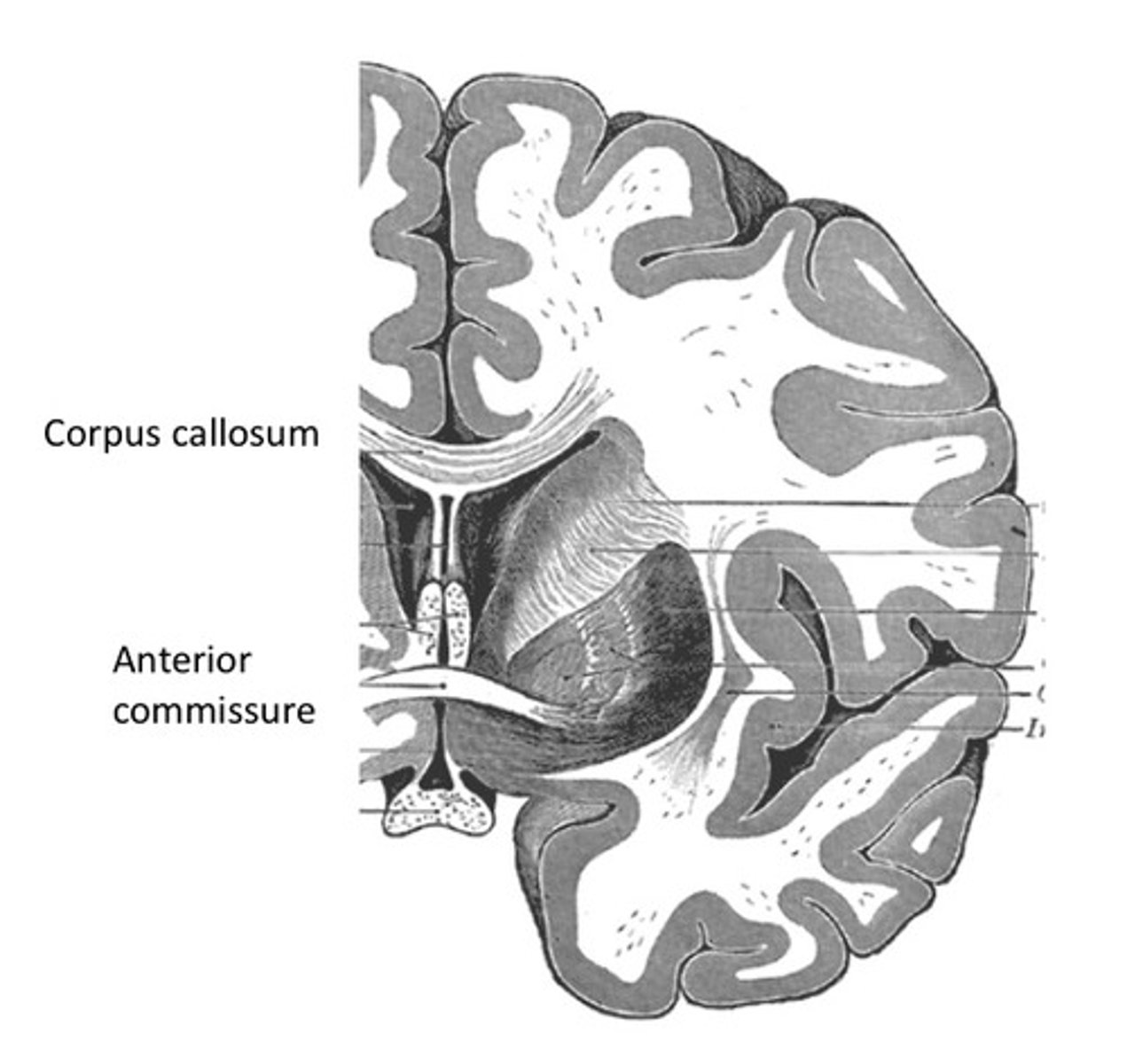
commissures (corpus callosum and anterior commissure) structure
commissures: left and right hemisphere connected by few white-matter tracts
largest is corpus callosum
brief history of split-brain research
1940s
- researchers show that epileptic discharge can spread from one hemisphere to another via corpus callosum in monkeys
- first limited callosotomy procedure in humans to control seizures in patients with intractable epilepsy (not successful)
1950s
- roger sperry study split-brain rats, cats, and monkeys to assess each hemisphere separately (noble prize)
1960s
- new, more complete commissurotomy performed → sucessfully controls epilepsy
- small # of people had corpus callosum surgically severed as a treatment for intractable epilepsy → hemisphere-hemisphere testing
- adaptation of animal techniques for us in human patients
visual field study
each hemisphere sees only the contralateral (opposite) visual field (some midline overlap)
by keeping eyes fixated, experimenters can present information to only one hemisphere
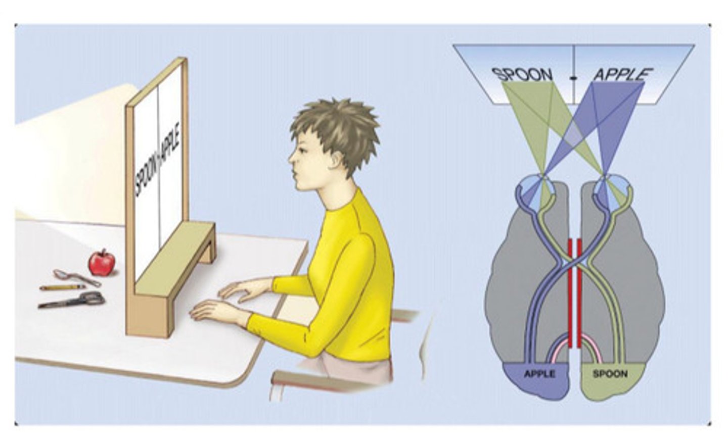
visual transfer following staged callosal section study
tasks present words or images to one side
cutting the posterior corpus callosum blocks sharing of sensory information but preserves semantic transfer, because meaning travels primarily through the anterior callosum
the hemispheres can still share meaning but not visual form, so the patient can describe the concept of the stimulus but cannot say the word
if it's fully split, patient verbally says they didn't see the stimulus (left hemisphere) but they are able to draw it (right hemisphere)
touch central processing
each hemisphere feels touch input from contralateral side of body → sensory input crosses at brainstem
left hand → right hemisphere (nonverbal)
right hand → left hemisphere (verbal)
if they don’t see it and don’t use cross-midline touch, researchers can present touch info into each hemisphere of split-brain patients
by placing an object in the left hand out of view, left-hand touch goes to the right hemisphere, which cannot access language, and can’t send information to the left hemisphere due to the severed corpus callosum → anomia (can’t name object)
subcortical and cortical functions
eye movements (saccades) are coordinated by subcortical structures, so both hemispheres can control them
some functions (e.g., judging line orientation, apparent motion) remain intact → rely on subcortical pathways, not the corpus callosum
*look at task performance in split-brain patients to make inferences about cortical vs. subcortical systems
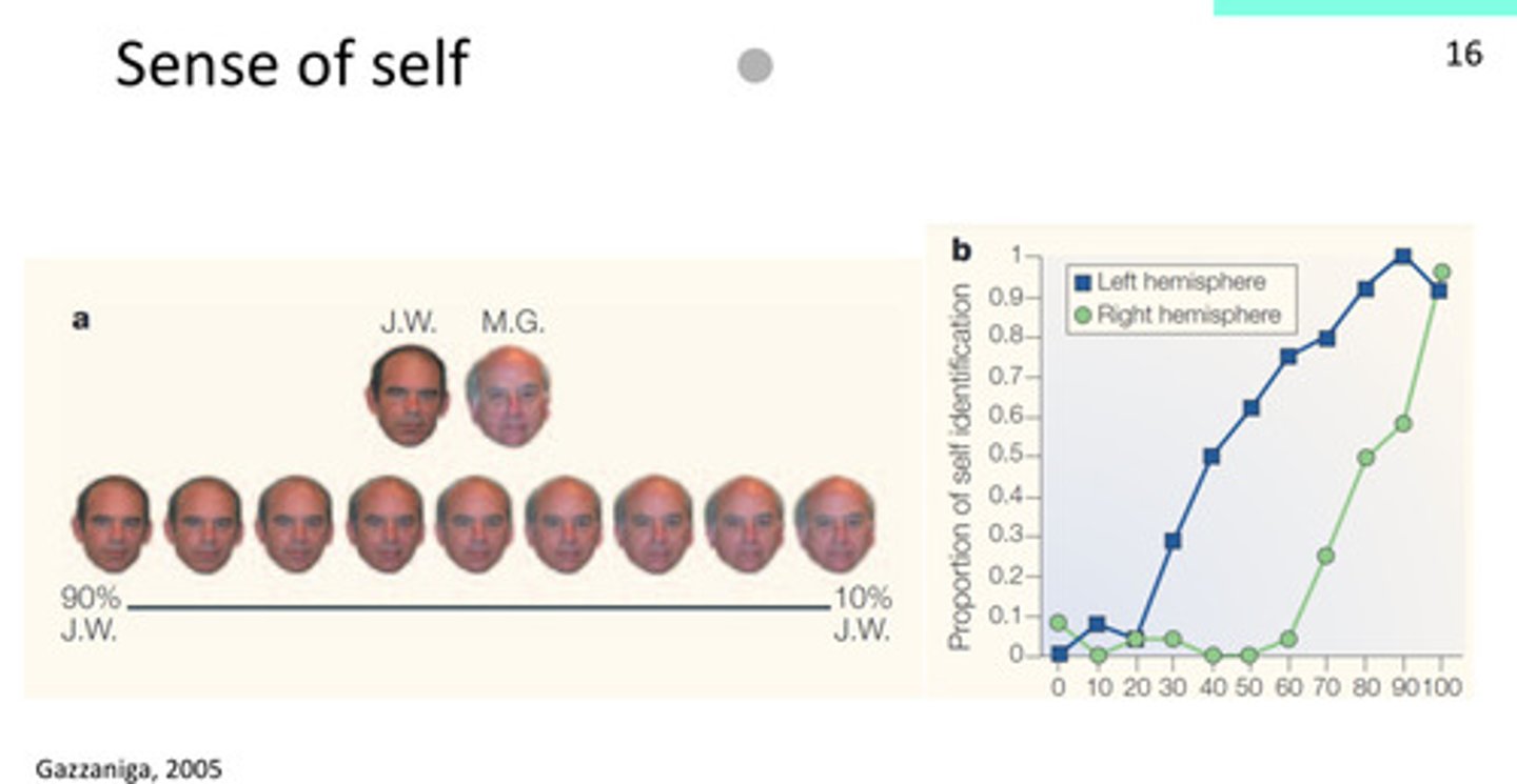
sense of self study
if it’s fully JW or MG, both hemispheres can say “yes that’s me” and “no that’s not me”
right hemisphere: conservative, doesn’t identify as self until it’s close to self
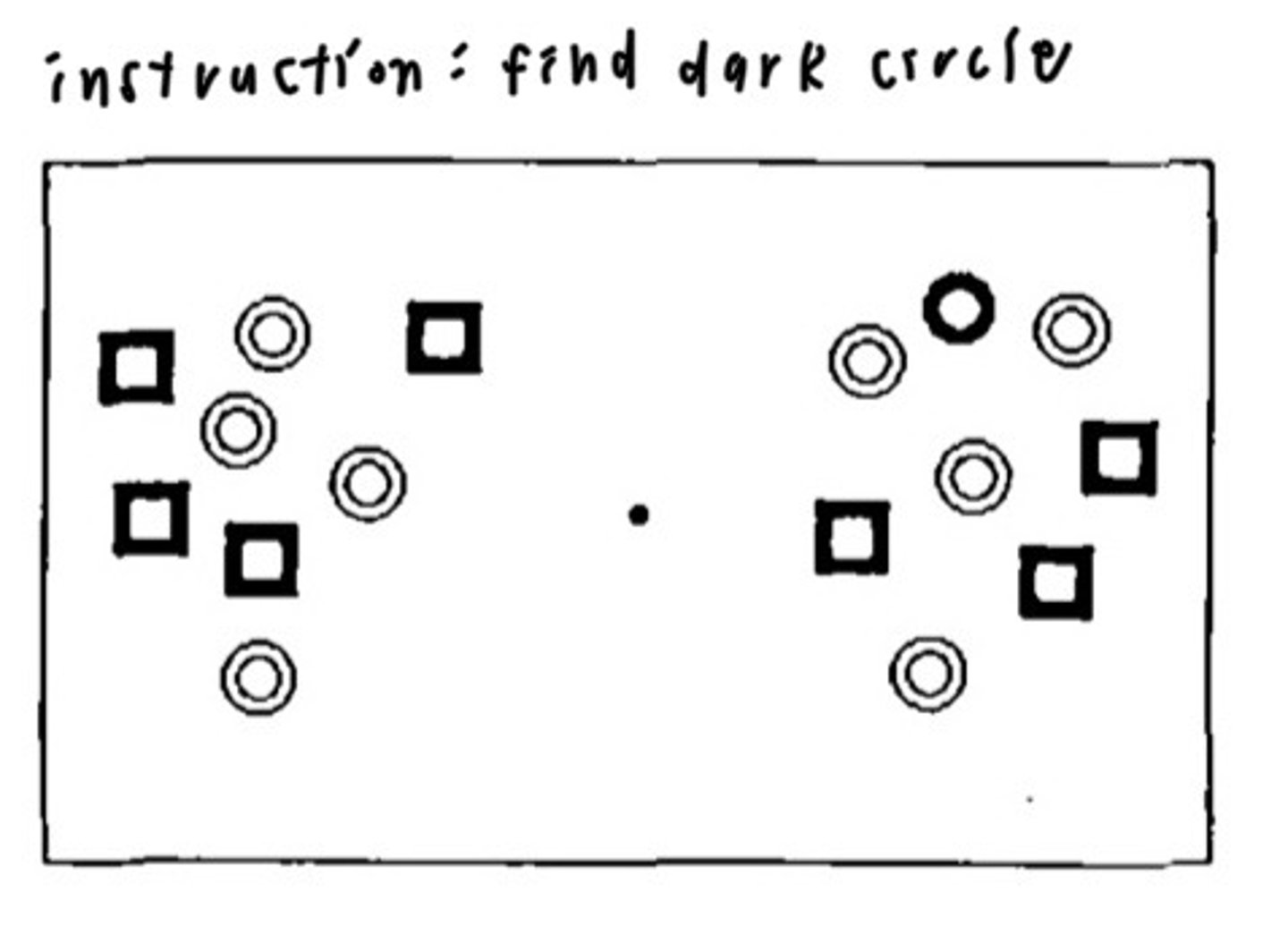
visual search study
in normal people: search time increases with number of distractors
in split-brain patients: each hemisphere searches its own side independently → search time rises half as much
left hemisphere uses more strategic search than right (e.g. immediately limit search to only dark items)
LH as interpreter study
when given mismatched cues (chicken claw + shovel), the left hemisphere invents a logical explanation for combined responses → shows interpreter (meaning-making) role of LH
coherence, fills in gaps

producing facial expressions study
LH produces voluntary facial expressions → right side of the face moves first
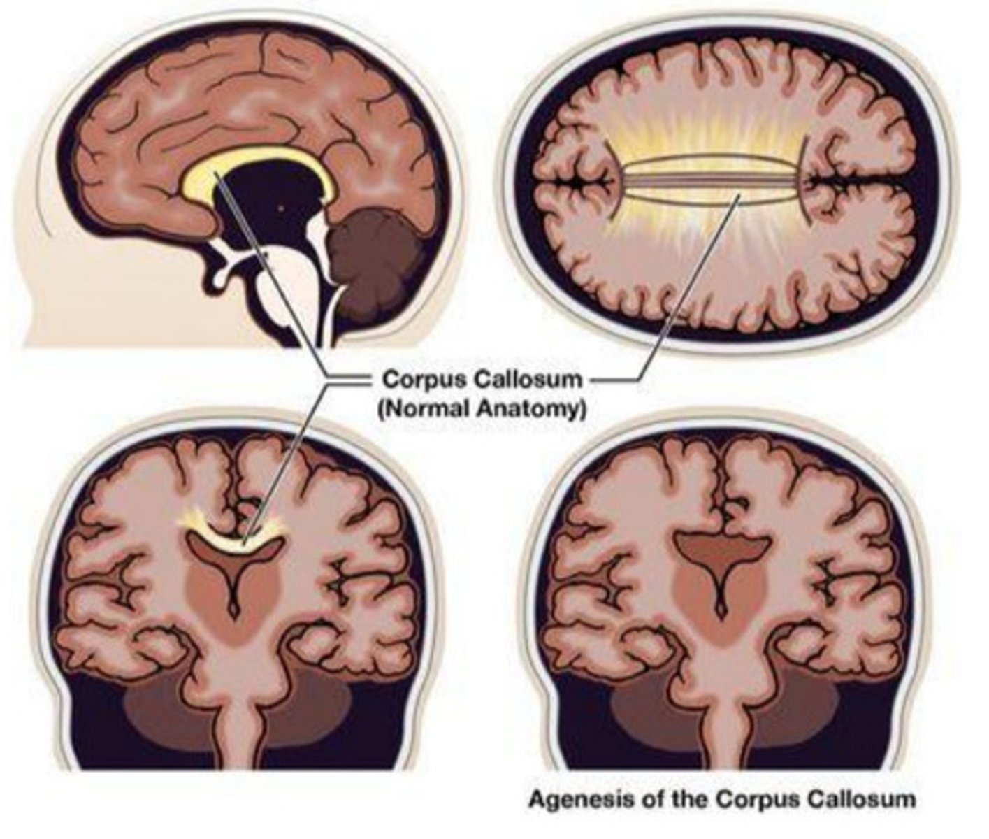
agenesis of corpus callosum
the congenital absence of corpus callosum which may be partial or complete
IQ and language normal unless other anomalies are present
minimal disconnection syndrome compared to adult split-brain cases → neuroplasticity allows alternative cross-hemispheric pathways (e.g., anterior commissure)
only tasks needing fast, complex interhemispheric integration show impairment